Practical Evaluation of Engraftment and Mixed Chimerism Using PCR Amplification of a Microsatellite in the Class II Eb Gene in Murine MHC-mismatched, Nonmyeloablative
Bone Marrow Transplantation
Sang-Young Roh1, Min Jung Park2, Hyunsil Park2, Seok Goo Cho2,3, So-Youn Min2, Jong Wook Lee3, Woo Sung Min3, Chun Choo Kim3,
Ho-Youn Kim2 and Hong Seok Chang4
1Division of Oncology, Department of Internal Medicine, 2Rhematism Research Center, Catholic Institute of Medical Sciences, 3Catholic Hematopoietic Stem Cell Transplantation Center, 4Department of Radiation Oncology,
The Catholic University of Korea, Seoul, Korea
Background: Although engraftment following murine allogeneic bone marrow transplantation (BMT) is most commonly confirmed by H2 typing using flow cytometry, recipient mice can be seriously injured during peripheral blood (PB) sampling. Therefore, we developed an alternative DNA-based assay that does not require the large volume of PB necessary for flow cytometry.
Methods: A minute volume of PB from the tail vein was used to evaluate the engraftment by PCR amplification of a microsatellite in the class II Eb gene. Dilution experiments were performed to evaluate the sensitivity of this assay for detecting donor cells in mixed cell populations compared with flow cy- tometry analysis.
Results: Early engraftment and mixed chimerism were confirmed, based on the length variation of the microsatellite in the class II Eb gene. The degree of donor chimerism in the donor-recipient cell mixture could be estimated semiquantitatively in a dilution experiment. The sensitivity of this assay by the naked eye approached 10% of the degree of donor chimerism.
Conclusion: PCR amplification of a microsatellite in the class II Eb gene can be a useful alternative to flow cytometry for evaluating early engraftment and mixed chimerism following murine nonmyel- oablative BMT. (Korean J Hematol 2007;42:91-97.)
Key Words: Mixed chimerism, Eb gene, Nonmyeloablative
91
Correspondence to:Seok Goo Cho, M.D., PhD
Catholic Hematopoietic Stem Cell Transplantation Center, St.
Marry's Hospital, The Catholic University of Korea 62, Yeouido-dong, Yeongdeungpo-gu, Seoul 150-713, Korea Tel: +82-2-3779-1130, Fax: +82-2-789-3132
E-mail: [email protected]
This work was supported by the Korea Science and Engineering Foundation (KOSEF) grant funded by the Korea government (MOST) (No. R11-2002-098-08004-0).
접수:2006년 12월 5일, 수정:2007년 3월 12일 승인:2007년 3월 15일
교신저자:조석구, 서울시 영등포구 여의도동 62
150-713, 가톨릭대학교 의과대학 여의도성모 병원 조혈모이식센터
Tel: 02-3779-1130, Fax: 02-789-3132 E-mail: [email protected]
INTRODUCTION
The mouse model of allogeneic bone marrow transplantation (BMT) has played an important role in extending our understanding of the biol- ogy of BMT.1) Recently, the intentional induction of a mixed chimerism following nonmyeloabla- tive conditioning has been developed in a mouse model as a strategy for reducing BMT-related toxicity, overcoming the major histocompatibility complex (MHC) barrier,2) and providing an im- munological platform for cell therapy,3) tumor vaccines,4) or organ transplantation.5) It is im- portant to confirm the state of mixed chimerism for integrating additive therapies into the post- BMT course without developing graft versus host disease (GVHD).4,6) In the murine model, H2 typing is most commonly performed using flow cytometry to evaluate early engraftment and the degree of donor chimerism.2) However, this meth- od is based on the presence or absence of hap- lotype-specific antigens and available antibodies.
Moreover, it is often limited by the need for ex- pensive equipment and by an inability to collect sufficient cells without animal sacrifice for serial assessment of BMT in mice.
We have established a unique murine model of a stable mixed chimerism in MHC-mismatched nonmyeloablative BMT2) and investigated its po- tential for clinical application.7-10) While per- forming that research, we sampled peripheral blood (PB) from the retro-orbital venous plexus to obtain sufficient uncoagulated PB for flow cy- tometry and evaluated the allogeneic mixed chi- meric state. Unfortunately, the recipient mice were sometimes injured seriously or died during PB sampling. These unexpected injuries had an adverse effect on our murine BMT-related exper- iments. Therefore, we sought to develop an alter- native, DNA-based assay that could detect en- graftment/mixed chimerism using a minute a- mount of PB without causing serious sampling injuries. Since a highly polymorphic microsatel-
lite in the class II Eb gene can be used to trace the MHC in mice,11) we used microsatellite poly- morphism in the class II Eb gene to evaluate en- graftment/mixed chimerism following mouse BMT.
MATERIALS AND METHODS 1. Mice
Female BALB/c (8 weeks old, H-2kd) and C57BL/6 (6 to 7 weeks old, H-2kb) mice were purchased from Samtago (Gwangju, Korea). The mice were maintained under specific pathogen- free conditions in an animal facility with con- trolled humidity (55±5%), light (12/12 h, light/
dark), and temperature (22±1oC). The air in the animal facility was passed through a high-effi- ciency particle arrestance (HEPA) filter system designed to exclude bacteria and viruses. The an- imals were allowed mouse chow and tap water ad libitum. The Animal Care and Use Committee of the Catholic University of Korea approved the protocols used in this study.
2. Preparation of mixed chimera
Allogeneic mixed chimeras were prepared us- ing established procedures.2) In brief, one day be- fore BMT, the recipient mice (8-week-old BALB/
c, H-2kd) were injected intraperitoneally with 200 μL of phosphate-buffered saline (PBS) contain- ing 40μL of reconstituted anti-asialoGM1 (anti- ASGM1; Wako Chemicals, Osaka, Japan). Reci- pient mice were exposed to a single x-ray dose of 500cGy from a Mevatron MXE-2 (Siemens, NY) with a focus-to-skin distance of 100cm and a rate of 200cGy/min. Donor (6- to 7-week-old C57BL/6, H-2kb) bone marrow cells were col- lected into Cedarlane cytotoxicity medium (RPMI 1,640 medium supplemented with 25mm HEPES buffer and 0.3% bovine serum albumin) by flush- ing the shafts of the femurs and tibias of C57BL/
6 mice. Resuspended bone marrow cells were de- pleted of T cells by incubation with anti-Thy-1.2 microbeads (Miltenyi Biotec, Auburn, CA) ac-
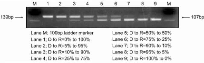
Fig. 1. Assessing the degree of mixed chimerism in an arbi- trary cell mixture using PCR amplification of a microsatellite in the class II Eb gene of the murine MHC. Donor (D, C57BL/6, H-2kb mice) and recipient (R, BALB/c, H-2kd) splenocytes were mixed in vitro in various proportions (D to R=0 to 100, 5 to 95, 10 to 90, 25 to 75, 50 to 50, 75 to 25, 90 to 10, 95 to 5, and 100 to 0) in dilution experiments. PCR amplification of the class II Eb gene and flow cytometry analysis were performed using the cell mixtures. The DNA fragments amplified from the donor and recipient were 107 and 139bp, respectively. The lanes show the results for artificial mixtures of donor and recipi- ent splenocytes.
cording to the manufacturer’s instructions. With- in 6 h after irradiation, 2×10⁷ T cell-depleted bone marrow cells, in a final volume of 0.2mL of PBS, were infused into recipient mice.
3. Flow cytometry analysis
Early engraftment and chimerism were de- termined using the relative ratio of peripheral blood lymphocytes (PBLs) expressing recipient MHC class I molecules to PBLs expressing donor molecules on day 21. Briefly, peripheral blood was collected from the retro-orbital vein using heparinized capillary tubes. Donor (H-2Kb) and recipient (H-2Kd) cells were distinguished during lymphoid gating by staining with fluorescein iso- thiocyanate-labeled anti-H-2Kb and phycoerythrin- labeled anti-H-2Kd antibodies (PharMingen, San Diego, CA), respectively. Samples of peripheral blood were stained with FITC-anti-H-2kb and PE-anti-H-2kd (PharMingen) or Ig isotype con- trols (PharMingen), followed by treatment with an RBC lysis kit (Optylase B, Immunotech- Coulter, Marseille, France). Stained cells were analyzed on a FACSCalibur flow cytometer using CellQuest software (both from Becton Dickinson, Mountain View, CA). The percentage of donor- derived cells was calculated by dividing the num- ber of donor cells by the total number of donor and recipient cells that showed positive stain- ing.2)
4. PCR amplification of a microsatellite in the class II Eb gene
We collected 20μL of peripheral blood from a tail vein 3 weeks after MHC-mismatched, T-cell- depleted, nonmyeloablative bone marrow trans- plantation according to our protocol.2) Genomic DNA was prepared using an extraction kit (Dr.
Gentle; Takara Bio, Otsu, Japan) according to the manufacturer’s instructions. A highly polymor- phic microsatellite containing tandem repeats (TRs) of two tetranucleotide units, TGGA and GGCA, located at the 3’ end of the second intron in the class II Eb gene was amplified. Genomic
DNA (50ng from a mixed chimeric mouse) was amplified using primers specific for the micro- satellite in the class II Eb gene, 5’-CGACTG- TAGAACCTTAGCCTG-3’ and 5’-TGGAGCTG- TCCTCCTTGTAG-3’, and yielded 107-bp (donor, C57BL/6, H-2b) and 139-bp (host, BALB/c, H-2d) fragments of the Eb TR allele.12) PCR amplifica- tion was performed under the following condi- tions: an initial denaturation at 95oC for 3 min, followed by 35 cycles of 94oC for 30 s, 65oC for 30 s, and 72oC for 30 s, and a final incubation at 72oC for 7 min in a thermal cycler (GeneAmp 9700, Perkin-Elmer, Foster City, CA, USA). All of the amplified DNA samples were electro- phoresed in a 2% agarose gel stained with ethi- dium bromide and photographed (Fig. 1).
5. Dilution experiment for the sensitivity of PCR amplification of a microsatellite in the class II Eb gene in the allogeneic mixed chimeric state
Spleen cells from both recipient (BALB/c, H-2kd) and donor (C57BL/6, H-2kb) mice were removed using ACK lysis buffer, washed, and re- suspended in complete culture medium (RPMI 1,640 medium supplemented with 10% heat-in- activated fetal calf serum, 1mM sodium pyruvate,
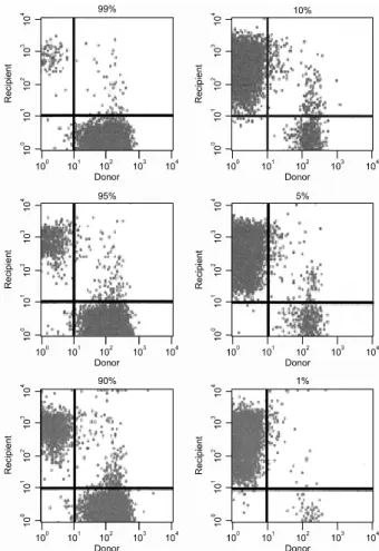
Fig. 2. Assessing the degree of mixed chimerism in arbi- trary cell mixtures using flow cytometry. Donor (C57BL/6, H-2kb mice) and recipient (BALB/c, H-2kd) splenocytes were mixed in vitro in various proportions (D to R=99 to 1, 95 to 5, 90 to 10, 10 to 90, 5 to 95, and 1 to 99) for dilution experiments. Donor (H-2Kb) and recipient (H-2Kd) cells were distinguished during lymphoid gating by stain- ing with fluorescein isothiocyanate-labeled anti-H-2Kb and phycoerythrin-labeled anti-H-2Kd antibodies (PharMingen, San Diego, CA), respectively. Stained cells were analyzed using CellQuest software on a FACSCalibur flow cytometer (both from Becton Dickinson, Mountain View, CA). The per- centage of donor-derived cells was calculated by dividing the number of donor cells by the total net number of donor plus recipient cells that showed positive staining.
5×10⁻⁵M 2-ME, 20mM HEPES, 100U/mL pen- icillin, and 100μg/mL streptomycin). Donor splenocytes were mixed with recipient spleno- cytes in various proportions for the dilution ex- periments. PCR amplification of the class II Eb gene and flow cytometry analysis were performed using the cell mixtures. The sensitivity of each method was assessed.
RESULTS
1. Sensitivity of PCR amplification of a micro- satellite in the class II Eb gene
Donor and recipient DNA could be distin- guished simply by PCR amplification of a micro- satellite in the class II Eb gene, even in the allo- geneic mixed chimeric state. A highly poly- morphic microsatellite made up of two tetranu- cleotide blocks, (TGGA)x and (GGCA)y, is lo- cated at the 3’ end of the second intron in the Eb gene. The number of repeats differs according to the H-2 haplotype.12) We investigated whether the length polymorphism of this satellite was ap- plicable to the characterization of H-2 haplotypes and could be used to determine engraftment and the degree of donor chimerism in murine MHC- mismatched nonmyeloablative BMT. As donors and recipients possessed alleles that differed by 32 bases, it was possible to analyze this trans- plant with ethidium bromide staining without the use of radioactivity. Dilution experiments in- dicated that residual donor or recipient DNA ex- tracted from a cell mixture could be detected at the 5 to 10% level (Fig. 1). By contrast, flow cy- tometry could detect donor or recipient cells at the 1% level (Fig. 2). Although the DNA-based assay using PCR amplification of a microsatellite in the class II Eb gene was slightly less sensitive than flow cytometry analysis at detecting engraft- ment/mixed chimerism, these observations in- dicated that it was equally useful for detecting engraftment and a donor-dominant mixed chi- meric state (>90% donor chimerism) in murine MHC-mismatched nonmyeloablative BMT.
2. Assessing engraftment/mixed chimerism us- ing a minute amount of peripheral blood from allogeneic mixed chimeric mice
Allogeneic BMT was performed according to our own protocol with the intentional induction of a mixed chimerism. Engraftment/mixed chi- merism was evaluated 3 weeks post-transplant
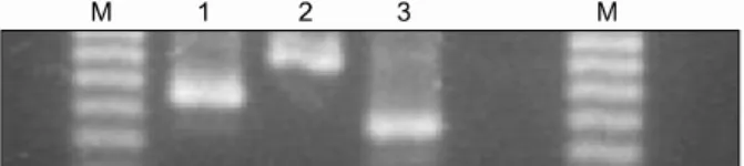
Fig. 3. Donor chimerism in peripheral blood of an alloge- neic mixed chimera following MHC-mismatched non- myeloablative BMT. Engraftment/mixed chimerism at 3 weeks post-BMT was evaluated using PCR amplification of a microsatellite in the class II Eb gene. Allogeneic mixed chimeric mice (lanes 1, 2, and 4) showed both the 107-bp (donor, C57BL/6, H-2b) and 139-bp (host, BALB/c, H-2d) fragments, whereas non-transplanted recipient mice as a negative control (lane 3) showed only the 107-bp (host, BALB/c, H-2d) fragment. The relative ratios of do- nor-derived cells in the PB from those mice were 76, 42, 0, and 89% of all lymphocytes, respectively. The DNA-based assay developed in this study roughly corre- lated with the results of the flow cytometry assay.
using PCR amplification of a microsatellite in the class II Eb gene with a minute PB sample.
The PCR products obtained from the PB of allo- geneic mixed chimeric mice (lanes 1, 2, 4) showed both 107-bp (donor, C57BL/6, H-2b) and 139-bp (host, BALB/c, H-2d) fragments, whereas the product from non-transplanted recipient mice used as a negative control (lane 3) showed only the 139-bp (host, BALB/c, H-2d) fragment on ethidium bromide-stained agarose gels (Fig.
3). To validate the results of the DNA-based as- say, we sacrificed the same mice and evaluated the degree of donor chimerism using a flow cy- tometry assay. The relative ratios of cells of do- nor origin in the respective PB samples were 76, 42, 0, and 89% of all lymphocytes. These ob- servations suggest that our DNA-based assay us- ing PCR amplification of a microsatellite in the class II Eb gene is a practical alternative to flow cytometry for assessing engraftment/mixed chi- merism following murine nonmyeloablative BMT.
DISCUSSION
The presence of engraftment/mixed chimerism or leukemic relapse in human BMT13,14) is usu- ally assessed using PCR of Y-chromosome-specif- ic sequences, short tandem repeat sequences (STRs), or microsatellites,15-17) whereas DNA-bas- ed assays have been used less frequently than flow cytometry analysis in mouse BMT. Conse- quently, there are few papers on DNA-based as- says in mice. O’Neil et al reported that the changes in donor chimerism following mouse BMT could be monitored using PCR amplifica- tion of 12 mouse STR markers.18) However, the resolution in their analysis of mixed chimerism using selected STR markers was poor, and there was no clear difference in allele size between do- nor and recipient.
Of various molecular markers in mice, micro- satellites in the class II Eb gene have an extra- ordinarily high level of polymorphism. Seven length variants are known in the 12 standard haplotypes of the H2 Eb gene (b, k, r, v, f, s, u, z, d, q, j, and p).11) We used the polymorphism of a microsatellite in the class II Eb gene of the murine major histocompatibility complex (MHC) to evaluate mixed chimerism/engraftment follow- ing mouse BMT, because it had not been used previously. We found that mixed chimerism was obviously detectable at a level approaching 10%
with this method.
Although PCR amplification of a microsatellite in the class II Eb gene was less sensitive than flow cytometry, it has several important advan- tages. First, this method can clearly distinguish donor- and recipient-derived cellular portions in as little as 10 to 20μL of PB, and it does not require the use of polyacrylamide gels, radioiso- topes, or expensive equipment. Second, sampling a small volume of blood from the tail vein does not have an adverse effect on the clinical out- come of murine allogeneic BMT, making it possi- ble to obtain serial samples from transplanted
Fig. 4. Assessing NOD/Scid mice using PCR amplification of a microsatellite in the class II Eb gene of the murine MHC. The presence of NOD/Scid-derived cells can be de- tected using PCR amplification of a microsatellite in the class II Eb gene of the murine MHC, although NOD/Scid mice do not express the H-2 haplotype antigen. The ampli- fied DNA fragments of the C57BL/6 (H-2b), BALB/c (H-2d), and NOD/Scid mice were 107, 139, and 80bp, respec- tively.
mice. Third, the semiquantitative assessment of engraftment or donor-dominant chimerism using this method makes it possible to conduct sub- sequent experiments, such as donor lymphocyte infusion (DLI)3,6) or tumor vaccine.4) Fourth, this method can be used in murine BMT experiments with NOD/Scid mice, which express few MHC antigens (Fig. 4).
We have provided evidence that PCR amplifi- cation of a microsatellite polymorphism of the mouse class II Eb gene is a practical method for evaluating mixed chimerism/engraftment follow- ing murine BMT in a single PCR amplification.
When cell therapy or the manipulation of trans- planted mice following engraftment is antici- pated, this method will prove convenient in the early post-transplant period.
요 약
배경: 생쥐에서 동종골수이식 후 공여자 조혈모 의 생착 확인을 위하여 흐름세포측정법(flow cy- tometry)을 이용한 H2 판별이 가장 흔히 사용되지 만, 말초 혈액을 채취하는 과정에서 이식받은 생쥐 에게 치명적인 손상을 줄 수 있다. 저자들은 생착 여부를 판별하는 방법으로 많은 양의 말초 혈액을 필요로 하는 흐름세포 측정법을 대체할 수 있는 DNA 관련 분석법을 개발하고자 하였다.
방법: 생쥐의 꼬리 정맥에서 채취한 극소량의 말 초 혈액에서 class II Eb 유전자 microsatellite의 중 합효소연쇄반응(PCR)을 이용한 증폭을 통하여 공
여자 조혈모세포의 생착 여부를 평가하였다. 이와 함께 공여자와 받는이 생쥐의 혼합 세포주에서 공 여자 세포의 검출에 대하여 저자들이 개발한 분석 법과 흐름세포측정법의 민감도를 비교하기 위하여 희석실험(dilution experiments)을 실시하였다.
결과: Class II Eb 유전자의 microsatellite 변이에 대한 PCR 측정법을 이용하여 조기 생착 및 혼합키 메리즘이 확인 가능하였다. 공여자와 받는이 생쥐 의 혼합세포주에서 공여자의 키메리즘 정도는 희석 실험을 통하여 반정량적으로 추정 가능하였다. 이 분석법은 공여자 키메리즘이 10% 이상을 차지하는 경우 키메리즘을 육안적으로도 식별할 수 있는 민 감도를 보였다.
결론: 생쥐에서 비골수억제성(non-myeloablative) 골수이식 후 조기 생착 및 혼합 키메리즘의 평가 방 법으로 class II Eb 유전자의 microsatellite에 대한 PCR 검사법은 흐름세포측정법을 대체하여 유용하 게 이용될 수 있다.
REFERENCES
1) Exner BG, Domenick MA, Bergheim M, Mueller YM, Ildstad ST. Clinical applications of mixed chimerism. Ann N Y Acad Sci 1999;872:377-85; dis- cussion 385-6.
2) Cho SG, Shuto Y, Soda Y, et al. Anti-NK cell treat- ment induces stable mixed chimerism in MHC-mis- matched, T cell-depleted, nonmyeloablative bone mar- row transplantation. Exp Hematol 2004;32:1246-54.
3) Mapara MY, Kim YM, Wang SP, Bronson R, Sachs DH, Sykes M. Donor lymphocyte infusions mediate superior graft-versus-leukemia effects in mixed com- pared to fully allogeneic chimeras: a critical role for host antigen-presenting cells. Blood 2002;100:1903-9.
4) Luznik L, Slansky JE, Jalla S, et al. Successful ther- apy of metastatic cancer using tumor vaccines in mixed allogeneic bone marrow chimeras. Blood 2003;101:1645-52.
5) Exner BG, Acholonu IN, Bergheim M, Mueller YM, Ildstand ST. Mixed allogeneic chimerism to induce tolerance to solid organ and cellular grafts. Acta Haematol 1999;101:78-81.
6) Mapara MY, Kim YM, Marx J, Sykes M. Donor lym- phocyte infusion-mediated graft-versus-leukemia ef- fects in mixed chimeras established with a non- myeloablative conditioning regimen: extinction of graft-versus-leukemia effects after conversion to full
donor chimerism. Transplantation 2003;76:297-305.
7) Sykes M, Preffer F, McAfee S, et al. Mixed lympho- haemopoietic chimerism and graft-versus-lymphoma effects after non-myeloablative therapy and HLA- mismatched bone-marrow transplantation. Lancet 1999;353:1755-9.
8) Li H, Kaufman CL, Boggs SS, Johnson PC, Patrene KD, Ildstad ST. Mixed allogeneic chimerism induced by a sublethal approach prevents autoimmune dia- betes and reverses insulitis in nonobese diabetic (NOD) mice. J Immunol 1996;156:380-8.
9) Nikolic B, Takeuchi Y, Leykin I, Fudaba Y, Smith RN, Sykes M. Mixed hematopoietic chimerism allows cure of autoimmune diabetes through allogeneic tol- erance and reversal of autoimmunity. Diabetes 2004;
53:376-83.
10) Cho SG, Min SY, Park MJ, et al. Immunoregulatory effects of allogeneic mixed chimerism induced by nonmyeloablative bone marrow transplantation on chronic inflammatory arthritis and autoimmunity in interleukin-1 receptor antagonist-deficient mice. Arth- ritis Rheum 2006;54:1878-87.
11) Saha BK. Typing of murine major histocompatibility complex with a microsatellite in the class II Eb gene.
J Immunol Methods 1996;194:77-83.
12) Saha BK, Shields JJ, Miller RD, Hansen TH, Shreffler DC. A highly polymorphic microsatellite in the class II Eb gene allows tracing of major histo- compatibility complex evolution in mouse. Proc Natl
Acad Sci U S A 1993;90:5312-6.
13) Gardiner N, Lawler M, O’Riordan J, DeArce M, Humphries P, McCann SR. Persistent donor chi- maerism is consistent with disease-free survival fol- lowing BMT for chronic myeloid leukaemia. Bone Marrow Transplant 1997;20:235-41.
14) Gardiner N, Lawler M, O’Riordan J, De’Arce M, McCann SR. Donor chimaerism is a strong indicator of disease free survival following bone marrow trans- plantation for chronic myeloid leukaemia. Leukemia 1997;11(Suppl 3):512-5.
15) Lawler M, Humphries P, McCann SR. Evaluation of mixed chimerism by in vitro amplification of dinu- cleotide repeat sequences using the polymerase chain reaction. Blood 1991;77:2504-14.
16) Park SJ, Min WS, Yang IH, et al. Effects of mixed chimerism and immune modulation on GVHD, dis- ease recurrence and survival after HLA-identical marrow transplantation for hematologic malignan- cies. Korean J Intern Med 2000;15:224-31.
17) Buno I, Nava P, Simon A, et al. A comparison of flu- orescent in situ hybridization and multiplex short tandem repeat polymerase chain reaction for quanti- fying chimerism after stem cell transplantation.
Haematologica 2005;90:1373-9.
18) O’Neill PA, Lawler M, Pullens R, et al. PCR amplifi- cation of short tandem repeat sequences allows serial studies of chimaerism/engraftment following BMT in rodents. Bone Marrow Transplant 1996;17:265-71.