Immunohistochemical c-fos Expression in Osteosarcoma
Yong-Koo Park, M.D., Hye Rim Park, M.D.*
Department of Pathology, College of Medicine, Kyung Hee University, Seoul, Korea Department of Pathology, College of Medicine, Hallym University, Chun Chon, Korea*
- Abstract -
The products of c-fos and c-jun proto-oncogenes form the heterodimeric complex activator protein 1 (AP-1), which plays an important part in the control of bone cell proliferation and dif- ferentiation, as well as in the development of bone tumors. The expression of c-fos protein was examined in 35 cases of human osteosarcomas as formalin-fixed paraffin-embedded tissue sec- tions using a monoclonal antibody. The expression of c-fos was restricted to bone-forming lesions, while low grade cartilaginous tumors were devoid of immunoreactivity. The highest levels of c-fos expression were detected in osteoblastic osteosarcoma (13 of 17 cases with grade one on two) while two chondroblastic osteosarcomas, one fibroblastic osteosarcoma, and two parosteal osteosarcomas were negative. Two cases of telangiectatic osteosarcomas were positive for c-fos protein. However, since there is a tendency of high c-fos protein expression at the higher histological grade, significant differences were not present in the expression of c-fos pro - tein. Thus c-fos expression may be implicated in the development of osteosarcomas, but they appear to have little or no relevance in the development of low grade cartilaginous neoplasms.
Key Words : Osteosarcoma, c-fos, Immunohistochemistry
※통신저자 : Hye Rim Park, M.D.
Department of Pathology, College of Medicine, Hallym University
#896 Pyongchon-dong, Dongan-ku, Anyang-si, Kyungki-do, 431-070, Korea Tel : 0343) 380-3935, Fax : 0343) 381-9646, E-mail : Hyerim@chollian.net J. of Korean Bone & Joint Tumor Soc.
Volume 5, Number 3, September, 1999
I N T R O D U C T I O N
c-fos and c-jun are part of a family of transcription factors that form dimers neces- sary for biding to regions of DNA termed AP-1 (activator protein 1) sites9 , 2 1 , 2 4 ).
Modifications in the levels of fos and jun family members that result in the presence of a specific subset of dimers within the cell influence the transcription of classes of
genes with appropriate AP-1 flanking r e g i o n s1 , 1 2 ). An upregulation and mainte- nance of c-fos expression has been reported to precede programmed cell death in vivo and observed in osteoblasts in vitro3 , 1 6 , 2 3 ).
The fos/jun family of transcription factors exhibit complex and functionally relevant changes in cellular representation during differentiation, as well as in transactivation capability. Striking developmental modifica- tions in expression of various fos and jun
proteins are found in osteoblasts reflected by both proteins and mRNA levels1 6 ). Antisense strategies demonstrate consequential effects of c-fos on development of mature osteoblast phenotypic properties and establishment of bone tissue organization.
Bone is physiologic target for the action of c-fos and c-jun. The expression of c-fos proto-oncogene has been demonstrated in developing bone and teeth, elevated levels of c-fos have been found in osteoblasts, osteo- cytes, osteoclasts, periosteal cells, articular and growth plate chondrocytes by mRNA in situ hybridization studies and immunohisto- c h e m i s t r y2 , 4 , 5 , 1 3 , 1 5 , 1 8 , 2 2 ). The role of c-fos proto- oncogene expression in skeletal development and remodeling processes has been investi- gated using in vivo approaches employing mice with both loss of function and gain of function of the gene. Mice lacing c-fos are affected by a severe form of osteopetrosis owing to lack of osteoclast activity1 1 , 3 1 ). Over expression of c-fos in transgenic mice results in increased formation of woven bone, increased resorption, and the development of o s t e o s a r c o m a s8 , 2 0 , 3 0 ).
Subsequently, an association between c-fos overexpression and human osteosarcoma has been postulated on the basis of the results of immunohistochemical studies showing signif- icantly higher oncoprotein expression in osteosarcoma than in normal tissues or nonosteosarcoma lesions3 2 ).
In order to clarify the possible role of c-fos expression in the development of skeletal neoplasms as well as to compare the histolog- ical grade, we analyzed the immunohisto- chemical expression of c-fos in osteosarcomas.
MATERIALS AND METHODS
We collected 35 cases of osteosarcomas
from the department of Pathology, Kyung Hee University and Hallym University.
There are 17 osteoblastic osteosarcomas, 7 chondroblastic osteosarcomas, 6 fibroblastic osteosarcomas, three parosteal osteosarcomas and two telangiectatic osteosarcomas.
Immunohistochemical c-fos expression c-fos protein expression was determined by the automated immunoperoxidase immuno- histochemical technique (Ventana 320 ES, Ventana Medical Systems, Tucson, AZ, USA) as recommendation of the manufac- ture. Briefly, formalin-fixed, paraffin- embedded 5-㎛ tissue block sections were deparaffinized in xylene and graded alco- hols. The deparaffinized sections were loaded onto the Ventana ES Automated Slide Stainer, and incubated with Protease 1 (8 minutes), 1 : 50 diluted c-fos primary anti- body (32 minutes, 37℃ with rabbit polyclon- al K-25; Santa Cruz), biotinylated secondary antibody, avidin-streptavidin-enzyme conju- gate, and chromogenic enzyme substate (8 minutes, 37℃ each) according to the auto- mated protocol. This was followed by appli- cation of a copper diaminobenzidine enhancer, hematoxylin counter staining, and liquid cover-slip as part of the automated process. All reagents and secondary antibody were obtained from Ventana. A negative control reaction was carried out with irrele- vant isotype-matched primary rabbit poly- clonal antibody. Positive controls included reactions with paraffin-embedded sections of normal tonsil with documented c-fos expres- sion. The sections were examined by light microscopy. c-fos expression in tumor- involved areas was graded as negative(-), weak(+), or strong positive(++).
R E S U L T S
Osteoblastic osteosarcoma
Among the 17 cases of osteoblastic osteosarcomas, 13 cases (76%) were positive for c-fos antigen. The staining intensity were varied from grade one to two.
Histological grading of these positive stain- ing cases ranged from grade 1 to grade 3.
One grade 1 osteosarcoma showed grade one staining intensity. Among the seven grade two osteosarcomas, four showed grade one and three showed grade two staining inten- sity. Among the five grade three osteosarco- mas, three revealed grade one and two revealed grade two staining intensity. There is no specific correlation between staining intensity and histological grading. The posi- tive staining cases showed tumor cell nuclear staining pattern(Fig. 1). Lots of giant cells among the tumor tissue showed also charac- teristic nuclear staining pattern(Fig 2). In some area, there are also cytoplasmic stain- ing pattern. Four cases were negative for c- fos immunostaining. The histological grading of these negative cases ranged from grade 1 to 4. Two cases showed heavily osteoid for- mation cases.
Chondroblastic osteosarcoma
We had seven cases of chondroblastic osteosarcomas. Five cases (71%) were posi- tive for c-fos antigen. Staining intensities of these cases ranged from grade + to grade ++.
Histological grade of the positive cases ranged from grade 2 to 3. There is no spe- cific correlation between histological grade and staining intensity. Most of the positive cases showed cytoplasmic staining pattern.
The positive cells were spindle cells peripher- al zone of the neoplastic chondroid tissue
(Fig. 3). Most of the chondroid tissue were negative and only some of the chondroid tis- sue were positive.
Fibroblastic osteosarcoma
There were six fibroblastic osteosarcomas.
Five cases (83%) were positive for c-fos anti- gen and one case was negative. Staining pat- tern of these five positive cases were cyto- plasmic staining of the tumor cells. Giant cells among the tumor tissue were also nuclear positive pattern. Histological grade of these positive cases ranged from grade 2 to 3.
One case of parosteal osteosarcoma showed positive for c-fos immunostaining. Spindle cells between the newly formed trabecular bones were grade + positive. The cartilage cap of the tumor tissue were consistenly negative for c-fos antigen. We had two cases of telangiectatic osteosarcomas. Tumor giant cells showed nuclear positive staining. High grade tumor cells were negative.
D I S C U S S I O N
The results of this study confirm that the c-fos gene is frequently overexpressed in human osteosarcomas6 , 3 2 ). We observed higher percentage (84%) of osteosarcomas with dif- fuse expression of c-fos than in the studies of Wu et al. or Franchi et al 6 , 3 2 ).
This is probably differences used sample.
We performed this investigation on forma- lin-fixed, paraffin-embedded materials, while Wu et al. used fresh tumor sections.
Also Frianchi et al. used paraffin tissue and they counted only nuclear staining. In our study, we counted not only nuclear staining but cytoplasmic staining as positive. Indeed, nearly exclusive nuclear staining has been obtained in studies on fresh tissue sections2 8 ), while both cytroplasmic and nuclear staining
has been observed in fixed material1 0 , 1 4 ). However, the study of bone-forming lesions is extremely difficult in fresh tissue sections owing to the presence of calcified matrix, which is particularly abundant in benign neoplasms and in some variants of osteosar- c o m a .
However, benign and low grade malignant cartilaginous tumors do not express immuno- histochemically detectable levels of c-fos in others, suggesting that these oncogenes are not primarily involved in the development of these neoplasms6 ). This is apparent contrast to the observations that ectopic expression of c-fos chimeric mice is associated with fre- quent development of cartilaginous tumors2 9 ). However, it should be noted that these experimentally induced tumors do not repro- duce the same histological profile of human chondrosarcomas exactly, as they also con- tain foci of bone-forming neoplastic cells and undifferentiated mesenchymal spindle cells2 9 ). In this present study, we observed seven chondroblastic osteosarcomas. More than 70% of the tumor showed positive staining either intranuclear as well as intracytoplas- mic c-fos reactivity. However these positive cells were mainly spindle cells periphery to the neoplastic chondroid tissue. Most of the chondroid lesions were negative for c-fos immunostaining. This kind of reaction could be also obtained in the parosteal osteosarco- mas. In parosteal osteosarcomas, there were low grade neoplastic cartilage cap. It clearly showed negative reaction for c-fos immunos- t a i n i n g .
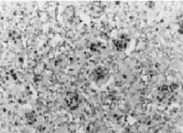
Elevated levels of c-fos has been described in several tumor types1 4 , 2 6 , 2 8 )and it has been suggested that elevated c-fos and c-jun expression is an important event in tumori- genesis, because it may determine an increased proliferation rate1 4 ). With specific Fig. 1. Photomicrograph of the spindle tumor cells show
intense grade ++ nuclear staining for c-fos anti- body (ABC, ×200).
Fig. 2. Lots of tumor giant cells show intense nuclear staining (ABC, ×200).
Fig. 3. Some spindle cells peripheral to the neoplastic cartilage show nuclear staining for c-fos anti- body (ABC, ×200).
reference to osteosarcomas, it is of interest that the expression of both c-fos and c-jun is significantly higher in high-grade osteosarco- mas (characterized by aggressive growth with tendency to systemic spread) than in low grade osteosarcomas (locally aggressive lesions with infrequent metastases), suggest- ing that these oncogenes may be involved in determining the clinical behavior of these n e o p l a s m s6 ). In this present studies, we could observe higher expression tendency of the high grade osteosarcomas than low grade osteosarcoma. However, there is no statisti- cally significant correlation between histolog- ical grade versus immunohistochemical expression of c-fos proto-oncogene.
The elevated levels of c-fos oncoproteins in high-grade osteosarcomas may be the result of the alteration of several pathways that ultimately control cell proliferation. First, c- fos and c-jun are under the regulation of other oncogenes, such as the retinoblastoma tumor suppressor gene (RB), whose product can down regulate c-fos transcription and AP-1 activity1 9 ). The RB gene is frequently altered in human osteosarcomas2 7 ), and loss of RB activity could be involved in deter- mining increased levels of c-fos and c-jun in these tumors. In addition, the expression of c-fos appears to be regulated by transform- ing growth factor beta, one of the major growth factors for bone tissue. Recent stud- ies have shown that TGF-βinduces an increase of c-fos mRNA levels in culture normal and transformed human osteoblast- like cells2 5 ), and this may be responsible for an increase in proliferative activity, since the uptake of antisense c-fos oligonucleotide abolishes the mitogenic effect of TGF-βo n osteoblast-like cells1 3 ). Franchi et al.’s study supports the existence of a strong direct relationship between the expression of c-fos
and TGF-βin human osteosarcomas, since high-grade osteosarcomas show significantly higher levels of c-fos and of TGF-β1 than low-grade lesions6 , 7 ). The observation that high--grade osteosarcomas have a signifi- cantly higher proliferative activity than low- grade osteosarcomas suggests that in these variants of osteosarcoma the elevated expression of TGF-β1 and c-fos may sustain a higher proliferative activity and may con- tribute substantially to establishment of an aggressive phenotype1 7 ). Conversely, in low- grade osteosarcomas lower levels of TGF-β1 and c-fos may result in lower proliferative activity and ultimately in less aggressive g r o w t h1 7 ). Taken together, these data indi- cate that the control pathways of the expression of c-fos and c-jun could play an important part in determining the clinical behaviour of osteosarcomas. Further studies with larger series are needed to determine whether the evaluation of c-fos and c-jun expression may be useful in predicting of clinical outcome in these neoplasms.
REFERENCES
01) Angel P and Karin M : The role of jun, fos and the AP-1 complex in cell proliferation and transforma- tion. Biochem Biophys Acta, 1072:129-1577, 1991.
02) Caubert JF and Bernaudin JF : Expression of the c-fos proto-oncogene in bone, cartilage and tooth forming tissues during mouse development. B i o l Cell, 64:101-104, 1988.
03) Colotta F, Polentarutti N, Sironi M and Mantovani A : Expression and involvement of c- fos and c-jun protooncogenes in programnmed cell death induced by growth factors deprivation in lym- phoid cell lines. J Biol Chem, 267:18278-18283, 1992.
04) De Togni P, Niman H, Raymond V, Sawchenko P and Verma IM : Detection of fos protein during osteogenesis by monoclonal antibodies. Mol Cell Biol, 8:2251-2256, 1988.
05) Dony C and Gruss P : Proto-oncogene c-fos expression in growth regions of fetal bone and mesodermal wet tissue. Nature, 328:711-714, 1987.
06) Franchi A, Calzolari A and Zampi G : Immunohistochemical detection of c-fos and c-jun expression in osseous and cartilaginous tumors of the skeleton. Virchows Arch, 432:515-519, 1998.
07) Franchi A, Arganini L, Baroni G, Calzolari A, Capanna R, Campanacci D, Caldora P, Masi L, Brandi ML and Zampi G : Expression of trans- forming growth factor b isoforms in osteosarcoma variants : association of TGF-b1 with high grade osteosarcomas. J Pathol (Lond), 185:284-289,1998.
08) Grigoriadis AE, Schellander K, Wang Z-Q and Wagner EF : Osteoblasts are target cells for trans- formation in c-fos transgenic mice. J Cell Biol, 122:685-701, 1993.
09) Hadman M, Loo M and Bos TJ : In vivo viral and cellular jun complexes exhibit differential interac- tions with a number of in vitro generated AP-1- and CREB-like target sequences. O n c o g e n e, 8:1895- 1903, 1993.
10) Hoyland J and Sharpe PT : Upregulation of c-fos protooncogene expression in pagetic osteoclasts. J Bone Miner Res, 9:1191-1194, 1994.
11) Johnson RS, Spiegelman BM and Papaioannu V : Pleiotropic effects of a null mutation in the c-fos proto-oncogene. Cell, 71:577-586, 1992.
12) Kovary K and Bravo R : Existence of different fos/jun complexes during G0-to-G1 transition and during exponential gorwth in mouse fibroblasts : differential role of fos proteins. Mol Cell Biol, 12:5015-5023, 1992.
13) Machwate M, Julienne A, Mouktar M and Marie PJ : Temporal variation of c-fos proto-onco- gene expression during osteoblast differentiation and osteogenesis in developing rat bone. J Cell Biochem, 57:62-70, 1995.
14) Magrisso IJ, Richmond RE, Carter JH, Pross CB, Gilfillen RA and Carter HW : Immunohistochemical detection of RAS, JUN, FOS, and p53 oncoprotein expression in human colorectal adenomas and carcino- mas. Lab Invest, 69:6774-681, 1993.
15) Mason DJ, Hillam RA and Skerry TM : Constitutive in vivo mRNA expression by osteo- cytes of b-actin, osteocalcin, connexin-43, IGH-1, c-fos and c-jun, but not TNF-a nor tartrate resistant acid phosphatase. J Bone Miner Res, 11:350-357, 1996.
16) McCabeLR, Lian JB, Stein JL and Stein GS : Selective expression of fos and jun related genes during osteoblast proliferation and differentiation.
Exp Cell Res, 218:255-262, 1995.
17) Oda Y, Wehrmann B, Radig K, Walter H, Rose I, Neumann W and Roessner A : Expression of growth factors and their receptors in human osteosarcomas. Gen Diagn Pathol, 141:97-103, 1995.
18) Ohta S, Yamamuro T, Lee K, Okumura H, Kasai R, Hiraki J, Ikeda T, Iwasaki R, Kijuchi H, Konishi J and Shigeno C : Fracture healing induces expression of the proto-oncogene c-fos in vivo. possible involvement of the Fos protein in osteoblastic differentiation. FEBS Lett, 284:42-45, 1991.
19) Robbins PD, Horowitz JM and Mulligan RC : Negative regulation of human c-fos expression by the retinoblastoma gene product. N a t u r e, 346:668- 671, 1990.
20) Ruther U, Komitowski D, Schubert FR and Wagner EF : c-fos expression induces bone tumors in transgenic mice. Oncogene, 4:861-865, 1989.
21) Ryseck R and Bravo R : c-jun JUN B and JUN D differ in their binding affinities to AP-1 and CRE consensus sequence : effect of FOS proteins.
Oncogene, 6:533-542, 1991.
22) Sandberg M, Vuorio T, Hirvonen H, Alitalo K and Vuorio E : Enhanced expression of TGF-b and c-fos mRNAs in the growth plates of develping human long bones. D e v e l o p m e n t, 102:461-470, 1988.
23) Smeyne RJ, Vendrell M, Hayward M, Baker SJ, Miao GG, Schilling K, Robertson LM, Curran T and Morgan JI : Continuous c-fos expression pre- cedes programmed cell death in vivo. N a t u r e, 363:166-169, 1993.
24) Sonnenberg JL, Rauscher FJ, Morgan JI and Curran T : Regulation of proenkephalin by Fos and Jun. Science, 246:1622-1625, 1989.
25) Subramaniam M, Oursler MJ, Rasmussen BL, Riggs BL and Spelberg TC : TGF-b regulation of nuclear proto-oncogenes and TGF-b gene expres- sion in normal human osteoblast-like cells. J Cell Biochem, 57:52-61, 1995.
26) Tiniakos DG, Scott LE, Borbett IP, Piggott NH and Horne CH : Studies of c-jun oncogene expres- sionin human breast using a new monoclonal anti- body, NCL-DK4. J Pathol (Lond), 172:19-26, 1994.
27) Wadayama B, Toguchida J, Shimizu T, Ishizaki K, Sasaki MS, Kotoura Y and Yamamuro T : Mutation spectrum of the retinoblastoma gene in osteosarcomas. Cancer Res, 54:3042-3048,1994.
28) Walker RA and Cowl J : The expression of c-fos protein in human breast. J Pathol (Lond), 163:323- 327, 1991.
29) Wang Z-Q, Grigoriadis AE, Mohle-Steinlein U and Wagner EF : A novel target cell for c-fos- induced oncogenesis : development of chondro- genic tumors in embryonic stem cell chimeras.
EMBO J, 10:2537-2450, 1991.
30) Wang Z-Q, Liang J, Schellander K, Wagner EF
and Grigoriadis AE : c-fos-induced osteosarcoma formation in transgenic mice : cooperativity with c- jun and the role of endogenous c-fos. Cancer Res, 55:6244-6251, 1995.
31) Wang Z-Q, Ovitt C, Grigoriadis AE, Mohle- Steinlein U, Ruther U and Wagner EF : Bone and haematopoietic defects in mice lacking c-fos.
Nature, 360:741-7745, 1992.
32) Wu J-X, Carpenter M, Gresens C, Keh R, Niman H, Morris JWS and Mercola D : The proto-oncogene c-fos is over-expressed in the majority of human osteosarcomas. O n c o g e n e, 5:989-1000, 1990.
= 국문초록 =
골육종의 c - fos 발현에 관한 면역조직화학적 검색
경희대학교 의과대학 병리학교실, 한림대학교 의과대학 병리학교실*
박 용 구・박 혜 림*
c - f o s와 c - j u n은 암 유전자의 하나이며, 이 유전자의 단백질 산물은 여러 가지의 다른 활성 화 단백 (activator protein 1, AP-1)으로 골 종양에서 골세포의 증식과 분화를 조절하는 중요한 역할을 하는 인자 중 하나로 알려져 있다.
본 연구에서는 단클론 항체를 이용하여 포르말린에 고정된 파라핀 포매조직을 이용하여 3 5 례의 사람 골육종에서 c-fos 단백의 발현을 연구하였다. c-fos의 발현은 골 형성 병변에서 주 로 발현되며, 저등급의 연골형성 병변에서는 발현이 관찰되지 않았다. 높은 빈도의 c-fos 단 백의 발현이 골아성 골육종에서 발현되었으나 ( 1 7례 중 1 3례에서 1등급 내지 2등급으로 발 현), 2례의 연골형성 골육종, 1례의 섬유아세포성 골육종, 2례의 방골성 골육종에서는 음성 으로 나타났다. 2례의 혈관확장성 골육종에서는 양성으로 c-fos 단백의 발현되었다. 비록 조 직학적으로 고등급의 골육종에서 면역조직화학적 염색상 고 빈도의 c-fos 단백의 발현이 관찰 되나, 조직학적 등급과, 면역염색상 발현사이에 통계적인 유의성은 관찰되지 않았다. 이상의 결과로 c-fos 단백이 골육종의 발생에 관여할 것으로 추론되며, 저등급의 연골형성 육종에 이 단백의 역할에는 추후 연구가 필요할 것으로 사료된다.
중심어 : 골육종, c-fos, 면역조직화학 검사