J Korean Surg Soc 2013;84:168-177 http://dx.doi.org/10.4174/jkss.2013.84.3.168
ORIGINAL ARTICLE
JKSS JKSS JKSS
Journal of the Korean Surgical Society pISSN 2233-7903ㆍeISSN 2093-0488
Received November 6, 2012, Revised December 11, 2012, Accepted January 31, 2013 Correspondence to: Young-Hoe Hur
Department of Surgery, Chonnam National University Hwasun Hospital, Chonnam National University Medical School, 322 Seoyang-ro, Hwasun 519-763, Korea
Tel: +82-61-379-7646, Fax: +82-61-379-7661, E-mail: [email protected]
cc Journal of the Korean Surgical Society is an Open Access Journal. All articles are distributed under the terms of the Creative Commons Attribution Non-Commercial License (http://creativecommons.org/licenses/by-nc/3.0/) which permits unrestricted non-commercial use, distribution, and reproduction in any medium, provided the original work is properly cited.
Impact of lymph node ratio as a valuable prognostic factor in gallbladder carcinoma, focusing on stage IIIB gallbladder carcinoma
Byung-Gwan Choi, Choong-Young Kim, Seung-Hyun Cho, Hee-Joon Kim, Yang-Seok Koh, Jung-Chul Kim, Chol-Kyoon Cho, Hyun-Jong Kim, Young-Hoe Hur
Department of Surgery, Chonnam National University Medical School, Gwangju, Korea
Purpose: It is increasingly being recognized that the lymph node ratio (LNR) is an important prognostic factor for gall- bladder carcinoma patients. The present study evaluated predictors of tumor recurrence and survival in a large, mono-in- stitutional cohort of patients who underwent surgical resection for gallbladder carcinoma, focusing specifically on the prog- nostic value of lymph node (LN) status and of LNR in stage IIIB patients. Methods: Between 2004 and 2011, 123 patients who underwent R0 radical resection for gallbladder carcinoma at the Chonnam National University Hwasun Hospital were re- viewed retrospectively. Patients were staged according to the American Joint Committee on Cancer 7th edition, and prog- nostic factors affecting disease free survival, such as age, sex, comorbidity, body mass index, presence of preoperative symp- toms, perioperative blood transfusion, postoperative complications, LN dissection, tumor size, differentiation, lymph-vas- cular invasion, perineural invasion, T stage, presence of LN involvement, N stage, numbers of positive LNs, LNR and im- plementation of adjuvant chemotherapy, were statistically analyzed. Results: LN status was an important prognostic factor in patients undergoing curative resection for gallbladder carcinoma. The total number of LNs examined was implicated with prognosis, especially in N0 patients. LNR was a powerful predictor of disease free survival even after controlling for compet- ing risk factors, in curative resected gallbladder cancer patients, and especially in stage IIIB patients. Conclusion: LNR is con- firmed as an independent prognostic factor in curative resected gallbladder cancer patients, especially in stage IIIB gall- bladder carcinoma.
Key Words: Lymph node ratio, Gallbladder, Carcinoma, Stage IIIB
INTRODUCTION
Adenocarcinoma of the gallbladder has historically been considered an incurable malignancy with dismal prognosis due to its propensity for early dissemination.
Complete surgical resection with tumor-free surgical mar- gins, especially in the early stages of the disease, offers the only hope for long-term survival and is considered a standard of care. Thus, accurate identification of the extent of disease is an important diagnostic consideration for
Fig. 1. Flow chart detailing surgical management of all the patients with gallbladder cancer.
therapeutic decision-making of gallbladder cancer.
The presence or absence of lymph node (LN) metastases is an established prognostic factor in patients with re- sected gallbladder carcinoma [1-3]. Patients with LN metastases have a significantly worse survival than pa- tients with node-negative disease [4]. Most Eastern sur- geons and even some Western surgeons have adopted rad- ical LN dissection for this disease and have reported the beneficial effect of nodal dissection on survival and some long-term survivors with nodal disease [5-10]. Thus, it ap- pears that radical LN dissection plays a critical role in the surgical management of gallbladder cancer.
Increasingly, the idea that an inadequate number of LNs examined may adversely influence on survival and lead to understaging of various gastrointestinal cancers is being recognized [11]. Rather, evidence in the literature that is based on other malignancies demonstrates that the actual number of LNs evaluated had an effect on survival [12-14].
In fact, most studies have demonstrated an inverse rela- tionship between the number of LNs examined and over- all survival. Moreover, the ratio between metastatic and examined LNs is an additional recent prognostic factor in different gastrointestinal malignancies [15-17]. Le Voyer et al. [14] analyzed data from patients with stage II and III co- lon cancer, and reported that the lymph node ratio (LNR) was an important prognostic factor even after adjusting for other parameters such as tumor stage, grade, and histology. Marchet et al. [18] and Pawlik et al. [19] also demonstrated that LNR was an independent predictor of survival in gastric cancer and pancreas cancer, respec- tively.
In the light of these considerations, the aim of the pres- ent study was to evaluate predictors of tumor recurrence and survival in a large, mono-institutional cohort of pa- tients who underwent surgical resection for gallbladder carcinoma, focusing specifically on the prognostic value of LN status and of LNR in stage IIIB patients.
METHODS
Patient population
Between 2004 and 2011, retrospective data was collected from 144 consecutive patients with gallbladder carcinoma who underwent laparotomy at Chonnam National Uni- versity Hwasun Hospital. Fifteen patients who underwent palliative surgery or a resection with microscopic or mac- roscopic residual tumor were excluded, as were six pa- tients who underwent exploratory laparotomy because of peritoneal seeding (Fig. 1). The remaining 123 patients who underwent a R0 radical resection for gallbladder car- cinoma were included. Demographics, operative details, and pathologic details of the 123 patients were recorded.
Operative details
The operative procedure basically consisted of chol- ecystectomy, regional lymphadenectomy, and resection of the gallbladder bed with a rim of the adjacent liver tissue.
Cholecystectomy included the removal of the entire con- nective tissue lying between the gallbladder and the liver parenchyma. Lymphadenectomy included en bloc clear- ance of cystic duct, pericholedochal, hepatic artery, portal
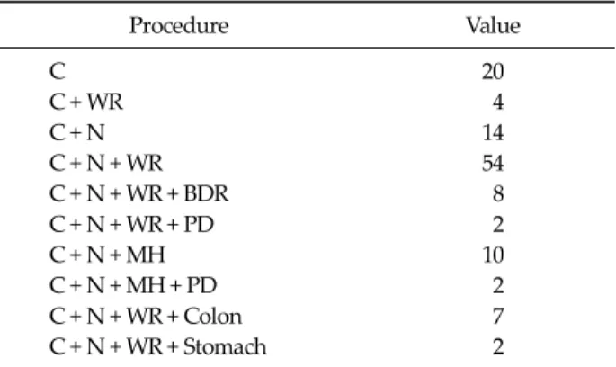
Table 1. Radical procedures in 123 patients with gallbladder carcinoma
Procedure Value
C 20
C + WR 4
C + N 14
C + N + WR 54
C + N + WR + BDR 8
C + N + WR + PD 2
C + N + MH 10
C + N + MH + PD 2
C + N + WR + Colon 7
C + N + WR + Stomach 2
C, cholecystectomy; WR, wedge resection of the gallbladder bed;
N, radical lymphadenectomy; BDR, extrahepatic bile duct resection; PD, pancreatico-duodenectomy; MH, major hepatec- tomy including sectionectomy, lobectomy and trisectionectomy.
vein LNs, and, if needed, periduodenal, peripancreatic, celiac, and aortocaval LNs. Wedge resection involved a 2-cm deep nonanatomical wedge resection of gallbladder fossa, with the guiding principle being attainment of neg- ative surgical margin while at the same time preserving the maximal amount of liver parenchyma. In all the pa- tients, frozen section analysis of cystic duct remnant was obtained to ensure negative margin. Bile duct resection was not performed routinely, except in case of positive cystic duct margin or definite bile duct invasion.
The choice of a radical resection procedure for each pa- tient depended on the extent of the tumor on preoperative cross sectional imaging. Some early-stage diseases re- quired less aggressive resection. Five patients with patho- logically proven T1a (confined within the lamina propria of the primary tumor) after laparoscopic cholecystectomy did not have an additional procedure. Although the tumor extent of T1b with relaparotomy after laparoscopic chol- ecystectomy is debatable, we did not perform more proce- dures in seven patients proven as T1b after cholecy- stectomy. Twelve patients underwent major hepatectomy depending on the extent of the tumor and two patients re- ceived additional pancreatico-duodenectomy: one be- cause of an extension of the tumor to the intrapancreatic bile duct and the other due to involvement of peripancre- atic LNs. Another two patients also received an additional pancreatico-duodenectomy without major hepatectomy
for similar reasons. Adjacent organs, such as colon and stomach, were resected in nine patients owing to direct in- vasion in the operative field (Table 1).
Pathologic examination
Primary tumor was examined to determine the size of the primary tumor, histologic type, histologic grade, depth of infiltration, lympho-vascular invasion, perineu- ral invasion, and tumor involvement of excised con- tiguous viscera and resection margins. Although the pathologic findings were staged using the 6th edition of the American Joint Committee on Cancer (AJCC) manual during the period of study, final description was done ac- cording to the 7th edition of the AJCC classification. The primary tumor status was pT1a in 11 patients, pT1b in 15, pT2 in 69, pT3 in 25, and pT4 in 2.
The histologic grade was determined based on the area of tumor with highest grade and was classified as well dif- ferentiated or not well differentiated (moderate or poor).
Adenocarcinoma was identified in 102 patients of primary gallbladder tumor, papillary adenocarcinoma in nine pa- tients, adenosquamous carcinoma in five patients, carci- nosarcoma in three patients, undifferentiated carcinoma in two patients, and combined adenocarcinoma and neu- roendocrine carcinoma in two patients.
Fibrofatty tissue containing LNs were retrieved from the specimen just after resection, and nodes were metic- ulously separated into individual node groups according to the location of the LNs. LN metastasis was defined as tu- mor cells detected on histopathologic examination using hematoxylin and eosin stain. The location and the number of positive LNs were used to assess the degree of lym- phatic spread. The location of positive LN was classified into three categories according to the 7th edition of AJCC classification: pathologic N0 (pN0), no regional LN meta- stasis; pathologic N1 (pN1), metastases to nodes along the cystic duct, common bile duct, hepatic artery, and/or por- tal vein; pathologic N2 (pN2), metastases to periaortic, pericaval, superior mesenteric artery, and/or celiac artery LNs. The number of total retrieved LNs and positive LNs were recorded for each patient. LNR was determined by dividing the total number of LNs harboring metastases by the total number of examined nodes. Patients were div-
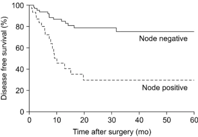
Fig. 2. Kaplan-Meier survival curves according to the presence or absence of regional nodal disease. Five-year disease free survival rate was 80.3% in patients without regional nodal disease, but the median survival time was not reached. The five-year disease free survival rate was 48.5% with a median survival time of 30 months in patients with regional nodal disease (P < 0.001).
ided into three different groups based on their LNR, in- cluding one group of patients with negative nodes (LNR = 0) and two groups with positive LNs as follows, based on sensitivity analyses that identified this cutoff value as po- tentially being the most discriminating: 0 < LNR ≤ 0.5, and LNR more than 0.5.
Follow-up
Follow-up comprised physical examination, laboratory tests including tumor markers (carcinoembryonic anti- gen, cancer antigen 19-9), and imaging study including ab- dominal ultrasonography and computed tomography.
Follow-up was carried out every 3 months for the first year and every 6 months thereafter. Follow-up tests were per- formed any time if there was suspicion of disease recurrence.
Adjuvant chemotherapy after resection was admini- stered at the discretion of the individual surgeons, though most patients with positive LNs were recommended.
Forty four patients received adjuvant chemotherapy with intravenous gemcitabine intend to receive 6 cycles. No pa- tient received adjuvant radiotherapy.
The sites of recurrence were recorded only when a le- sion was visible at imaging and was classified as loco-re- gional (liver resection margin, hepaticojejunostomy, or re- gional LNs), distant (liver, peritoneum, or systemic), or both. Disease-free survival (DFS) was calculated from the day of surgery to the time when recurrent disease was first detected. Death of patients was defined as disease related or due to other causes, in order to evaluate disease specific survival.
Statistical analyses
Medical records and survival data were obtained for all 123 patients. Continuous data are presented as median (interquartile range). Categorical variables were com- pared using chi square or Fisher’s test. When comparing two groups, normally distributed continuous variables were compared using a two-sample Student’s t-test, while the Mann-Whitney U test was used for non-normally dis- tributed variables. Survival was defined as the time from diagnosis to death. The primary outcome parameter of in- terest was DFS and is expressed as median ± standard error. Survival probability was estimated according to the
Kaplan-Meier method, whereas the log-rank test was used for univariable comparison between groups. A Cox pro- portional hazards regression model using a step-back- ward fitting procedure was applied to determine sig- nificant influence on survival. Risk factors associated with disease recurrence were determined by multivariate logis- tic regression. The hazard ratio (HR) with 95% confidence intervals (CI) was reported as an estimate of the risk of dis- ease specific death. P < 0.05 was considered statistically significant. Statistical analysis was performed with the PASW ver. 18.0 (IBM Co., Armonk, NY, USA).
RESULTS
At a median follow-up of 22.8 months (range, 3.1 to 100.7 months), the overall cumulative DFS rates of the 123 patients who underwent radical procedure for gall- bladder carcinoma were 65% at 3 years and 63% at 5 years.
The overall cumulative survival rates were 67% at 3 years and 60% at 5 years. There was no perioperative mortality.
The median DFS time and overall survival time were 16.4 months and 22.8 months respectively.
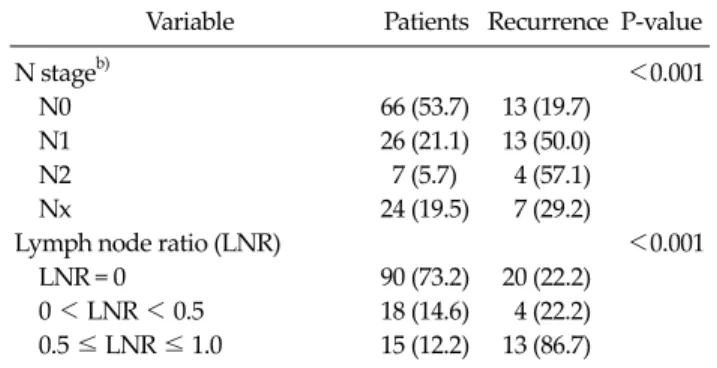
Sixty six patients (67%) were node negative patients and positive LNs were found in 33 patients (33%) among the 99 patients who underwent LN harvest. Twenty six patients were included in the pN1 group and seven patients were
Table 2. Univariate analysis of factors associated with disease-free survival after curative intent resection for gallbladder cancer
Variable Patients Recurrence P-value
Age (yr) 0.111
<65 63 (51.2) 15 (23.8)
≥65 60 (48.8) 22 (36.7)
Sex 0.712
Male 60 (48.8) 19 (28.6)
Female 63 (51.2) 18 (31.7)
Comorbidity 0.475
Yes 71 (57.7) 23 (32.4)
No 52 (42.3) 14 (26.9)
Body mass index (kg/m2) 0.687
≥25 44 (35.8) 12 (27.3)
<25 79 (64.2) 25 (31.6)
Symptom 0.075
Yes 69 (56.1) 24 (34.8)
No 54 (43.9) 13 (24.1)
Perioperative blood transfusion 0.294
Yes 19 (15.4) 3 (15.8)
No 104 (84.6) 34 (32.7)
Postoperative complication 0.905
Yes 24 (19.5) 6 (25.0)
No 99 (80.5) 31 (31.3)
Lymph node dissection 0.451
Yes 99 (80.5) 30 (30.3)
No 24 (19.5) 7 (29.2)
Tumor size (cm) 0.188
≥3 49 (39.8) 17 (34.7)
<3 74 (60.2) 20 (27)
Differentiation group <0.001
Well 71 (57.7) 12 (16.9)
Etca) 52 (42.3) 25 (48.1)
Lympho-vascular invasion 0.001
Yes 22 (20.0) 11 (50)
No 88 (80.0) 22 (25)
Perineural invasion 0.001
Yes 28 (25.5) 14 (50)
No 82 (74.5) 19 (23.2)
T stageb) <0.001
T1–T2 95 (77.2) 22 (23.2)
T3–T4 28 (22.8) 15 (33.6)
Lymph node involvement <0.001
Yes 33 (26.8) 17 (51.5)
No 90 (73.2) 20 (22.2)
Table 2.Continued
Variable Patients Recurrence P-value
N stageb) <0.001
N0 66 (53.7) 13 (19.7)
N1 26 (21.1) 13 (50.0)
N2 7 (5.7) 4 (57.1)
Nx 24 (19.5) 7 (29.2)
Lymph node ratio (LNR) <0.001
LNR = 0 90 (73.2) 20 (22.2)
0 < LNR < 0.5 18 (14.6) 4 (22.2) 0.5 ≤ LNR ≤ 1.0 15 (12.2) 13 (86.7) Values are presented as number (%).
a)Included moderate differentiation, poor differentiation and undifferentiation. b)According to the American Joint Committee on Cancer 7th edition.
in pN2 group with positive celiac and paraaortic nodal disease. Patients with node negative disease had a median survival of 22.7 months compared with a median survival of 7.3 months for patients with positive LNs (P < 0.001) (Fig. 2). The number of dissected regional LNs was ex- tremely high in patients who underwent pancreatico-
duodenectomy or stomach resection. The mean number of harvested and positive regional LNs was 5.31 and 0.73, respectively.
Factors influencing DFS in patients with radically resected gallbladder carcinoma
On univariate analysis, factors associated with worse DFS included nonwell differentiated histologic group, presence of lymph-vascular invasion, presence of peri- neural invasion, T-stage more than T3, LN involvement, higher N stage, and higher LN ratio (Table 2). The sig- nificant univariate variables were entered into Cox re- gression multivariate analysis; high T-stage group and LN ratio remained as independently significant variables.
Patients in the T3–T4 group had a median survival of 6.97 months compared with a median survival of 21.4 months for patients in the T1–T2 group. The T3–T4 patients had a nearly 3-fold increased risk of recurrence (HR, 2.998; 95%
CI, 1.392 to 6.458; P = 0.005) compared with patients in the T1–T2 group. LNR was also associated strongly with prognosis. Patients with a LNR exceeding 0.5 had an al- most 5-fold increased risk of recurrence rate (HR, 5.998;
95% CI, 2.576 to 13.962; P < 0.001) compared with patients who had a LNR of 0 (Table 3).
Factors influencing DFS in patients with node- negative gallbladder carcinoma
Patients without metastasis to adjacent LNs (n = 66) had a 5-year DFS of 77.8%. Univariate analysis of risk factors
Table 3. Multivariate analysis of factors associated with disease free survival after curative intent resection for gallbladder cancer
Variable Hazard ratio 95% CI P-value T stage groupa)
T1–T2 1.000
T3–T4 2.998 1.392–6.458 0.005
Lymph node ratio (LNR)
LNR = 0 1.000
0 < LNR < 0.5 1.093 0.318–3.754 0.888 0.5 ≤ LNR ≤ 1.0 5.998 2.576–13.962 <0.001 CI, confidence interval.
a)According to the American Joint Committee on Cancer 7th edition.
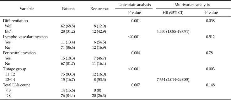
Table 4. Factors predictive of disease free survival in patients with node-negative gallbladder cancer after radical resection
Variable Patients Recurrence Univariate analysis Multivariate analysis
P-value HR (95% CI) P-value
Differentiation 0.001 0.038
Well 62 (68.8) 8 (12.9)
Etca) 28 (31.2) 12 (42.9) 4.550 (1.085–19.091)
Lympho-vascular invasion <0.001 0.512
Yes 11 (13.4) 6 (54.5)
No 71 (86.6) 12 (16.9)
Perineural invasion 0.004 0.78
Yes 15 (18.3) 7 (46.7)
No 67 (81.7) 11 (16.4)
T stage group <0.001 0.003
T1–T2 75 (83.3) 12 (16.0)
T3–T4 15 (16.7) 8 (53.3) 7.654 (2.014–29.085)
Total LNs count 0.087 0.148
≥8 14 (15.6) 0 (0)
<8 76 (84.4) 20 (26.3)
Values are presented as number (%).
HR, hazard ratio; CI, confidence interval; LN, lymph node.
a)Includes moderate differentiation, poor differentiation and undifferentiation.
Fig. 3. Kaplan-Meier survival curves comparing patients in node negative disease with fewer than eight lymph nodes (LNs) examined to those with eight or more LNs examined. All the patients with more than eight LNs examined were not recurred, whereas patients with fewer than eight LNs examined displayed 5-year disease free survival rate of 73.7% (P = 0.087).
for tumor recurrence revealed T3–T4 tumor stage, poor tu- mor differentiation, lymphovascuar invasion, and peri- neural invasion as significantly associated with tumor recurrence. On multivariate analysis, T3–T4 tumor stage was significantly associated with poor disease free surviv- al (HR, 7.654; 95% CI, 2.014 to 29.085; P = 0.003). Tumor grade worse than well differentiation had statistical sig- nificance (HR, 4.550; 95% CI, 1.085 to 19.091; P = 0.038) (Table 4). Total number of harvested LNs was not asso- ciated with DFS. Patients with N0 disease were stratified in two groups according to the total number of LNs
evaluated. Because eight LNs tended to determine the greatest actuarial survival difference between the result- ing groups, we used this value as the cutoff. N0 patients who had at least eight LNs evaluated had superior DFS than patient with fewer number of LNs examined, but the difference was not significant (P = 0.087) (Fig. 3).
Table 5. Factors predictive of disease free survival in patients with stage IIIB gallbladder cancer after radical resection
Variable Patients Recurrence Univariate analysis Multivariate analysis
P-value HR (95% CI) P-value
Comorbidity 0.047
Yes 16 (61.5) 10 (62.5)
No 10 (38.5) 3 (30.0)
Positive LN number 0.502
1 11 (42.3) 4 (36.4)
2–3 11 (42.3) 7 (63.6)
More than 4 4 (15.4) 2 (50.0)
Lymph node ratio (LNR) 0.003 0.007
0 < LNR < 0.5 16 (61.5) 4 (25.0) 1.000
0.5 ≤ LNR ≤ 1.0 10 (38.5) 9 (90.0) 5.231 (1.586–17.255)
Values are presented as number (%).
HR, hazard ratio; CI, confidence interval; LN, lymph node.
Fig. 4. Kaplan-Meier survival curves according to the lymph node ratio in patients with stage IIIB gallbladder cancer. Five-year disease free survival rate was 75% in patients with a lymph node ratio (LNR) < 0.5, but the median survival time was not reached.
The 5-year disease free survival rate was only 10% with a median survival time of 8.5 months in patients with a LNR > 0.5 (P = 0.003).
Factors influencing DFS in patients with stage IIIB gallbladder carcinoma after radical resection
All N1 gallbladder cancer patients with T1–T3 tumor depth were classified as stage IIIB in the AJCC 7th edition.
According to the classification, T4 and N2 were included in stage IV disease meaning poor prognosis like distant metastasis. Thus, in this study, we analyzed prognostic factors of stage IIIB patients, akin to the analysis of node positive patients in other studies. Age, sex, comorbidity, body mass index (BMI), presence of preoperative symp- tom, perioperative blood transfusion, postoperative com-
plication, tumor size, differentiation, lymph-vascular in- vasion, perineural invasion, T stage, numbers of positive LNs, LNR, and implementation of adjuvant chemo- therapy were identified as prognostic factors that influ- enced DFS. In univariate analysis, presence of comorbidi- ty and LNR ≥0.5 were associated with significantly worse DFS in stage IIIB gallbladder cancer patients after curative resection. However, subsequent multivariate analysis re- vealed only LNR ≥0.5 to be an independent predictor of DFS in those patients (HR, 5.231; 95% CI, 1.586 to 17.255; P
= 0.007) (Table 5, Fig. 4).
DISCUSSION
Complete surgical resection is the mainstay of treat- ment in patients with localized gallbladder cancer. In pa- tients with early gallbladder carcinoma, in which invasion was limited to the muscularis propria layer, favorable out- come has been achieved [20-22]. Although, there has been little controversy concerning the operative strategy of ear- ly gallbladder carcinoma limited to the lamina propria layer (T1a), but there are still many debates about the oper- ative methods in gallbladder carcinoma limited to the muscularis propria (T1b). Since Glenn and Hays [23] pro- posed ‘radical cholecystectomy’ for gallbladder carcino- ma involving the en bloc excision of the gallbladder bed with a rim of the adjacent liver tissue and the lymphatic tis-
sue within hepatoduodenal ligament in 1954 [1], a favor- able outcome has been achieved for advanced gallbladder cancer. Also extended resection including extended LN dissection, extended hepatectomy, pancreaticoduodenec- tomy, and combined vascular resection has been per- formed in far advanced gallbladder carcinoma [24,25].
Although mortality and mobidity rates have been high, many surgeons have reported a number of long-term sur- vivors [5-9,24]. This suggests that extended resection is ef- fective for select cases of advanced gallbladder carcinoma.
At diagnosis, accurate and uniform gallbladder cancer staging is essential for prognosis, planning treatment, and comparing treatment outcomes. Currently, gallbladder cancer is staged by two main staging systems: the AJCC tu- mor-node-metastasis staging system (7th edition) [2] and the Japanese Society of Biliary Surgery (JSBS) staging sys- tem [3]. In the AJCC 7th edition, the nodal status has been divided into three groups based on the presence of pos- itive LNs and the anatomic location of positive LNs: N0 (no regional LN metastasis), N1 (metastases to nodes along the cystic duct, common bile duct, hepatic artery, and/or portal vein), and N2 (metastases to periaortic, peri- caval, superior mesenteric artery, and/or celiac artery LNs). The JSBS subdivides the nodal status of gallbladder carcinoma into four categories, according to the anatomic location of positive LNs: N0, N1, N2, or N3. We adopted the AJCC staging system because it is the most accepted system worldwide, and also JSBS requires extended LN dissection to acquire sufficient knowledge on the LN status.
The present study revealed that the survival rates of pa- tients with node positive gallbladder carcinoma are worse than those of node negative, although LN metastasis itself had insignificant prognostic power in multivariate analy- sis. LN metastasis is consistently one of the strongest pre- dictors of poor outcome in patients with advanced gall- bladder carcinoma [4,26-28]. Few Western reports have described long-term, disease-free survivor who had lym- phatic metastases [26]. In contrast, Japanese reports showed that the prognosis of patients who had LN metastasis lim- ited to the LN within the hepatoduodenal ligament is im- proved by LN dissection [5,27]. In our study, 5-year surviv- al of node negative patients was 80.3%, whereas that of
node positive patients was 48.5% (P < 0.001). Among the node positive patients, the N2 group belongs to stage IVB according to the AJCC 7th edition, and has little chance of long-term survival. Whereas patients in N1 group, espe- cially in stage IIIB with T1–T3, are expected to survive for a relatively long time, so it is important to select long-term survivors among the patients with stage IIIB, and to ana- lyze the prognostic factors.
Recent studies have also demonstrated the influence of retrieved LN counts, metastatic LN counts, and LNR on survival of patients with gallbladder carcinoma [29,30].
Presently, the survival of N0 patients who had fewer than eight LNs evaluated at the time of operation tended to be shorter than the survival of N0 patients who had at least eight LNs examined (Fig. 3). Negi et al. [29] also reported that N0 patients with fewer than six LNs had worse prognosis. The total number of LNs examined in pre- sumed N0 patients, therefore, appears to be an important prognostic variable. Specifically, N0 patients who have fewer than six to eight LNs examined may be understaged due to an inadequate LN evaluation. Despite the lack of statistical significance in the current study, the strong trend was seen in both the current study and the study by Negi et al. [29], as well as the established biologic plausi- bility of this concept based on other disease sites.
In contrast to N0 patients, in N1 patients, the total num- ber of LNs evaluated was not an important prognostic variable. The presence of LN metastases was such a strong prognostic factor that it probably outweighed any prog- nostic effect of the total number of LNs evaluated.
Using the presence of nodal disease or absolute number of affected LNs may introduce bias due to the inevitable possibility of incomplete lymphadenectomy or inade- quate histopathologic examination. To overcome the po- tential problems associated with nodal staging and to de- fine better the prognostic role of nodal disease, several studies have used the LNR as a prognostic parameter for various gastrointestinal tumors. LNR, rather than total node count or total number of positive LNs, may have ben- efit for a number of different reasons. Firstly, LNR pro- vides a denominator that accounts for some degree of ad- equacy of the LN dissection. Consequently, LNR is less prone to “stage migration.” Bando et al. [12] and Inoue et
al. [13] also have demonstrated the superiority of LNR for gastric cancer as compared to routine staging of LNs with regard to stage migration. Secondly, LNR provides for a more clinically distinct comparison of patients with sim- ilar counts of total positive LNs. For example, patients with two positive LNs, but different numbers of retrieved LNs (4 and 40) have different LNR (0.5 and 0.05).
Comparison between the patients with positive LN counts may not be appropriate. Thus, LNR provides a more dis- criminatory tool to stratify patients based on both the ex- tent of their disease as well as the adequacy of their lymph- adenectomy.
As our data demonstrated, LNR and T-stage affect DFS in patients with radically resected gallbladder carcinoma;
in particular, a LNR exceeding 0.5 had the highest HR. In N1 patients, especially in stage IIIB patients of gallbladder carcinoma, LNR was the most potent predictor of prog- nosis when we adjusted for many competing risk factors, such as age, sex, comorbidity, BMI, presence of preopera- tive symptoms, perioperative blood transfusion, post- operative complications, tumor size, differentiation, lym- ph-vascular invasion, perineural invasion, T stage, num- bers of positive LNs, and implementation of adjuvant chemotherapy. Thus, LNR was confirmed as an in- dependent prognostic factor in N1 patients, especially in stage IIIB gallbladder carcinoma.
In conclusion, LN status is an important prognostic fac- tor in patients undergoing curative resection for gall- bladder carcinoma. The total number of LNs examined may be associated with prognosis, especially in N0 pa- tients. In patients with stage IIIB disease, a high LNR ap- pears to be a better prognostic predictor than simply the total number of positive LNs. LNR remained a powerful predictor of DFS even after controlling for competing risk factors, in curative resected gallbladder cancer patients, and especially in stage IIIB patients. LNR should be con- sidered as a prognostic factor and stratification tool in fu- ture trials evaluating adjuvant treatments in radically re- sected gallbladder carcinoma patients.
CONFLICTS OF INTEREST
No potential conflict of interest relevant to this article was reported.
REFERENCES
1. Shirai Y, Wakai T, Hatakeyama K. Radical lymph node dis- section for gallbladder cancer: indications and limitations.
Surg Oncol Clin N Am 2007;16:221-32.
2. Edge SB, Byrd DR, Compton CC, Fritz AG, Greene FL, Trotti A. AJCC cancer staging manual. 7th ed. New York:
Springer; 2010.
3. Japanese Society of Biliary Surgery. Classification of biliary tract carcinoma. 2nd ed. Tokyo: Kanehara; 2004.
4. Benoist S, Panis Y, Fagniez PL. Long-term results after cu- rative resection for carcinoma of the gallbladder. French University Association for Surgical Research. Am J Surg 1998;175:118-22.
5. Tsukada K, Kurosaki I, Uchida K, Shirai Y, Oohashi Y, Yokoyama N, et al. Lymph node spread from carcinoma of the gallbladder. Cancer 1997;80:661-7.
6. Kondo S, Nimura Y, Hayakawa N, Kamiya J, Nagino M, Uesaka K. Regional and para-aortic lymphadenectomy in radical surgery for advanced gallbladder carcinoma. Br J Surg 2000;87:418-22.
7. Endo I, Shimada H, Tanabe M, Fujii Y, Takeda K, Morioka D, et al. Prognostic significance of the number of positive lymph nodes in gallbladder cancer. J Gastrointest Surg 2006;10:999-1007.
8. Sasaki R, Itabashi H, Fujita T, Takeda Y, Hoshikawa K, Takahashi M, et al. Significance of extensive surgery in- cluding resection of the pancreas head for the treatment of gallbladder cancer: from the perspective of mode of lymph node involvement and surgical outcome. World J Surg 2006;30:36-42.
9. Yokomizo H, Yamane T, Hirata T, Hifumi M, Kawaguchi T, Fukuda S. Surgical treatment of pT2 gallbladder carcino- ma: a reevaluation of the therapeutic effect of hepatectomy and extrahepatic bile duct resection based on the long- term outcome. Ann Surg Oncol 2007;14:1366-73.
10. D'Angelica M, Dalal KM, DeMatteo RP, Fong Y, Blumgart LH, Jarnagin WR. Analysis of the extent of resection for ad- enocarcinoma of the gallbladder. Ann Surg Oncol 2009;16:
806-16.
11. Smith DD, Schwarz RR, Schwarz RE. Impact of total lymph node count on staging and survival after gastrectomy for gastric cancer: data from a large US-population database. J Clin Oncol 2005;23:7114-24.
12. Bando E, Yonemura Y, Taniguchi K, Fushida S, Fujimura T, Miwa K. Outcome of ratio of lymph node metastasis in gas- tric carcinoma. Ann Surg Oncol 2002;9:775-84.
13. Inoue K, Nakane Y, Iiyama H, Sato M, Kanbara T, Nakai K,
et al. The superiority of ratio-based lymph node staging in gastric carcinoma. Ann Surg Oncol 2002;9:27-34.
14. Le Voyer TE, Sigurdson ER, Hanlon AL, Mayer RJ, Macdonald JS, Catalano PJ, et al. Colon cancer survival is associated with increasing number of lymph nodes ana- lyzed: a secondary survey of intergroup trial INT-0089. J Clin Oncol 2003;21:2912-9.
15. Nitti D, Marchet A, Olivieri M, Ambrosi A, Mencarelli R, Belluco C, et al. Ratio between metastatic and examined lymph nodes is an independent prognostic factor after D2 resection for gastric cancer: analysis of a large European monoinstitutional experience. Ann Surg Oncol 2003;10:
1077-85.
16. Berger AC, Sigurdson ER, LeVoyer T, Hanlon A, Mayer RJ, Macdonald JS, et al. Colon cancer survival is associated with decreasing ratio of metastatic to examined lymph nodes. J Clin Oncol 2005;23:8706-12.
17. Mariette C, Piessen G, Briez N, Triboulet JP. The number of metastatic lymph nodes and the ratio between metastatic and examined lymph nodes are independent prognostic factors in esophageal cancer regardless of neoadjuvant chemoradiation or lymphadenectomy extent. Ann Surg 2008;247:365-71.
18. Marchet A, Mocellin S, Ambrosi A, Morgagni P, Garcea D, Marrelli D, et al. The ratio between metastatic and exam- ined lymph nodes (N ratio) is an independent prognostic factor in gastric cancer regardless of the type of lymphade- nectomy: results from an Italian multicentric study in 1853 patients. Ann Surg 2007;245:543-52.
19. Pawlik TM, Gleisner AL, Cameron JL, Winter JM, Assumpcao L, Lillemoe KD, et al. Prognostic relevance of lymph node ratio following pancreaticoduodenectomy for pancreatic cancer. Surgery 2007;141:610-8.
20. Shimada H, Endo I, Togo S, Nakano A, Izumi T, Nakaga- wara G. The role of lymph node dissection in the treatment of gallbladder carcinoma. Cancer 1997;79:892-9.
21. Yamaguchi K, Chijiiwa K, Saiki S, Nishihara K, Takashima M, Kawakami K, et al. Retrospective analysis of 70 oper- ations for gallbladder carcinoma. Br J Surg 1997;84:200-4.
22. Miyakawa S, Ishihara S, Horiguchi A, Takada T, Miyazaki M, Nagakawa T. Biliary tract cancer treatment: 5,584 re- sults from the Biliary Tract Cancer Statistics Registry from 1998 to 2004 in Japan. J Hepatobiliary Pancreat Surg 2009;16:1-7.
23. Glenn F, Hays DM. The scope of radical surgery in the treatment of malignant tumors of the extrahepatic biliary tract. Surg Gynecol Obstet 1954;99:529-41.
24. Miyazaki M, Itoh H, Ambiru S, Shimizu H, Togawa A, Gohchi E, et al. Radical surgery for advanced gallbladder carcinoma. Br J Surg 1996;83:478-81.
25. Kondo S, Nimura Y, Hayakawa N, Kamiya J, Nagino M, Uesaka K. Extensive surgery for carcinoma of the gallbladder. Br J Surg 2002;89:179-84.
26. Fong Y, Wagman L, Gonen M, Crawford J, Reed W, Swanson R, et al. Evidence-based gallbladder cancer stag- ing: changing cancer staging by analysis of data from the National Cancer Database. Ann Surg 2006;243:767-71.
27. Kondo S, Takada T, Miyazaki M, Miyakawa S, Tsukada K, Nagino M, et al. Guidelines for the management of biliary tract and ampullary carcinomas: surgical treatment. J Hepatobiliary Pancreat Surg 2008;15:41-54.
28. Jensen EH, Abraham A, Jarosek S, Habermann EB, Al- Refaie WB, Vickers SA, et al. Lymph node evaluation is as- sociated with improved survival after surgery for early stage gallbladder cancer. Surgery 2009;146:706-11.
29. Negi SS, Singh A, Chaudhary A. Lymph nodal involve- ment as prognostic factor in gallbladder cancer: location, count or ratio? J Gastrointest Surg 2011;15:1017-25.
30. Shirai Y, Sakata J, Wakai T, Ohashi T, Ajioka Y, Hatakeyama K. Assessment of lymph node status in gallbladder cancer:
location, number, or ratio of positive nodes. World J Surg Oncol 2012;10:87.