Letter to the Editor
Vol. 27, No. 5, 2015 643
Received September 17, 2014, Revised January 5, 2015, Accepted for publication February 4, 2015
Corresponding author: Hyojin Kim, Department of Dermatology, Busan Paik Hospital, Inje University College of Medicine, 75 Bokji-ro, Busanjin-gu, Busan 47392, Korea. Tel: 82-51-890-6135, Fax: 82-51- 897-6391, E-mail: [email protected]
This is an Open Access article distributed under the terms of the Creative Commons Attribution Non-Commercial License (http://
creativecommons.org/licenses/by-nc/4.0) which permits unrestricted non-commercial use, distribution, and reproduction in any medium, pro- vided the original work is properly cited.
REFERENCES
1. Valeyrie L, Bastuji-Garin S, Revuz J, Bachot N, Wechsler J, Berthaud P, et al. Adverse cutaneous reactions to imatinib (STI571) in Philadelphia chromosome-positive leukemias: a prospective study of 54 patients. J Am Acad Dermatol 2003;
48:201-206.
2. Brouard M, Saurat JH. Cutaneous reactions to STI571. N Engl J Med 2001;345:618-619.
3. Park HJ, Kim HS, Kim HJ, Lee JY, Cho BK, Lee AW, et al.
Immunohistochemical characterization of cutaneous drug eruptions by STI571. J Dermatol Sci 2005;38:9-15.
4. Hsiao LT, Chung HM, Lin JT, Chiou TJ, Liu JH, Fan FS, et al.
Stevens-Johnson syndrome after treatment with STI571: a case report. Br J Haematol 2002;117:620-622.
5. Lee JH, Chung JY, Jung MY, Kim CR, Park JH, Park JH, et al.
Lichenoid drug eruption after low-dose imatinib mesylate treatment. Ann Dermatol 2013;25:500-502.
http://dx.doi.org/10.5021/ad.2015.27.5.643
A Giant, Deep, Benign Fibrous Histiocytoma with a Palisading Pattern
Jeong Nan Kang, Wonkyung Lee, So Young Jung
1, Se Won Jung, Jung Eun Seol, Hyojin Kim, Ho Suk Sung
Department of Dermatology, Busan Paik Hospital, 1Department of Dermatology, Haeundae Paik Hospital, Inje University College of Medicine, Busan, Korea
Dear Editor:
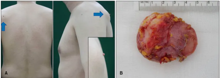
A 42-year-old male patient presented to our clinic with a skin-colored mass that had been present for 2 years; his medical and family histories were otherwise unremarkable.
Physical examination revealed a soft, movable, skin-col- ored mass on his upper left back, with a brown, bean-sized pedunculated nodule on the overlying skin (Fig. 1A). Exci- sional biopsy was performed. The tumor, which was well demarcated and localized in the subcutaneous layer, measured 5.4×5.1×2.1 cm (Fig. 1B). The histopathological examination indicated that the lesion was an encapsulated mass, located in subcutaneous tissue with a central cavity.
The tumor was well-circumscribed by a fibrous capsule, and no honeycomb pattern was observed in the marginal area. In the tumor periphery, large blood vessels with a di- lated, branching appearance were observed (Fig. 2A). The tumor cells were arranged in a palisading and partially
storiform pattern, with focal myxoid change (Fig. 2B).
Under high magnification, the tumor cells were spindle shaped with elongated nuclei and an ill-defined cytoplasm;
individual cells were infiltrating between collagen bundles and arranged in a palisading pattern, similar to Verocay bodies (Fig. 2C). Cellular atypia and mitotic bodies were not detected. Immunohistochemical analysis revealed that the cells were strongly positive for CD34 (Fig. 2D); focally positive for factor XIIIa; and negative for S-100 (Fig. 2E), desmin, actin, and CD68. A diagnosis of giant, deep, be- nign fibrous histiocytoma (BFH), with a palisading pattern, was rendered, with no evidence of recurrence at 15 months after excision.
BFH, which is characterized by several histological sub- types, is among the most-common soft tissue skin tumors.
Since Fletcher’s1 description in 1990 of 21 cases of deep BFH (DBFH), several more cases have been reported1-3.
Letter to the Editor
644 Ann Dermatol
Fig. 2. (A) Scanning view of the histopathological examination of the lesion showing an encapsulated mass located from the lower dermis through the subcutaneous tissue with a central cavity. The tumor was well circumscribed by a fibrous capsule, and no honeycomb pattern was detected in the marginal area. Hemangiopericytoma-like dilated vessels were seen in the periphery of the tumor (H&E,
×5). (B) Tumor cells arranged in a palisading pattern, similar to that of Verocay bodies, with a focal storiform pattern and partial myxoid change (H&E, ×40). (C) Spindle-shaped tumor cells with elongated nuclei and an ill-defined cytoplasm. Neither cellular atypia nor mitotic bodies were detected (H&E, ×400). Immunohistochemical staining showed (D) a strong positivity to CD34 (×40) and (E) negativity to S-100 (×40).
Fig. 1. (A) A skin-colored mass on the back (arrows) with a coincident overlying skin tag (inset: close-up view). (B) The excised 5×5×2 cm well-encapsulated erythematous oval mass.
DBFH, which accounts for <1%∼2% of all BFH cases, is larger and arises lower in the subcutis1, and confers an in- creased risk of local recurrence and distant metastasis3. Similar to BFH, giant fibrous histiocytoma (>5 cm), which represents a rare variant of fibrous histiocytoma, is benign, and no local recurrence after surgical excision has been reported to date4.
Palisading BFH is a rare variant of BFH, first described by
Schwob and Santa Cruz5 in 1986. On histopathological examination, palisading BFH resembles a schwannoma;
the lack of neural cells and S-100 negativity could facili- tate the differential diagnosis5. Although several cases of palisading BFH have been reported2,5, only one case of palisading DBFH has been documented2.
In our patient, the tumor cells were strongly positive for CD34; therefore, it is difficult to exclude dermatofibro-
Letter to the Editor
Vol. 27, No. 5, 2015 645 sarcoma protuberance. However, Gleason and Fletcher3
reported that 40% of DBFH cases are positive for CD34.
The histopathological findings of a well-circumscribed cap- sule, low cell density, and a lack of atypical cells and mi- totic bodies also suggest a benign nature; therefore, the fi- nal diagnosis was an uncommon case of giant DBFH.
REFERENCES
1. Fletcher CD. Benign fibrous histiocytoma of subcutaneous and deep soft tissue: a clinicopathologic analysis of 21 cases.
Am J Surg Pathol 1990;14:801-809.
2. Fukunaga M. Palisading subcutaneous fibrous histiocytoma.
Pathol Int 2004;54:360-363.
3. Gleason BC, Fletcher CD. Deep "benign" fibrous histiocytoma:
clinicopathologic analysis of 69 cases of a rare tumor in- dicating occasional metastatic potential. Am J Surg Pathol 2008;32:354-362.
4. Requena L, Fariña MC, Fuente C, Piqué E, Olivares M, Martín L, et al. Giant dermatofibroma. A little-known clinical variant of dermatofibroma. J Am Acad Dermatol 1994;30:
714-718.
5. Schwob VS, Santa Cruz DJ. Palisading cutaneous fibrous histiocytoma. J Cutan Pathol 1986;13:403-407.