Received May 12, 2014, Revised July 15, 2014, Accepted for publication July 21, 2014
Corresponding author: Burhan Engin, Cerrahpaşa Medical Faculty, Department of Dermatology, İstanbul University, 34098 İstanbul, Turkey. Tel: 90-212-414-30-00, Fax: 90-212-414-30-00, E-mail:
This is an Open Access article distributed under the terms of the Creative Commons Attribution Non-Commercial License (http://
creativecommons.org/licenses/by-nc/3.0) which permits unrestricted non-commercial use, distribution, and reproduction in any medium, provided the original work is properly cited.
ORIGINAL ARTICLE
Evaluation of Advanced Oxidation Protein Products, Prooxidant-Antioxidant Balance, and Total Antioxidant Capacity in Untreated Vitiligo Patients
Gülcan Güntaş, Burhan Engin1, Özlem Balcı Ekmekçi2, Zekayi Kutlubay1, Hakan Ekmekci2,
Abdullah Songür1, Tuğba Kevser Üstünbaş Uzunçakmak1, Hayriye Ertem Vehid3, Server Serdaroğlu1, Yalçın Tüzün1, Hafize Uzun2
Kırklareli University, School of Health, Kırklareli, 1Cerrahpaşa Medical Faculty, Department of Dermatology, İstanbul University,
2Cerrahpaşa Medical Faculty, Department of Medical Biochemistry, İstanbul University, 3Cerrahpaşa Medical Faculty, Department of Medical Biostatistics, İstanbul University, İstanbul, Turkey
Background: Vitiligo is a chronic, common disease of un- known etiology, and oxidative stress is suggested to have a role in its etiopathogenesis. Objective: Advanced oxidation protein products (AOPPs), prooxidant-antioxidant balance (PAB), and ferric-reducing antioxidant power (FRAP) were evaluated regarding their role in the pathogenesis of vitiligo as well as their relationship with clinical presentation and disease severity, and these parameters were compared with those of healthy controls. Methods: The study included 53 patients with vitiligo and 20 healthy volunteers as the control group. AOPP level, PAB, and FRAP were determined by col- orimetric methods. Results: PAB and FRAP level were sig- nificantly higher in patients with vitiligo than in healthy con- trols (p<0.001). The AOPP levels in vitiligo patients were not statistically significantly higher than those in healthy controls. The Vitiligo Area Scoring Index positively corre- lated with disease duration (rs: 0.531, p<0.001). Conclusion:
To the best of our knowledge, this is the first report of AOPP and PAB status in vitiligo. PAB may be used as an indicator for oxidative stress in the etiopathogenesis of vitiligo. Our re- sults show that these parameters may play a major role in the
melanocyte damage observed in vitiligo. Further studies are required to confirm the mechanisms underlying this effect.
(Ann Dermatol 27(2) 178∼183, 2015) -Keywords-
Advanced oxidation protein products, Ferric-reducing anti- oxidant power, Prooxidant-antioxidant balance, Vitiligo
INTRODUCTION
Vitiligo is a chronic, asymptomatic, usually acquired pig- mentation disorder characterized by depigmented white areas on the skin and mucosal surfaces1. Depigmentation is caused by melanocyte destruction that results in ab- sence of pigment production in these areas2. It affects ap- proximately 0.1%∼8.8% of the population2. The etiology remains unclear, but the theories regarding its patho- genesis include genetic, neural, cytotoxic, viral, growth fac- tor, autoimmune, and oxidant-antioxidant theories. However, none of these theories has been proven3.
Recently, oxidative stress has been extensively investigated in melanocyte destruction. This reaction is a result of over- production of reactive oxygen species (ROS) or impaired clearance of these species and characterized by an im- balance in the prooxidant and antioxidant systems4. To evaluate prooxidant-antioxidant balance (PAB), the deter- mination of both the oxidant status and the antioxidant status is often necessary. The majority of currently avail- able methods that separately determine oxidative balance measure the total prooxidant and antioxidant capacities.
Consequently, these methods are difficult, time-consum- ing, expensive, and imprecise5. Here, we evaluated the PAB in vitiligo patients using a simple, rapid, and inex- pensive PAB assay. Despite the well-documented role of oxidative stress in vitiligo, the determination of PAB is not a routine clinical laboratory test, mainly because of the lack of a universally accepted method6,7.
Some investigators have reported increased total anti- oxidant levels in serum, while others have reported nor- mal levels or decreased levels of these biomarkers2,8-10. In recent years, a simple test measuring the ferric-reducing ability of plasma, the ferric-reducing antioxidant power (FRAP) assay, has been presented as a new method for evaluating antioxidant capacity11. The total antioxidant ca- pacity (TAC) can be defined as the cumulative action of all antioxidants present in the plasma and body fluids, thus providing a composite parameter rather than the sum of measurable antioxidants12.
Clinically, the most important indicators of oxidative stress are lipid peroxidation and protein oxidation. Lipid perox- idation is particularly important in the vascular damage and melanocyte destruction in vitiligo. The addition of carbonyl groups, which are the result of proteolysis and breakage of protein side chains, are the most important components of protein oxidation13,14. Many of the pub- lished studies involve the detection of lipid peroxidation production and antioxidant levels in serum2,8-10. Studies regarding protein oxidation levels in patients with vitiligo are lacking. Recently, a new marker of protein oxidation, advanced oxidation protein products (AOPPs), has been considered for oxidant-mediated protein damage15. AOPPs are proteins that are damaged by oxidative stress, most no- tably albumin and its aggregates. AOPPs contain abundant dityrosines, which allow cross-linking, disulfide bridges, and carbonyl groups. AOPPs are formed primarily by chlorinated oxidants, including hypochloric acid and chloramines, which result from myeloperoxidase activity16. Although several studies show the importance of oxidative stress in the pathogenesis of vitiligo, we were unable to find data in the literature regarding the effects of im- mune-mediated oxidative stress on AOPPs and PAB in viti- ligo patients. Therefore, PAB, AOPP levels, and FRAP were evaluated regarding their role in the pathogenesis of vitiligo as well as their relationship with clinical pre- sentation and disease severity, and these parameters were compared with those of healthy controls.
MATERIALS AND METHODS
Subjects
A total of 73 individuals (53 vitiligo patients, mean age:
36.87±11.09 years; 20 controls, mean age: 48.00±4.81 years) were included in this study. None of the patients had been treated for vitiligo. A general dermatological ex- amination was performed for all patients. The location of vitiligo lesions was recorded. Clinical subtypes of vitiligo were defined as segmental or nonsegmental. The severity and extent of disease were calculated using the palmar unit measurement (one palmar unit represents the depig- mented lesion size of the palm of the hand). According to the history provided by the patients, the Vitiligo Disease Activity Score (VIDA) was used to determine either active or stable disease. Active disease refers to an expansion of lesions and appearance of new lesions in the previous year. All patients and controls were of Turkish descent. All participants were informed about the survey and freely signed and dated the consent form. The protocol was ap- proved by the ethics committee of the Cerrahpaşa Medical Faculty and was conducted in accordance with the Decla- ration of Helsinki.
Inclusion criteria
Eligibility was based on the following inclusion criteria: >18 years old; either sex; not pregnant; no smoking habit; no history of an autoimmune disease, concomitant dermato- logical disease, or thyroid dysfunction; and no history of smoking, alcohol intake, vitamin intake, or use of anti-in- flammatory or other drugs. In addition, none of the pa- tients had undergone any therapy for 3 months prior to enrolment.
Exclusion criteria
The control group consisted of 20 healthy subjects se- lected from the hospital staff with no personal or family history of diabetes or dyslipidemia, and had normal thy- roid, hepatic, and renal function. The 2-h glucose toler- ance test was used to rule out impaired glucose regulation in the healthy controls. Smokers and healthy subjects who were using drugs that were known to affect carbohydrate and lipid metabolism or oxidant and antioxidant status were also excluded from the control group.
Laboratory analysis
1) Sample collection and preparation
None of the patients and controls used any drugs for at least 24 h prior to blood collection. Clinical parameters, including routine biochemical parameters, were measured using standard protocols. Peripheral blood samples from patients with vitiligo were obtained from the Department of Dermatology in the Cerrahpaşa Medical School. Normal peripheral blood samples were obtained from the clin-
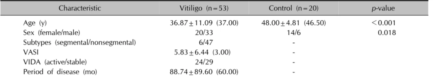
Table 1. Demographic and clinical characteristics of vitiligo patients and controls
Characteristic Vitiligo (n=53) Control (n=20) p-value
Age (y) 36.87±11.09 (37.00) 48.00±4.81 (46.50) <0.001
Sex (female/male) 20/33 14/6 0.018
Subtypes (segmental/nonsegmental) 6/47 -
VASI 5.83±6.44 (3.00) -
VIDA (active/stable) 24/29 -
Period of disease (mo) 88.74±89.60 (60.00) -
Values are presented as mean±standard deviation (median). VASI: Vitiligo Area Scoring Index, VIDA: Vitiligo Disease Activity Score.
ically healthy individuals in our medical school and in our departments. Blood samples were collected in EDTA-con- taining tubes and anticoagulant-free tubes after an over- night fast. After immediate centrifugation (3,000 g) for 10 min at 4oC, plasma and serum were separated in Eppendorf tubes and frozen immediately at −80oC until analysis.
2) Measurements of plasma advanced protein oxidation product levels
Spectrophotometric determinations of AOPP levels were performed using a modification of the method by Gelisgen et al.17 Samples were prepared as follows: 200 μl of plas- ma was diluted 1 : 5 in PBS, followed by the addition of 10 μl of 1.16 M KI to each tube and, finally, 20 μl of ab- solute acetic acid 2 min later. The absorbance of the re- action mixture was immediately read at 340 nm against a blank containing 2,000 μl of PBS, 200 μl of acetic acid, and 100 μl of KI. The linear range of chloramine-T ab- sorbance at 340 nm occurs between 0 and 100 μmol/L.
AOPP concentrations are expressed in μmol/L of chlor- amine-T equivalents. The coefficients of intra- and in- ter-assay variation were 3.1% (n=15) and 3.6% (n=15), respectively.
3) Measurement of serum ferric-reducing antioxidant power
The FRAP assay was performed according to the protocol of Benzie and Strain11, with minor modifications. To pre- pare the FRAP solution, 10 ml of acetate buffer (300 mM, pH=3.6) was mixed with 1 ml of 20 mM FeCl3 dissolved in distilled water and 1 ml of 10 mM 2,4,6-tri (2-pyridyl)-s-tri- azine dissolved in 40 mM HCl. Next, 50 μl of serum was added to 1.5 ml of a freshly prepared FRAP solution and measured over a period of 4 min at 593 nm. Different concentrations of uric acid (0.09∼9.00 mM) were used to obtain a calibration curve on each day of the experiment.
Uric acid is the primary contributor of the antioxidant ca- pacity of serum17 and was thus used as a standard. The co- efficients of intra- and inter-assay variations were 4.2%
(n=15) and 6.1% (n=15), respectively.
4) Measurement of serum prooxidant-antioxidant balance The PAB assay was performed according to the method of Alamdari et al.7, with minor modifications. Tetramethyl- benzidine 3, 3′, 5.5′ (TMB) and TMB cations were used as oxidation-reduction indicators, due to their optical and electrochemical properties. With this approach, it is possi- ble to measure the PAB simultaneously in one experiment by two different reaction types: one enzymatic reaction where the chromogen TMB is oxidized to a color cation by peroxides and one chemical reaction where the TMB cation is reduced to a colorless compound by antioxidants.
The absorbance was measured using an enzyme-linked immunosorbent assay plate reader (ELX 800 UV; BioTek Instruments, Winooski, VT, USA) at 450 nm, with a refer- ence wavelength of 620 or 570 nm. A standard curve was determined using the values obtained from the standard samples. PAB are expressed in arbitrary units, which cor- respond to the percentage of H2O2 in the standard solution. The coefficients of intra- and inter-assay variation were 5.3% (n=15) and 6.6% (n=15), respectively.
5) Statistical analysis
Data analysis was conducted using IBM SPSS Statistics 21.0 (IBM Co., Armonk, NY, USA). The results are re- ported as mean±standard deviation or median. Sex dis- tribution was compared between the groups using Pearson chi-square analysis. Because the data were not normally distributed (Shapiro-Wilk test, p<0.05), nonparametric statistics (Mann-Whitney U tests) were used to evaluate AOPP levels and FRAP. Parametric statistics (Student t-tests) were used to evaluate PAB and age. Spearman correlation tests were used to determine correlations. Statistical sig- nificance was determined using p<0.05.
RESULTS
The demographic and clinical characteristics of the groups are shown in Table 1. According to the Pearson chi-square analysis, the distribution of sex was significantly different
Table 2. Oxidative stress parameters of vitiligo patients and controls
Parameter Vitiligo (n=53) Control (n=20) p-value
AOPPs (μmol/L chloramine T) 55.16±27.59 (42.09) 47.37±13.32 (46.04) 0.853
FRAP (mmol/L uric acid) 0.17±0.05 (0.16) 0.13±0.03 (0.12) <0.001
PAB (AU) 41.79±18.79 (42.95) 17.29±12.80 (19.51) <0.001
Values are presented as mean±standard deviation (median). AOPPs: advanced protein oxidation products, FRAP: ferric-reducing antioxidant power, PAB: prooxidant–antioxidant balance, AU: arbitrary units that correspond to the percentage of H2O2 in the standard solution.
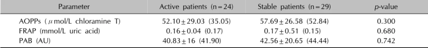
Table 3. Oxidative stress parameters of vitiligo patients with active or stable disease
Parameter Active patients (n=24) Stable patients (n=29) p-value
AOPPs (μmol/L chloramine T) 52.10±29.03 (35.05) 57.69±26.58 (52.84) 0.300
FRAP (mmol/L uric acid) 0.16±0.04 (0.17) 0.17±0.51 (0.15) 0.680
PAB (AU) 40.83±16 (41.90) 42.56±20.65 (44.44) 0.742
Values are presented as mean±standard deviation (median). AOPPs: advanced protein oxidation products, FRAP: ferric-reducing antioxidant power, PAB: prooxidant–antioxidant balance, AU: arbitrary units correspond to the percentage of H2O2 in the standard solution.
Fig. 1. Relationship between the Vitiligo Area Scoring Index (VASI) and disease duration in patients with vitiligo.
between the groups (p=0.018).
The oxidative stress parameters in the groups are shown in Table 2. FRAP and PAB in patients with vitiligo were sig- nificantly higher than those in the control group (p<0.001).
However, the AOPP levels in the patient group were not significantly higher than those in the control group be- cause of the high median values in the control group.
AOPP levels, FRAP, and PAB did not correlate with age, period of disease, vitiligo subtypes, the Vitiligo Area Scoring Index (VASI), or VIDA (Table 3) in the patient group. However, there was a significant positive correla- tion (rs: 0.531, p<0.001) between the disease duration and VASI (Fig. 1).
DISCUSSION
Human skin serves as an interface between the environ- ment and the body. It is constantly exposed to a broad ar- ray of physical, chemical, and biological agents, many of which either are inherent oxidants or catalyze the gen- eration of ROS. Oxidative stress is believed to be involved in the pathogenesis of inflammatory skin diseases18. The present study demonstrated that FRAP and PAB were sig- nificantly higher in patients with vitiligo than in healthy controls. Although the AOPP levels were higher in the pa- tient group, this was not statistically significant. These re- sults indicate that increased FRAP, PAB, and AOPP levels are likely associated with oxidative stress in vitiligo.
Melanocyte damage in vitiligo pathogenesis remains unclear.
A single mechanism is not responsible for all cases of mel- anocyte damage in vitiligo, and oxidative stress is consid- ered a possible pathogenic event in melanocyte loss6. PAB is important for establishing the health status of the body19. Decreased antioxidant levels and increased prooxidant levels may play important roles in the damage of melano- cytes observed in vitiligo patients20. PAB was higher in vi- tiligo patients in the present study. One possible ex- planation for this might be impaired PAB. A shift in this equilibrium likely contributes to the pathogenesis of vitiligo.
The PAB assay is a simple, rapid, and inexpensive method that can measure the prooxidant burden and the anti- oxidant capacity in one assay. This ratio can be used as an additive marker of oxidative stress in patients with vitiligo as in patients with Behçet disease (BD)19. Based on our re-
sults, PAB may be the initial pathogenic event in melano- cyte degeneration. Nevertheless, we suggest that the PAB assay may be useful as a risk predictor in diseases such as vitiligo and could help to identify patients with high oxi- dative stress to facilitate early preventive interventions for melanocyte damage.
Several investigators have assessed the capacity of anti- oxidative defense systems in vitiligo21,22. However, the re- sults are controversial. TAC has been reported to be low- er21 or higher22, depending on the study. Akoglu et al.21 reported that patients with vitiligo had significantly lower TAC than controls. Boisseau-Garsaud et al.22 reported the blood antioxidant status of African American patients with active generalized vitiligo. The total blood antioxidant lev- el was significantly higher in vitiligo patients than in sex- and age-matched controls. The FRAP in patients with vitili- go was also significantly higher than that in the control group. Our results are similar to those of Boisseau-Garsaud et al.22 Perhaps, antioxidant molecules cannot be used ac- tively within the white areas. Clinically, it is also difficult to determine the active period of vitiligo. In contrast, we found that both PAB and FRAP were higher, but not corre- lated, in vitiligo patients. It is unclear whether high FRAP indicates a resource of unused antioxidants or up-regu- lated antioxidant production as a response to oxidative stress. We consider oxidative stress as a causative factor in patients with vitiligo. However, further studies are re- quired to confirm the mechanisms underlying this effect.
AOPPs have been proposed as a possible marker of oxida- tive injury that originate as a result of the action of free radicals on proteins. Although AOPP levels were higher, this was not statistically significant. AOPP levels have been reported to be significantly increased in active peri- ods of patients with BD compared with those in healthy controls and in the remission periods of patients with BD19. Previous studies (both human and experimental) have demonstrated that older people have higher AOPP levels19. Despite an older age in the control group, AOPP levels were higher (statistically insignificant) in patients with vitiligo in the present study. This might partially sup- port the role of oxidative stress in vitiligo pathogenesis. In addition, high AOPP levels are indicative of an increase in oxidative stress and can cause direct oxidative damage to proteins in vitiligo patients. These oxidized proteins may partially contribute to the development of melanocyte loss.
As a disease characterized with oxidative stress-auto- immunity-mediated melanocyte loss, the pathogenesis of vitiligo is unclear. Our results indicate that oxidative stress might play a role in the etiopathogenesis of vitiligo, be- cause of the increased serum PAB and AOPP. PAB may be especially useful in this regard because of its measurement
is rapid, easy, and cost-effective in laboratories. Thus, we recommend that PAB measurement be more widely used in clinical practice. FRAP should be reevaluated in future clinical trials in the early stage of the disease. Also, FRAP is important in treated patients with vitiligo due to changes in the antioxidant capacity as a result of treatment.
REFERENCES
1. Denli Y, Acar MA, Sönmezoğlu Maraklı S, Yücel A. Vitiligo.
Disorders of pigmentation. In: Tüzün Y, Gürer MA, Ser- daroğlu S, Oğuz O, Aksungur V, editors. Dermatology. 3rd ed. İstanbul: Nobel Publishing, 2008:1465-1490.
2. Kutlubay Z, Uzuncakmak TK, Engin B, Tüzün Y. Vitiligo and oxidative stress. J Turk Acad Dermatol 2011;5:1154r1.
3. Schallreuter KU, Bahadoran P, Picardo M, Slominski A, Elassiuty YE, Kemp EH, et al. Vitiligo pathogenesis: autoi- mmune disease, genetic defect, excessive reactive oxygen species, calcium imbalance, or what else? Exp Dermatol 2008;17:139-140; discussion 141-160.
4. Karaca Ş, Güder H. Antioxidant system in dermatology.
Turk J Dermatol 2009;3:32-39.
5. Parizadeh SM, Azarpazhooh MR, Moohebati M, Nematy M, Ghayour-Mobarhan M, Tavallaie S, et al. Simvastatin therapy reduces prooxidant-antioxidant balance: results of a placebo- controlled cross-over trial. Lipids 2011;46:333-340.
6. Laddha NC, Dwivedi M, Mansuri MS, Gani AR, Ansarullah M, Ramachandran AV, et al. Vitiligo: interplay between oxidative stress and immune system. Exp Dermatol 2013;
22:245-250.
7. Alamdari DH, Ghayour-Mobarhan M, Tavallaie S, Parizadeh MR, Moohebati M, Ghafoori F, et al. Prooxidant-antioxidant balance as a new risk factor in patients with angiographically defined coronary artery disease. Clin Biochem 2008;41:
375-380.
8. Arican O, Kurutas EB. Oxidative stress in the blood of patients with active localized vitiligo. Acta Dermatovenerol Alp Pannonica Adriat 2008;17:12-16.
9. Jain A, Mal J, Mehndiratta V, Chander R, Patra SK. Study of oxidative stress in vitiligo. Indian J Clin Biochem 2011;26:
78-81.
10. Koca R, Armutcu F, Altinyazar HC, Gürel A. Oxidant- antioxidant enzymes and lipid peroxidation in generalized vitiligo. Clin Exp Dermatol 2004;29:406-409.
11. Benzie IF, Strain JJ. The ferric reducing ability of plasma (FRAP) as a measure of "antioxidant power": the FRAP assay.
Anal Biochem 1996;239:70-76.
12. Karacay O, Sepici-Dincel A, Karcaaltincaba D, Sahin D, Yalvaç S, Akyol M, et al. A quantitative evaluation of total antioxidant status and oxidative stress markers in preeclampsia and gestational diabetic patients in 24-36 weeks of gestation.
Diabetes Res Clin Pract 2010;89:231-238.
13. Breusing N, Grune T. Biomarkers of protein oxidation from a chemical, biological and medical point of view. Exp Gerontol 2010;45:733-737.
14. Dalle-Donne I, Rossi R, Giustarini D, Milzani A, Colombo
R. Protein carbonyl groups as biomarkers of oxidative stress.
Clin Chim Acta 2003;329:23-38.
15. Zhou Q, Wu S, Jiang J, Tian J, Chen J, Yu X, et al.
Accumulation of circulating advanced oxidation protein products is an independent risk factor for ischaemic heart disease in maintenance haemodialysis patients. Nephrology (Carlton) 2012;17:642-649.
16. Cakatay U, Kayali R, Uzun H. Relation of plasma protein oxidation parameters and paraoxonase activity in the ageing population. Clin Exp Med 2008;8:51-57.
17. Gelisgen R, Genc H, Kayali R, Oncul M, Benian A, Guralp O, et al. Protein oxidation markers in women with and without gestational diabetes mellitus: a possible relation with paraoxonase activity. Diabetes Res Clin Pract 2011;
94:404-409.
18. Glassman SJ. Vitiligo, reactive oxygen species and T-cells.
Clin Sci (Lond) 2011;120:99-120.
19. Ozyazgan S, Andican G, Erman H, Tuzcu A, Uzun H, Onal B, et al. Relation of protein oxidation parameters and disease activity in patients with Behçet's disease. Clin Lab 2013;59:819-825.
20. Schallreuter KU. Successful treatment of oxidative stress in vitiligo. Skin Pharmacol Appl Skin Physiol 1999;12:132-138.
21. Akoglu G, Emre S, Metin A, Akbas A, Yorulmaz A, Isikoglu S, et al. Evaluation of total oxidant and antioxidant status in localized and generalized vitiligo. Clin Exp Dermatol 2013;
38:701-706.
22. Boisseau-Garsaud AM, Garsaud P, Lejoly-Boisseau H, Robert M, Quist D, Arveiler B. Increase in total blood antioxidant status and selenium levels in black patients with active vitiligo. Int J Dermatol 2002;41:640-642.