INTRODUCTION
Gastrointestinal stromal tumors (GISTs) represent 1% of all gastrointestinal tract neoplasms and have an approximate in- cidence of 10 to 20 cases per million individuals per year [1]. In the United States, approximately 4,500 new cases of GIST are diagnosed each year [1]. The average age of patients at pre-
sentation is 60 years, and there is no gender predilection [1- 3]. GISTs may arise from the stomach (50-60% of GISTs), small bowel (20-30%), large bowel (10%), and esophagus (5%), or elsewhere in the abdominal cavity (e.g., omentum, mesentery) (5%) [4].
GISTs were initially described in the 1940s by Stout, who suggested that these tumors arose from the smooth muscle and therefore referred to them as leiomyomas or leiomyo- sarcomas [5]. The concept that these tumors arise from the smooth muscle lasted until the advent of electron microscopy and immunohistochemistry. In 1983, Mazur and Clark [6]
coined the term “GIST” to describe stromal tumors that had no differentiation into smooth muscle or into Schwann cells. In
Gastrointestinal stromal tumors as an incidental finding in patients with a presumptive diagnosis of ovarian cancer
Mario Muñoz1, Pedro T. Ramirez2, Carolina Echeverri3, Luis Guillermo Álvarez4, Maria Alejandra Palomino5, Luis René Pareja6
1Department of General Surgery, Instituto de Cancerologia, Clínica las Américas, Medellin, Colombia; 2Department of Gynecologic Oncology, The University of Texas MD Anderson Cancer Center, Houston, TX, USA; 3Department of Pathology, Clínica Las Américas, Medellin, Colombia; 4Department of Radiology, Hospital General de Medellin, Medellin, Colombia; 5Department of Gynecologic Oncology, Clinica Comfamiliar Risaralda, Pereira, Colombia; 6Department of Gynecologic Oncology, Instituto de Cancerologia, Clínica las Américas, and Department of Gynecology and Obstetrics, Universidad Pontificia Bolivariana, Medellin, Colombia
Received Sep 28, 2011, Revised Oct 31, 2011, Accepted Dec 13, 2011 Correspondence to Luis Rene Pareja
Department of Gynecologic Oncology, Instituto de Cancerologia, Clínica las Américas, Carrera 80 Diagonal 75 B # 2 A 80-140 Medellin, Colombia. Tel:
57-4-340-9383, Fax: 57-4-340-9370, E-mail: [email protected]
Objective: To report the clinical presentation and oncologic outcomes of a series of patients who presented with an abdominal or pelvic mass and were diagnosed with a gastrointestinal stromal tumor (GIST).
Methods: Data were obtained on all patients who presented with an abdominal or pelvic mass between September 2007 and June 2010 and who were ultimately diagnosed with a GIST. The patients’ medical records were reviewed. A literature review was also conducted.
Results: Six patients were identified who met the inclusion criteria. All six patients had a tumor in the intestinal tract arising from the small bowel. The mean tumor size was 12 cm (range, 6 to 22 cm). A complete resection was achieved in five of the six patients. There were no intraoperative complications; one patient had a postoperative complication. Two patients were treated with imatinib after surgery. The mean follow-up time was 32 months (range, 0.3 to 40 months). At the last follow-up, five of the six patients were without any evidence of disease. One patient died of an unrelated hepatic encephalopathy. The incidence in our institution is 3%.
Conclusion: GISTs are uncommon; however, they should be considered in the differential diagnosis of patients presenting with an abdominal or pelvic mass.
Keywords: Adnexal mass, Gastrointestinal stromal tumor, Incidental finding
the late1990s, several groups noted a similarity between the neoplastic cells of GISTs and the interstitial cells of Cajal [7-9].
GISTs typically express the receptor tyrosine kinase c-kit, also known as CD117 [4]. The prognosis of patients with a GIST is based on tumor size, mitotic rate, and organ of origin.
The published literature on GISTs in patients who present with an abdominal or pelvic mass is limited to isolated case reports and small case series [7-14]. The goal of this study was to report on a series of patients who presented with a presumptive diagnosis of ovarian cancer and who were ulti- mately found to have a GIST. We describe the clinical presen- tation, surgical management, adjuvant treatment, and overall outcome in these patients. In this report, we also summarize the published literature on GISTs presenting as ovarian cancer.
MATERIALS AND METHODS
After Institutional Review Board approval was received from Instituto de Cancerologia, Clinica las Americas, Medellin, Co- lombia, data were collected on patients who presented to In- stituto de Cancerologia with an abdominal or pelvic mass be- tween September 2007 and June 2010. Data were abstracted from the patients’ existing medical records on age, presenting symptoms, tumor size on physical examination, findings on imaging studies, serum CA-125 levels, intraoperative findings, primary site of disease, pathologic diagnosis, mitotic rate, ad- juvant therapy, and disease status at last follow-up.
Patients were included in this study if they met the following criteria: complete medical records, radiologic imaging show- ing a pelvic or abdominopelvic mass, CA-125 levels recorded, and a final pathology diagnosis of a GIST. CA-125 levels were measured by microparticle enzymatic immunoassay, and nor- mal values were defined as <35 U/mL. Pathologic diagnosis was confirmed by conventional methods and through immu- nohistochemistry using the following markers: c-Kit (CD117), desmin, smooth muscle actin, CD34, and S-100 protein.
We also performed a literature review on PubMed database, limited to the English language, using the following terms:
“gastrointestinal stromal tumor,” “adnexal mass,” and “inciden- tal finding.”
RESULTS
Six patients met the inclusion criteria and are the subject of this report. The mean age was 59 years (range, 42 to 86 years).
All patients were referred to the Instituto de Cancerologia with a presumptive diagnosis of ovarian cancer. The presentations
of these patients were as follows: lower abdominal pain with duration ranging from 2 weeks to 3 months prior to consulta- tion (4 patients), abnormal uterine bleeding with duration of 2 weeks (1 patient), and incidental finding of an adnexal mass on screening examination (1 patient).
On physical examination, all six patients had a palpable firm abdominal mass. On pelvic examination, four patients had a painful palpable mass and one patient had no abnormal find- ings; one patient did not allow pelvic examination. The mean size of the masses was 12 cm (range, 6 to 22 cm). On imaging studies, four patients had an ovarian mass visualized on both computed tomography and ultrasonography. The other two patients had a mass documented on computed tomogra- phy only (one patient) or ultrasonography only (one patient).
The mean serum CA-125 level for all patients was 41.2 U/mL (range, 1.6 to 156 U/mL) and the mean level was 20.05. Only one patient presented with a CA-125 level greater than 35 U/
mL (156 U/mL).
All patients underwent an exploratory laparotomy and had a preoperative diagnosis of ovarian malignancy. At the time of surgery, three patients were found to have a mass arising from the jejunum, and the other three patients were found to have a mass arising from the ileum. Two patients had inci- dental findings not described on the preoperative computed tomography or ultrasonography: hepatic metastatic nodules in one patient and a cirrhotic liver and ascites in the other pa- tient. All patients underwent intestinal resection with end-to- end anastomosis. Only one patient had residual disease. This was the patient who was found to have hepatic metastatic nodules during the intraoperative assessment. There were no intraoperative complications. The postoperative course was complicated in one patient, who developed a postoperative ileus that resolved after 6 days of conservative management.
The mean hospital stay for all patients was 6 days (range, 5 to 8 days).
On final pathology review, all patients had immunohis- tochemical analysis and all stained positive for CD117, two patients had disease considered to be intermediate risk for recurrence and four had disease considered as high risk.
The clinical oncologist reviewed all cases. The patients with intermediate-risk disease did not receive adjuvant therapy due their low mitosis rate. Two of the patients with high-risk disease were treated with imatinib (400 mg daily) after sur- gery. One of these two patients had a high rate of mitosis (28 per 50 high-power fields [HPF]) and underwent therapy with imatinib for a year; the other patient had evidence of residual disease in the liver and is still on imatinib treatment to date.
One of the patients with high-risk disease did not receive adjuvant therapy because of the low mitotic rate of the tu-
mor. The other patient with high-risk disease died soon after surgery from encephalopathy associated with cirrhosis. At a mean follow-up time of 32 months (range, 0.3 to 40 months), all five patients who were still alive were without any evidence of disease. As there were only 6 patients, this would not allow for appropriate statistical analysis. A total of approximately 200 patients with ovarian cancer were operated at Instituto de Cancerologia Las Americas during the time span of the study.
Based on this information the incidence is approximately 3%.
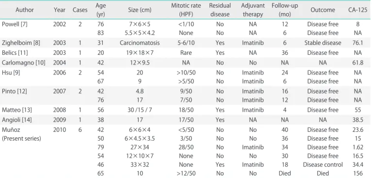
As a result of our literature review, we found eight reports including 11 patients who presented with an abdominal or pelvic mass and were ultimately diagnosed with a GIST. The details on these patients as well as the six patients in the cur- rent series are presented in Table 1. The mean age of the 11 previously reported patients was 53 years (range, 20 to 83 years). Most patients presented complaining of an abdominal mass. The diagnostic images usually showed irregular lesions with areas of necrosis of heterogeneous content ranging in size from 5 cm to giant or even carcinomatosis. All 11 patients underwent surgical resection, and six (54%) of them were treated with the tyrosine kinase inhibitor imatinib. The mean reported follow-up time was 12.2 months (range, 4 to 36 months). No patients were reported to have relapsed during follow-up; one patient had carcinomatosis at the time of sur- gery, progression before starting imatinib, but stable lesions at 6 months [8].
DISCUSSION
GISTs are rare tumors and may present a diagnostic dilem- ma. Complete surgical resection is considered the standard treatment for localized tumors. Segmental intestinal resection is often necessary as these tumors present with an exophytic (bulky growth) rather than infiltrative pattern. When segmen- tal resection is not possible, a wide, en bloc resection should be performed [10,15]. For metastatic and recurrent disease, surgery remains the first choice of therapy [1,15].
Approximately 95% of GISTs express the marker c-kit (CD117) [16]. Detection of receptor tyrosine kinase c-kit status is critical in the management of GISTs since tyrosine kinase inhibitors such as imatinib (Gleevec), sunitinib (Sutent), sorafenib (Nexavar), dasatinib, IPI504, oblimersen (Genasense), and perifosine have been shown to be effective treatments [4,17]. The use of such tyrosine kinase inhibitors is considered the standard therapy in patients with metastatic or unresectable disease [15,17].
The preferred imaging modality in the evaluation of patients with a suspected GIST is enhanced computed tomography.
The predominant pattern is a heterogeneously enhancing exophytic mass. Gastric lesions are characterized by a homo- geneous enhancement, while small bowel lesions are more heterogeneous. Since most GISTs grow exophytically and do not compromise the bowel concentrically, findings consis- tent with intestinal obstruction are not very common even
Table 1. Patient characteristics
Author Year Cases Age
(yr) Size (cm) Mitotic rate (HPF)
Residual disease
Adjuvant therapy
Follow-up
(mo) Outcome CA-125
Powell [7] 2002 2 76
83
7×6×5 5.5×5×4.2
<1/10 None
No No
NA NA
12 6
Disease free Disease free
8 NA
Zighelboim [8] 2003 1 31 Carcinomatosis 5-6/10 Yes Imatinib 6 Stable disease 76.1
Belics [11] 2003 1 20 19×18×7 Rare Yes NA 36 Disease free NA
Carlomagno [10] 2004 1 42 12×9.5 NA No No NA NA 61.8
Hsu [9] 2006 2 54
67
20 9
>10/50
>5/50
No No
Imatinib Imatinib
24 6
Disease free Disease free
NA NA
Pinto [12] 2007 2 42
76
4.8 17
9/50 7/50
No No
Imatinib Imatinib
16 12
Disease free Disease free
NA NA
Matteo [13] 2008 1 56 30 /15 / 7 18/50 Yes Imatinib 4 Disease free 55
Angioli [14] 2009 1 38 17 17/50 Yes NA NA NA 38.5
Muñoz (Present series)
2010 6 42
50 79 54 46 65
6×6×4 6×4.5×3.5
27×34 12×10×7
33×32 10
<5/50 3/50 28/50 None None
>12/50
No No No No Yes No
No No Imatinib
No Imatinib
No
40 36 34 30 18 Died
Disease free Disease free Disease free Disease free Disease control
Died
23.6 15 1.62 16.5 34.4 156 HPF, high-power fields; NA, not available.
with very large masses. Mesenteric extension may occur with encasement of adjacent organs [18-21]. Distal small bowel tumors and those arising from the sigmoid colon and rectum may be erroneously identified as gynecologic cancers [22].
Gross histopathologic findings typically include a fish-flesh appearance of soft consistency. Tumors can be necrotic or present with cystic degeneration. Approximately 70% are composed of spindle cells, 20% of epithelioid cells, and 10%
have a mixed phenotypic appearance [22-24]. Other markers expressed by these tumors are CD34 (70% of tumors), smooth muscle actin (30%), h-caldesmon (60%), and S-100 (5%) [24,25]. The DOG1 (discovered on GIST) marker is a fragment of cDNA encoding a protein whose function is unknown. This marker may be helpful when a GIST is negative for c-kit; DOG1 has high sensitivity (94.4%) in the diagnosis of GISTs and is positive in 30% of those that are negative for c-kit [26,27].
The prognosis of patients with a GIST is based on tumor size and number of mitoses. Tumors smaller than 2 cm with fewer than 5 mitoses per 50 HPF are considered very low risk for re- currence. Tumors measuring 2 to 5 cm with fewer than 5 mi- toses per 50 HPF are considered low risk. Tumors smaller than 5 cm with 6 to 10 mitoses per 50 HPF and tumors measuring 5 to 10 cm with fewer than 5 mitoses per 50 HPF are considered intermediate risk. High-risk tumors are those measuring 5 to 10 cm with 6 to 10 mitoses per 50 HPF, any tumor larger than 10 cm, and any tumor with more than 10 mitoses per 50 HPF [2,12,28].
All patients diagnosed with a GIST that is considered inter- mediate risk or high risk should be treated with a tyrosine kinase inhibitor—either imatinib 400 mg per day (first choice) or sunitinib 50 mg per day for 4 weeks on and 2 weeks off (second choice) either of them for at least 1 year. These regi- mens have been shown to improve disease-free survival and overall survival [1,16,17,26,27,29]. In patients treated with ty- rosine kinase inhibitors, studies have documented a relapse- free survival rate of 97% at 1 year, a 5-year disease-free survival rate of 65%, and a 5-year disease-specific survival rate of 68%
[4,29].
Unlike tyrosine kinase inhibitors, systemic chemotherapy has minimal effect on GISTs and thereby is not recommended for either neoadjuvant or adjuvant therapy [1]. Complete resec- tion is curative in patients with low-risk tumors, even without lymph node resection; lymphadenectomy does not improve survival since lymph node metastases are rare [1,16], and there is no need for adjuvant therapy in both very low risk and low risk patients . In patients with unresectable GISTs, neoadju- vant therapy with tyrosine kinase inhibitors can lead to a less extensive surgical procedure and decrease the risk of tumor rupture and bleeding during resection [1,17,18,29]. As cited
by several authors, tumors arising in the small intestine have been shown to be more aggressive and are associated with a poorer prognosis and higher rate of progressive disease than those arising from the stomach [16,30,31].
In summary, the diagnosis of a GIST should be entertained in all patients diagnosed with an abdominal or pelvic mass.
All patients with intermediate and high-risk GISTs should be treated postoperatively with tyrosine kinase inhibitors.
CONFLICT OF INTEREST
No potential conflict of interest relevant to this article was reported.
REFERENCES
1. Schnadig ID, Blanke CD. Gastrointestinal stromal tumors:
imatinib and beyond. Curr Treat Options Oncol 2006;7:
427-37.
2. Joensuu H. Current perspectives on the epidemiology of gastrointestinal stromal tumours. Eur J Cancer Suppl 2006;
4:4-9.
3. Schubert ML, Moghimi R. Gastrointestinal stromal tumor (GIST). Curr Treat Options Gastroenterol 2006;9:181-8.
4. Quek R, George S. Gastrointestinal stromal tumor: a clinical overview. Hematol Oncol Clin North Am 2009;23:69-78.
5. Stout AP. Bizarre smooth muscle tumors of the stomach.
Cancer 1962;15:400-9.
6. Mazur MT, Clark HB. Gastric stromal tumors: reappraisal of histogenesis. Am J Surg Pathol 1983;7:507-19.
7. Powell JL, Kotwall CA, Wright BD, Temple RH Jr, Ross SC, White WC. Gastrointestinal stromal tumor mimicking ova- rian neoplasia. J Pelvic Surg 2002;8:117-9.
8. Zighelboim I, Henao G, Kunda A, Gutierrez C, Edwards C.
Gas trointestinal stromal tumor presenting as a pelvic mass.
Gynecol Oncol 2003;91:630-5.
9. Hsu S, Chen SS, Chen YZ. Gastrointestinal stromal tumors presenting as gynecological tumors. Eur J Obstet Gynecol Reprod Biol 2006;125:139-40.
10. Carlomagno G, Beneduce P. A gastrointestinal stromal tu- mor (GIST) masquerading as an ovarian mass. World J Surg Oncol 2004;2:15.
11. Belics Z, Csapo Z, Szabo I, Papay J, Szabo J, Papp Z. Large gastrointestinal stromal tumor presenting as an ovarian tumor: a case report. J Reprod Med 2003;48:655-8.
12. Pinto V, Ingravallo G, Cicinelli E, Pintucci A, Sambati GS, Mari- naccio M, et al. Gastrointestinal stromal tumors mimi cking
gynecological masses on ultrasound: a report of two cases.
Ultrasound Obstet Gynecol 2007;30:359-61.
13. Matteo D, Dandolu V, Lembert L, Thomas RM, Chatwani AJ.
Unusually large extraintestinal GIST presenting as an abdo- mino-pelvic tumor. Arch Gynecol Obstet 2008;278:89-92.
14. Angioli R, Battista C, Muzii L, Terracina GM, Cafa EV, Sereni MI, et al. A gastrointestinal stromal tumor presenting as a pelvic mass: a case report. Oncol Rep 2009;21:899-902.
15. Blay JY, Bonvalot S, Casali P, Choi H, Debiec-Richter M, Dei Tos AP, et al. Consensus meeting for the management of gas tro intestinal stromal tumors: report of the GIST Con - sen sus Conference of 20-21 March 2004, under the au spi- ces of ESMO. Ann Oncol 2005;16:566-78.
16. Sarlomo-Rikala M, Kovatich AJ, Barusevicius A, Miettinen M. CD117: a sensitive marker for gastrointestinal stromal tumors that is more specific than CD34. Mod Pathol 1998;
11:728-34.
17. Kubota T. Gastrointestinal stromal tumor (GIST) and imati- nib. Int J Clin Oncol 2006;11:184-9.
18. Levy AD, Remotti HE, Thompson WM, Sobin LH, Miettinen M. Gastrointestinal stromal tumors: radiologic features with pathologic correlation. Radiographics 2003;23:283-304.
19. Sandrasegaran K, Rajesh A, Rydberg J, Rushing DA, Akisik FM, Henley JD. Gastrointestinal stromal tumors: clinical, radiologic, and pathologic features. AJR Am J Roentgenol 2005;184:803-11.
20. Gong JS, Zuo M, Yang P, Zang D, Zhang Y, Xia L, et al. Value of CT in the diagnosis and follow-up of gastro intesti nal stro- mal tumors. Clin Imaging 2008;32:172-7.
21. Levy AD, Remotti HE, Thompson WM, Sobin LH, Miettinen M. Anorectal gastrointestinal stromal tumors: CT and MR imaging features with clinical and pathologic correlation.
AJR Am J Roentgenol 2003;180:1607-12.
22. Miettinen M, Monihan JM, Sarlomo-Rikala M, Kovatich AJ, Carr NJ, Emory TS, et al. Gastrointestinal stromal tumors/
smooth muscle tumors (GISTs) primary in the omentum and mesentery: clinicopathologic and immuno histoche mi cal study of 26 cases. Am J Surg Pathol 1999;23:1109-18.
23. Hasegawa T, Matsuno Y, Shimoda T, Hirohashi S. Gastro- in testinal stromal tumor: consistent CD117 immuno stai- ning for diagnosis, and prognostic classi fication based on tumor size and MIB-1 grade. Hum Pathol 2002;33:669-76.
24. Fletcher CD, Berman JJ, Corless C, Gorstein F, Lasota J, Longley BJ, et al. Diagnosis of gastrointestinal stromal tu mors: A con- sensus approach. Hum Pathol 2002;33:459-65.
25. Liegl B, Hornick JL, Corless CL, Fletcher CD. Monoclonal anti body DOG1.1 shows higher sensitivity than KIT in the diagnosis of gastrointestinal stromal tumors, including un u- sual subtypes. Am J Surg Pathol 2009;33:437-46.
26. Demetri GD, Benjamin RS, Blanke CD, Blay JY, Casali P, Choi H, et al. NCCN Task Force report: management of patients with gastrointestinal stromal tumor (GIST)--update of the NCCN clinical practice guidelines. J Natl Compr Canc Netw 2007;5(Suppl 2):S1-29.
27. Raut CP, DeMatteo RP. Prognostic factors for primary GIST:
prime time for personalized therapy? Ann Surg Oncol 2008;
15:4-6.
28. Hassan I, You YN, Shyyan R, Dozois EJ, Smyrk TC, Okuno SH, et al. Surgically managed gastrointestinal stromal tumors:
a comparative and prognostic analysis. Ann Surg Oncol 2008;15:52-9.
29. Trent JC, Benjamin RS. New developments in gastrointesti- nal stromal tumor. Curr Opin Oncol 2006;18:386-95.
30. Silberhumer GR, Hufschmid M, Wrba F, Gyoeri G, Schopp- mann S, Tribl B, et al. Surgery for gastrointestinal stromal tumors of the stomach. J Gastrointest Surg 2009;13:1213-9.
31. Das A, Wilson R, Biankin AV, Merrett ND. Surgical therapy for gastrointestinal stromal tumours of the upper gastro- intestinal tract. J Gastrointest Surg 2009;13:1220-5.