Journal of Cerebrovascular and Endovascular Neurosurgery
pISSN 2234-8565, eISSN 2287-3139, https://doi.org/10.7461/jcen.2018.20.1.28
Case Report
Bilateral Infarction of the Recurrent Arteries of Heubner Following Clipping of an Anterior Communicating Artery Aneurysm
Sang Hyub Lee, Chul Hee Lee, In Sung Park, Jong Woo Han
Department of Neurosurgery, Gyeongsang National University Hospital, Gyeongsang National University School of Medicine, Jinju, Korea
A 50-year-old woman reported to the emergency department with thun- derclap headache and vomiting. Non-enhanced brain computed tomog- raphy (CT) showed a subarachnoid hemorrhage of Hunt-Hess Grade II and Fisher Grade III. Brain angiography CT and transfemoral cerebral an- giography (TFCA) revealed an aneurysm of the anterior communicating artery. A direct neck clipping was performed using the pterional approach. The post-operation CT was uneventful. Six days postoperatively, the patient became lethargic. The mean velocity (cm/s) of the middle cer- ebral artery peaked at 173 cm/s on the right side and 167 cm/s on the left. A TFCA revealed decreased perfusion in both recurrent arteries of Heubner (RAH), but no occlusion in either. Intra-arterial nimodipine in- jection was administered. On the 7th postoperative day, CT demonstrated a newly developed low-density lesion in the RAH territory bilaterally. The cause of the infarction was attributed to decreased perfusion caused by cerebral vasospasm. The patient was discharged with no definite neuro- logic deficit except for mild cognitive disorder.
J Cerebrovasc Endovasc Neurosurg.
2018 March;20(1):28-34 Received : 17 January 2018 Revised : 6 March 2018 Accepted : 15 March 2018 Correspondence to Chul Hee Lee
Department of Neurosurgery, Gyeongsang National University Hospital, Gyeongsang National University School of Medicine, 79 Gangnam-ro, Jinju 52727, Korea
Tel : 82-55-750-8112 Fax : 82-55-750-8737 E-mail : [email protected]
ORCID : https://orcid.org/0000-0001-8024-4192
This is an Open Access article distributed under the terms of the Creative Commons Attribution Non- Commercial License (http://creativecommons.org/li- censes/by-nc/3.0) which permits unrestricted non- commercial use, distribution, and reproduction in any medium, provided the original work is properly cited.
Keywords Aneurysm, Subarachonid hemorrhage, Heubner artery infarction, Infarction
INTRODUCTION
Infarction of the recurrent artery of Heubner (RAH) is a rare complication following clipping of an ante- rior communicating artery (ACoA) aneurysm. The cause of the infarction is usually complication result- ing from a subarachnoid hemorrhage (SAH), such as an operative maneuver or vasospasm. Bilateral RAH infarction following clipping of an ACoA aneurysm is relatively rare. Here, we report our experience with the territorial infarction of bilateral RAH following clipping of an ACoA aneurysm.
CASE REPORT
A 50-year-old woman presented to the emergency department with thunderclap headache and vomiting.
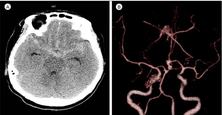
She showed no neurologic deficit except for neck stiffness. Non-enhanced brain computed tomography (CT) showed a subarachnoid hemorrhage of Hunt-Hess Grade II and Fisher Grade III. Brain an- giography CT revealed an aneurysm of the ACoA.
The aneurysm was left dominant, directed inferiorly, and multi-lobulated (Fig. 1). On the transfemoral cere- bral angiography (TFCA) performed pre-operatively,
SANG HYUB LEE ET AL
A B
Fig. 1. A 50-year-old woman reported at the emergency department with thunderclap headache and vomiting. (A) A non-enhanced brain computed tomography (CT) showed a subarachnoid hemorrhage in the suprasellar cistern, both sylvian and interhemispheric fissure. (B) Brain angiography CT revealed an aneurysm of the anterior communicating artery. The aneurysm was left dominant, in- ferior in direction, and multi-lobulated.
the right A1 segment was hypoplastic and the right A2 segment was perfused by the left A1 segment. The left A1 segment supplied both RAH. The mean diam- eter of the right (Rt.) and left (Lt.) RAH was 0.8 mm and 0.4 mm, respectively. Both RAH originated from the junction of the anterior cerebral artery (ACA) and ACoA (Fig. 2). Direct neck clipping was performed using the pterional approach. During the operation, neither RAH was sacrificed. A temporary clip was ap- plied five times to the left A1 segment. The duration of the first temporary clipping was 5 minutes and 48 seconds. The second was 6 minutes and 18 seconds;
the third, 7 minutes and 57 seconds; the fourth, 5 mi- nutes and 30 seconds; and the fifth, 1 minute and 12 seconds in duration. The total temporary clipping time was 26 minutes and 45 seconds. Intra-operative Doppler was performed, and the blood flow of the RAH was found to be intact. The post-operative CT was uneventful. The patient stayed in the neuro-in- tensive care unit for 4 days following surgery. She showed no neurological deficit while there. Our cere-
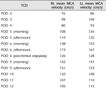
brovascular center conducted daily transcranial Doppler for 14 days following surgery to detect any elevation in the mean velocity (cm/s) of the middle cerebral artery (MCA) resulting from cerebral vasospasm. The initial mean MCA velocity was 76 cm/s on the right side and 86 cm/s on the left. At that time, there were no definite neurological symptoms. However, 3 days postoperatively, the mean MCA velocity started to increase, and the pa- tient reported gradually started to feel drowsy. The MCA velocity continued to increase each day. Even though induced hypertension, hemodilution, and hy- pervolemic therapy were performed to treat cerebral vasospasm, the patient became increasingly lethargic.
On the 6th postoperative day, the mean MCA velocity peaked at 173 cm/s on the right side and 167 cm/s on the left (Table 1). We then decided to perform a chemical angioplasty by intra-arterial nimodipine injection. For chemical angioplasty, TFCA was performed. We found angiographic vasospasm, not only in the M1 and A1 segments, but also in both
INFARCTION AFTER CLIPPING OF ANEURYSM
TCD Rt. mean MCA
velocity (cm/s) Lt. mean MCA velocity (cm/s)
POD 2 76 86
POD 3 98 100
POD 4 80 93
POD 5 (morning) 108 126
POD 5 (afternoon) 119 125
POD 6 (morning) 138 153
POD 6 (afternoon) 173 167
POD 6 (post-chemical angioplasty) 126 128
POD 9 (morning) 155 147
POD 9 (afternoon) 151 123
POD 10 132 108
POD 11 107 110
POD 12 105 115
MCA = middle cerebral artery; TCD = transcranial Doppler; Rt. = right; Lt. = left; POD = post-operative day.
Table 1. Mean MCA velocity measured by TCD
A B
Fig. 2. A transfemoral cerebral angiography was performed pre operatively. (A, B) TThe recurrent artery of Heubner (RAH) (arrows) originated bilaterally from the junction of the anterior cerebral artery and the anterior communicating artery. The mean diameter of right and left RAH was 8 mm and 4 mm, respectively.
RAH. Preoperatively, the perfusion of both RAH was normal on the cerebral angiogram. However, on the TFCA before chemical angioplasty, the Rt. RAH was blurred due to decreased perfusion and the Lt. RAH
was not visible (Fig. 3). Clip placement was a good to exclude the aneurysm from circulation without oc- cluding the parent artery. In order to ameliorate the angiographic vasospasm, an intra-arterial nimodipine injection was administered at both the M1 and Lt. A1 segments. The total volume of nimodipine ad- ministered was 5.5 mg. The Rt. M1 segment was se- lected using micro-catheter and 3 mg nimodipine was injected 30 times in 0.1 mg increments at pulsatile heart rate velocity. Then, the Lt. M1 and A1 segments were selected using a micro-catheter and 2.5 mg ni- modipine was injected 25 times in 0.1 mg increments, as was performed for the Rt. M1 segment. After the injections of 5.5 mg nimodipine, angiography was performed. Perfusion of both the M1 and A1 seg- ments and the Rt. RAH improved, and perfusion of the Lt. RAH became visible. The symptoms improved after chemical angioplasty, and the mean MCA veloc- ity decreased to 126 cm/s on the right side and 128 cm/s on the left. On the 7th postoperative day, rou- tine follow-up brain CT demonstrated a newly devel-
SANG HYUB LEE ET AL
A B
C
Fig. 3. (A) Both the recurrent artery of Heubner (RAH) (arrows) were evidently visible on the pre-operative transfemoral cerebral an- giography (TFCA). (B) On the TFCA performed 6 days postoperatively before chemical angioplasty, a blurred right RAH (arrowhead) was seen due to decreased perfusion. The left RAH (arrow) was not visible. (C) On the TFCA performed after chemical angioplasty, the perfusion of right RAH (arrowhead) improved and left RAH (arrow) was visible.
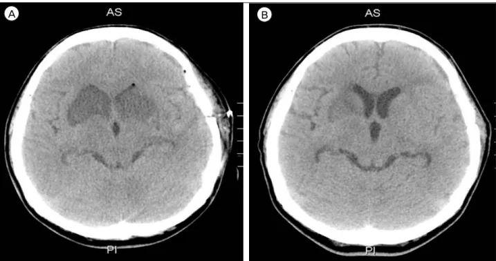
oped low-density lesion in the RAH territory bi- laterally, the caudate nucleus, and the anterior portion of the basal ganglia and internal capsule (Fig. 4).
There were no symptoms of bilateral RAH infarction such as motor weakness, aphasia, dysarthria, or aki- netic mutism. However, mild cognitive disorder was
INFARCTION AFTER CLIPPING OF ANEURYSM
A B
Fig. 4. (A) A 7-day postoperative follow-up brain computed tomography (CT) showed a newly developed low density lesion in the recurrent artery of Heubner (RAH) territory bilaterally, caudate nucleus, anterior portion of basal ganglia, and internal capsule. (B) The 14-day postoperative follow-up brain CT showed no progression of the low density lesion. The low density lesion in both of the RAH territory was blurred.
present. On the 14th postoperative day, routine fol- low-up brain CT showed neither a low-density lesion nor progression in any RAH territory infarction. The patient was discharged with just mild cognitive dis- order and no other definite neurological deficits. On examination at an outpatient department 6 months later, the patient’s cognition had improved.
DISCUSSION
The RAH exhibited a mean outer diameter of 0.8 ± 0.04 mm (range, 0.2-1.5) and a mean length of 23.4 ± 1.1 mm (range, 12-38). The origin of the RAH occurs most commonly at the junction of the ACA and ACoA, then from the proximal A2 segment, and fi- nally from the distal A1 segment.4)7)9) The RAH sup- plies parts of the caudate nucleus, basal ganglia, and anterior limb of the internal capsule.9)
There are only a few papers in the literature that specifically describe the territory affected by RAH
infarction.1)3) RAH occlusion can manifest as various symptoms, including weakness of the contralateral face and arm without sensory loss, transient aphasia, dysarthria, cognitive and behavioral abnormalities, abulia, agitation, hyperactivity, contralateral neglect, obsessive-compulsive disorder, and memory dysfunc- tion (i.e., poor ability to perform declarative and pro- cedural memory tasks). However, most RAH in- farctions are accompanied by only transient neuro- logical deficits, if any.1)2)10-12) The possible causes of cerebral infarction following SAH are cerebral vaso- spasm, microthrombo-embolism, cortical spreading is- chemia, spasm in the microcirculation and impaired cerebral autoregulation. Operative maneuvers that can lead to blood flow disturbance and consequent in- farction include inappropriate aneurysmal neck clip- ping, temporary occlusion of the parent artery, direct injury, retraction with a spatula, or trapping of the parent artery.12) In this case, multiple factors are con- sidered to have caused infarction of the bilateral RAH
SANG HYUB LEE ET AL
territory, including the SAH corresponding to a Fisher Grade III, placement five times of temporary clips on the A1 segment, hypotension during surgery (systolic blood pressure less than 100 mmHg), and a mean ar- terial pressure of 65 mmHg. Cerebral vasospasm was also a significant causative factor in this case. The mechanism of vasospasm remains poorly understood.
However, in the last 3 decades, certain mechanisms have been identified that might play a role in its development. Factors such as calcium-dependent and -independent vasoconstriction, breakdown of products in the blood of subarachnoid space, and imbalance between vasoconstrictor and vasodilator substances derived from the endothelium may be major causes of vasospasm.8) Cerebral vasospasm post-SAH may in- volve the complete arterial system from the circle of Willis up to the distal vessel segments.15) A high vol- ume of cisternal hemorrhage on an admission CT scan in a patient with Fisher Grade III aneurysmal SAH was found to be highly associated with delayed ischemic neurological deficit (DIND) due to cerebral vasospasm.6) When the subarachnoid blood went un- detected or was distributed diffusely, severe vaso- spasm was almost never encountered; however, in the presence of a large amount of SAH, severe vasospasm almost always occurred.5) Approximately 11-20% of episodes of DIND are characterized by cerebral infarction.13)14) Cerebral infarction mostly occurs in pa- tients who develop angiographic vasospasm.14) Our patient was alert during her stay in the neuro-in- tensive care unit. As the mean MCA velocity in- creased, the patient became lethargic; this was attrib- uted to DIND. Angiographic vasospasm was found in the preoperative and postoperative TFCA. Thus it was deduced that the decreased perfusion of both RAH due to cerebral vasospasm was a major cause of the bilateral RAH territorial infarction. In addition to cer- ebral vasospasm, the other causes of bilateral RAH in- farction were assumed to be the repeated temporary clipping on the Lt. A1 segment and the presence of hypotension during surgery. The prolonged tempo-
rary clipping time of 26 minutes, 45 seconds and hy- potension (systolic blood pressure less than 100 mmHg for 115 minutes and mean arterial pressure of 65 mmHg for 70 minutes) during surgery likely sig- nificantly lowered cerebral blood perfusion and cere- bral perfusion pressure. These conditions would have been sufficient to cause bilateral RAH infarction.
During the temporary clipping, the systolic blood pressure was always below 100 mmHg; the mean ar- terial blood pressure was below 65 mmHg for half of the clipping time. Temporary clipping of the main cerebral artery can lead to infarcts of the perforating artery, and temporary clipping of the A1 segment can lead to infarction of the RAH.12) Sasaki et al.12) re- ported nine patients of RAH infarction following aneurysm clipping, all of whom had ACoA aneurysm, in a population of 1,045 patients with treated aneur- ysms and 238 with ACoA aneurysms. The incidence of the causes of RAH infarctions: A1 temporary occlu- sion (four), retraction (three) and injury of RAH (two).
In this case, vasospasm was considered to be a more likely cause of RAH infarction than temporary occlu- sion or hypotension. This inference was made based on the fact that immediately following the operation the patient had no neurologic deficit or abnormal mental status. The patient’s reduced mental status ap- peared alongside the increase in the mean MCA velocity. There were no abnormal findings in the post-operative brain CT. An intra-operative Doppler was applied and intact flow of the RAH was observed.
CONCLUSION
It is important to keep in mind that the clipping of an ACoA aneurysm can cause infarction of the RAH.
In cases of RAH infarction following aneurysm sur- gery, the aneurysm is almost always in the ACoA.12) When a patient has DIND with an elevated mean MCA velocity after clipping of an ACoA aneurysm, neurosurgeons should consider the possibility of an
INFARCTION AFTER CLIPPING OF ANEURYSM
RAH infarction caused by decreased perfusion due to cerebral vasospasm.
Disclosure
The authors have no conflict of interest concerning the materials or methods used in this study or the findings specified in this paper.
REFERENCES
1. Berman SA, Hayman LA, Hinck VC. Correlation of CT cerebral vascular territories with function: I. Anterior cerebral artery. AJR Am J Roentgenol. 1980 Aug;135(2):
253-7.
2. Caplan LR, Schmahmann JD, Kase CS, Feldmann E, Baquis G, Greenberg JP, et al. Caudate infarcts. Arch Neurol. 1990 Feb;47(2):133-43.
3. Critchley M. Syndromes of the anterior cerebral artery.
Proc R Soc Med. 1930 Mar;23(5):630-2.
4. El Falougy H, Selmeciova P, Kubikova E, Haviarová Z.
The variable origin of the recurrent artery of Heubner:
an anatomical and morphometric study. Biomed Res Int.
2013;2013:873434.
5. Fisher CM, Kistler JP, Davis JM. Relation of cerebral vasospasm to subarachnoid hemorrhage visualized by computerized tomographic scanning. Neurosurgery. 1980 Jan;6(1):1-9.
6. Friedman JA, Goerss SJ, Meyer FB, Piepgras DG, Pichelmann MA, McIver JI, et al. Volumetric quantifica- tion of Fisher Grade 3 aneurysmal subarachnoid hemor- rhage: a novel method to predict symptomatic vaso- spasm on admission computerized tomography scans. J
Neurosurg. 2002 Aug;97(2):401-7.
7. Gomes F, Dujovny M, Umansky F, Ausman JI, Diaz FG, Ray WJ, et al. Microsurgical anatomy of the recurrent artery of Heubner. J Neurosurg. 1984 Jan;60(1):130-9.
8. Kolias AG, Sen J, Belli A. Pathogenesis of cerebral vaso- spasm following aneurysmal subarachnoid hemorrhage:
putative mechanisms and novel approaches. J Neurosci Res. 2009 Jan;87(1):1-11.
9. Loukas M, Louis RG Jr, Childs RS. Anatomical examina- tion of the recurrent artery of Heubner. Clin Anat. 2006 Jan;19(1):25-31.
10. Miller SP, O'Gorman AM, Shevell MI. Recurrent artery of Heubner infarction in infancy. Dev Med Child Neurol. 2000 May;42(5):344-6.
11. Mizuta H, Motomura N. Memory dysfunction in cau- date infarction caused by Heubner's recurring artery occlusion. Brain Cogn. 2006 Jul;61(2):133-8.
12. Sasaki T, Kodama N, Matsumoto M, Suzuki K, Konno Y, Sakuma J, et al. Blood flow disturbance in perforat- ing arteries attributable to aneurysm surgery. J Neurosurg. 2007 Jul;107(1):60-7.
13. Schmidt JM, Wartenberg KE, Fernandez A, Claassen J, Rincon F, Ostapkovich ND, et al. Frequency and clinical impact of asymptomatic cerebral infarction due to vaso- spasm after subarachnoid hemorrhage. J Neurosurg.
2008 Dec;109(6):1052-9.
14. Vergouwen MD, Ilodigwe D, Macdonald RL. Cerebral infarction after subarachnoid hemorrhage contributes to poor outcome by vasospasm-dependent and -independent effects. Stroke. 2011 Apr;42(4):924-9.
15. Weidauer S, Lanfermann H, Raabe A, Zanella F, Seifert V, Beck J. Impairment of cerebral perfusion and infarct patterns attributable to vasospasm after aneurysmal sub- arachnoid hemorrhage: a prospective MRI and DSA study. Stroke. 2007 Jun;38(6):1831-6.