INTRODUCTION
Breast cancer is the most common cause of cancer-related deaths in women globally and the second most common ma- lignancy in Korean women, accounting for 14.8% of all malig- nancies newly diagnosed in 2011 [1]. The incidence of inva- sive breast cancer (IBC) has increased continuously over time, reaching 58 cases per 100,000 women in 2011 [1]. Although early cancer detection and current standard treatments specif- ic for individual tumor characteristics have increased the sur- vival of patients with breast cancer, there remains a subset of
patients displaying unresponsiveness to standard therapy. The selection and control of these patients are problems awaiting solutions in oncology.
Epithelial-mesenchymal transition (EMT) of epithelial cells is defined as the loss of epithelial characteristics and acquisi- tion of a mesenchymal phenotype. Tumor cells undergoing EMT acquire migratory potential, stem cell-like features, and chemoresistance to conventional and targeted therapeutics [2- 4]. A variety of biomarkers, including transcription factors (zinc finger factor SNAIL [Snail], snail2 [slug], zinc finger E- box-binding homeobox 1 [ZEB1], twist, and forkhead box protein 2 [FOXC2]), extracellular matrix proteins (fibronectin and laminin), cell surface proteins (E-cadherin and N-cad- herin), and cytoskeletal proteins (vimentin and α-smooth muscle actin), have been used to define EMT phenotypes in cancer tissue; however, no biomarker has been accepted for EMT signatures [5].
E-cadherin is a transmembrane glycoprotein that has an
Epithelial-Mesenchymal Transition Phenotype Is Associated with
Clinicopathological Factors That Indicate Aggressive Biological Behavior and Poor Clinical Outcomes in Invasive Breast Cancer
Young Kyung Bae, Jung Eun Choi1, Su Hwan Kang1, Soo Jung Lee1
Departments of Pathology and 1Surgery, Yeungnam University College of Medicine, Daegu, Korea ORIGINAL ARTICLE
Purpose: Cancer tissue may display a wide spectrum of ex- pression phenotypes of epithelial-mesenchymal transition (EMT)-related proteins. The purpose of this study was to in- vestigate the clinical significance of EMT phenotypes in breast cancer. Methods: We evaluated the expression pattern of the EMT-related proteins E-cadherin and fibronectin in samples from 1,495 patients with invasive breast carcinoma (IBC) on tis- sue microarrays using immunohistochemistry to investigate the clinical significance of EMT phenotypes in IBC. EMT pheno- types were divided into complete type (E-cadherin-negative/fi- bronectin-positive), incomplete type (hybrid type, E-cadherin- positive/fibronectin-positive; null type, E-cadherin-negative/fi- bronectin-negative), and wild-type (E-cadherin-positive/fibro- nectin-negative). We analyzed the correlation of EMT phenotype with clinicopathological factors and patient survival. Results:
Loss of E-cadherin was observed in 302 patients (20.2%), and fibronectin was expressed in the cancer cells of 354 patients
(23.7%). In total, 64 (4.3%), 290 (19.4%), 238 (15.9%), and 903 (60.4%) samples were categorized as complete, hybrid, null, and wild-type, respectively. The complete EMT phenotype ex- hibited significant associations with young age (p=0.017), ad- vanced pT (p<0.001) and pN (p<0.001) stages, higher histo- logical grade (p<0.001), lymphovascular invasion (p<0.001), and triple negativity (p<0.001). Patients with complete and hy- brid EMT phenotypes had poorer overall survival (OS) and dis- ease-free survival (DFS) than those with the wild-type pheno- type (OS, p=0.001; DFS, p<0.001). In multivariate analysis, the hybrid EMT phenotype was an independent prognostic factor for DFS in patients with IBC (p=0.032). Conclusion: EMT pheno- types exhibited significant associations with clinicopathological factors indicating aggressive biologic behavior and poor out- come in patients with IBC.
Key Words: Breast neoplasms, Epithelial-mesenchymal transition, Prognosis
Correspondence to: Young Kyung Bae
Department of Pathology, Yeungnam University College of Medicine, 170 Hyeonchung-ro, Nam-gu, Daegu 42415, Korea
Tel: +82-53-640-6755, Fax: +82-53-622-8432 E-mail: [email protected]
This study was supported by the 2014 Yeungnam University Research Grants.
Received: April 28, 2015 Accepted: July 22, 2015
Cancer
important role in epithelial cell adhesion. Loss of E-cadherin expression has been considered a hallmark of EMT [4]. Fibro- nectin is a large heterodimeric glycoprotein that can exist in an insoluble, cellular form or soluble form in plasma. It plays a role in cell-matrix and cell-cell adhesion, cell migration, dif- ferentiation, morphogenesis, and oncogenic transformation [6]. A variety of benign and malignant epithelial and mesen- chymal cells can produce fibronectin, and upregulation of fi- bronectin occurs during the EMT process of epithelial tumors [7]. Fibronectin has been used to detect EMT phenotypes in human cancers [4,8,9].
Earlier studies reported that the EMT phenotype defined by patterns of EMT-related protein expression was an indepen- dent prognostic factor in gastrointestinal cancers involving the esophagus, stomach, or small intestine [8-10]. However, little is known about the clinical significance of EMT pheno- types in IBC. In this study, investigated whether EMT pheno- types could be used to select a distinct breast cancer subgroup with a worse clinical outcome, as demonstrated for other tu- mors [7-9]. For this purpose, we assessed the expression of E- cadherin and fibronectin in tumor samples from patients with IBC and defined EMT phenotypes based on the combined expression patterns of these markers. The correlation of EMT phenotypes with clinicopathological factors and patient sur- vival was analyzed to evaluate of the clinical significance of EMT phenotypes in patients with IBC.
METHODS
Tissue samples and tissue microarray construction
Two sets of tissue microarrays (TMAs) of IBCs were used [11,12]. Samples for a total of 1,596 patients with primary IBC who underwent surgical resection between January 1995 and December 2007 were collected retrospectively from the ar- chives of the Department of Pathology at Yeungnam Universi- ty Hospital, Daegu, Korea. Patients who could tolerate treat- ment received standard radiotherapy or adjuvant systemic therapy (hormone therapy or chemotherapy) after surgery ac- cording to a medical insurance program administered by the Ministry of Health and Welfare of Korea. For TMA construc- tion, we reviewed hematoxylin and eosin-stained slides of all patients and selected a representative tumor block for each pa- tient. The first set consisted of 13 TMA blocks containing 594 samples (obtained between 1995 and 2003). The method for TMA construction was described in our previous study [11].
The second set comprised 43 TMA blocks constructed with 1,002 samples (obtained between 2004 and 2007) using a Quick-Ray® Manual Tissue Microarrayer (Unitma, Seoul, Korea) and Quick-Ray® recipient blocks of 2 mm cores (Unitma) [12].
Patient and tumor characteristics, including age at the time of the initial diagnosis, pathologic tumor stage (pT), pathol- ogic lymph node stage (pN), histological grade [13], the pres- ence or absence of lymphovascular invasion, and follow-up information, were obtained from the pathology reports and patients’ medical records. Information on the presence and cause of death of each patient was also obtained using the mi- crodata service system provided by Statistics Korea (http://
mdss.kostat.go.kr). Overall survival (OS) was defined as the interval between the date of surgical resection and the date of disease-specific death or the last follow-up. Disease-free sur- vival (DFS) was expressed as the number of months from the date of surgical resection to the date of documented relapse, including locoregional recurrence and distant metastasis. This study was approved by the Institutional Review Board of Yeungnam University Hospital (YUH-12-0344-O20), and the requirement for informed consent was waived.
Immunohistochemistry and scoring
For molecular classification of the patients, because the staining conditions and interpretation criteria changed over the study period (1995−2007), we repeated immunohisto- chemistry for estrogen receptor (ER), progesterone receptor (PR), and human epidermal growth factor receptor 2 (HER2) and interpreted the results according to the current guidelines for ER/PR and HER2 testing [14,15].
TMA sections measuring 4 μm in thickness were used for immunohistochemistry. Immunohistochemical staining for ER (SP1, prediluted; Ventana Medical Systems, Tucson, USA), PR (1E2, prediluted; Ventana Medical Systems), HER2 (CONFIRMTM anti-HER2/neu (4B5) rabbit monoclonal; Ventana Medical Systems), E-cadherin (clone 4A2C7, 1:200 dilution; Invitrogen, Carlsbad, USA), and fibronectin (clone F14, prediluted; Biogenex, San Ramon, USA) was performed in the automated Benchmark® platform (Ventana Medical Systems) and labeled using an UltraViewTM universal DAB detection kit (Ventana Medical Sys- tems), as described in previous studies [9,16]. Silver-enhanced in situ hybridization for HER2 using an INFORM® HER2 DNA probe (Ventana Medical Systems) was performed for equivocal cases in HER2 staining [12].
Regarding the interpretation of E-cadherin and fibronectin, staining intensity and the proportion of tumor cells were eval- uated as described previously [9]. Staining intensity was clas- sified as follows: 1, weak; 2, moderate; and 3, strong. Positive cells were quantified as a percentage of the total number of tu- mor cells, and the value was categorized as follows: 0, ≤5%; 1,
>5% and ≤25%; 2, >25% and ≤50%; 3, >50% and ≤75%; 4,
>75%. The sum of the tumor area of two consecutive tumor cores was regarded as the total tumor area (100%) instead of
scoring the immunostaining results in each tumor core sepa- rately. The percentage of epithelial cell positivity and staining intensity were multiplied to generate an immunoreactivity score (IS) for each sample, which ranged from 0 to 12. The Kaplan-Meier method with the log-rank test was used to se- lect a cutoff point for designating immunopositivity for each marker that was most meaningful with respect to prognosis [8]. Using this method, samples with an E-cadherin IS ≥8 were considered positive for E-cadherin, and those with a fi- bronectin IS ≥2 were regarded as positive for fibronectin.
IBCs were classified according to the following types of EMT by modifying the proposal provided by Sung et al. [8]:
complete type (E-cadherin-negative/fibronectin-positive), in- complete type (hybrid type, E-cadherin-positive/fibronectin- positive; null type, E-cadherin-negative/fibronectin-negative), and wild-type (E-cadherin-positive/fibronectin-negative).
Based on their hormone receptor (HR, ER or PR) and HER2 statuses, the IBC samples were divided into four mo- lecular subtypes: HR+/HER2−, HR+/HER2+, HR−/HER2+, and HR−/HER2−.
Statistical analysis
Statistical analysis was performed using SPSS version 21.0 for Windows (IBM Corp., Armonk, USA). The chi-square test was used to evaluate the significance of correlations between EMT phenotypes and patient characteristics. Univariate and multivariate analyses were performed to assess the effect of EMT phenotypes on patient survival (OS and DFS). Survival curves were plotted using the Kaplan-Meier method and the log-rank test was used to test the significance of survival dif- ferences. Significant variables identified in univariate analyses were further analyzed using a Cox regression proportional hazard model. Adjusted hazard ratios and associated 95%
confidence intervals (CIs) were estimated for each variable. A p-value of <0.05 was considered statistically significant.
RESULTS
Patient demographics and tumor characteristics
Of the 1,596 patients, 101 were excluded from the analysis because of a failure to obtain immunohistochemical results as a result of noninformative cores (no invasive tumor) or a loss of cores while performing immunohistochemistry. Therefore, 1,495 patients were included in this study. Patients ranged in age from 20 to 86 years (median, 46 years; mean, 48 years).
Among these patients, 878 (58.7%) underwent mastectomy, and 617 (41.3%) underwent breast-conserving surgery. Senti- nel node biopsy or axillary dissection was performed in 1,492 patients. Concerning adjuvant chemotherapy, 1,010 patients
(67.6%) received anthracycline-based chemotherapy, 259 pa- tients (17.3%) received nonanthracycline chemotherapy, and the remaining 226 patients (15.1%) did not receive chemo- therapy. A total of 1,025 patients (68.6%) received hormone therapy. The median follow-up period was 62 months for OS and 60 months for DFS (range, 1−202 months).
Of the 1,495 patient samples, 1,008 (67.4%) were positive for ER, and 871 (58.3%) were positive for PR. Protein overexpres- sion or amplification of HER2 was observed in 293 samples (19.6%). Regarding molecular subtypes, 893 tumors (59.7%) were HR+/HER2−, 131 (8.8%) were HR+/HER2+, 162 (10.8%) were HR−/HER2+, and 309 (20.7%) were triple-negative.
Positive expression for E-cadherin and fibronectin was ob- served in 1,193 (79.8%) and 354 (23.7%) samples, respectively.
The characteristics of the tumors are shown in Table 1.
Classification of EMT phenotype and its correlation with clinicopathological features and patient survival
Classification of the 1,495 patients with IBC according to EMT phenotypes resulted in the identification of 64 patients (4.3%) with the complete EMT type, 903 patients (60.4%) with the wild-type, and 528 patients (35.3%) with the incom- plete EMT type (hybrid type, 290 cases; null type, 238 cases) (Figure 1). The EMT phenotype displayed significant associa- tions with age (p=0.015), pT (p<0.001), pN (p<0.001), his- tological grade (p<0.001), lymphovascular invasion (p=
0.035), and molecular phenotype (p<0.001) (Table 1). The complete EMT phenotype was observed more frequently in patients with a younger age (<50 years) (Figure 2), higher pT and pN stages, histologic grade 3, or triple negativity. Lym- phovascular invasion was observed more frequently in pa- tients with complete or hybrid phenotypes than in those with null or wild-type phenotypes. In total, 52 (81.3%), nine (14.1%), and three (4.7%) tumors with the complete EMT phenotype were invasive (ductal) carcinoma, no special type, invasive lobular carcinoma, and mixed type, respectively.
Concerning other histologic subtypes, including invasive mi- cropapillary, mucinous, tubular, medullary, metaplastic, and invasive papillary carcinomas, none of the tumors displayed the complete EMT phenotype (p<0.001). Patients who un- derwent mastectomy had higher rates of complete and hybrid EMT phenotypes than those who underwent breast-conserv- ing surgery (p<0.001). Patients with the complete EMT phe- notype received adjuvant chemotherapy more frequently compared to those with the wild-type phenotype (p<0.001).
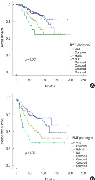
In the comparison of survival differences according to the EMT phenotype of patients with IBC, there was a significant survival difference among the four EMT subgroups (OS, p=0.001; DFS, p<0.001) (Figure 3). Survival differences were
observed between the different EMT phenotypes (complete vs. wild-type, OS, p=0.004, DFS, p<0.001; hybrid vs. wild- type, OS, p =0.002, DFS, p =0.001; hybrid vs. null, OS, p=0.026, DFS, p=0.018; complete vs. null, OS, p=0.014, DFS, p=0.003), but not between wild-type and the null type (OS, p=0.896; DFS, p=0.956) or between hybrid and com-
plete types (OS, p=0.407; DFS, p=0.203).
In addition to the EMT phenotype, pT (p <0.001 and p<0.001), lymph node status (p<0.001 and p<0.001), histo- logic grade (p<0.001 and p=0.001), lymphovascular invasion (p<0.001 and p<0.001), hormone receptor (p=0.019 and p=0.892) and HER2 status (p=0.02 and p=0.014) predicted Table 1. Correlation between epithelial-mesenchymal transition phenotype and clinicopathologic parameters in 1,495 patients with invasive breast carcinoma
Parameter Total
(n=1,495)
EMT phenotype, No (%)
p-value
Complete Incomplete
Hybrid Null Wild
Age (yr) 0.015
<50 948 48 (75.0) 164 (56.6) 150 (63.0) 586 (64.9)
≥50 547 16 (25.0) 126 (43.4) 88 (37.0) 317 (35.1)
Histological type <0.001
Ductal 1,325 52 (81.3) 269 (92.8) 180 (75.6) 824 (62.2)
Lobular 45 9 (14.1) 0 30 (12.6) 6 (0.7)
Mixed 33 3 (4.7) 6 (2.1) 5 (2.1) 19 (2.1)
Micropapillary 31 0 11 (3.8) 1 (0.1) 19 (2.1)
Mucinous 27 0 0 11 (4.6) 16 (1.8)
Tubular 15 0 0 0 15 (1.7)
Medullary 8 0 0 8 (3.4) 0
Metaplastic 7 0 3 (1) 3 (1.3) 1 (0.1)
Papillary 4 0 1 (0.3) 0 3 (0.3)
Tumor size <0.001
pT1 804 18 (28.1) 138 (47.6) 111 (46.6) 537 (59.5)
pT2 637 40 (62.5) 143 (49.3) 115 (48.3) 339 (37.5)
pT3 & pT4 54 6 (9.4) 9 (3.1) 12 (5.0) 27 (3.0)
Lymph node status <0.001
pN0 825 20 (31.3) 125 (43.1) 152 (64.4) 528 (58.5)
pN1 411 19 (29.7) 94 (32.4) 58 (24.6) 240 (26.6)
pN2 110 11 (17.2) 29 (10) 8 (3.4) 62 (6.9)
pN3 146 14 (21.9) 42 (14.5) 18 (7.6) 72 (8.0)
Histologic grade <0.001
1 272 0 33 (11.4) 31 (13.0) 208 (23.0)
2 424 11 (17.2) 71 (24.5) 62 (26.1) 280 (31.0)
3 799 53 (82.8) 186 (64.1) 145 (60.9) 415 (46.0)
Lymphovascular invasion <0.001
Absent 751 23 (35.9) 115 (39.7) 146 (61.3) 467 (51.7)
Present 744 41 (64.1) 175 (60.3) 92 (38.7) 436 (48.3)
Molecular subtype <0.001
HR+/HER2- 893 21 (32.8) 149 (51.4) 116 (48.7) 607 (67.2)
HR+/HER2+ 131 5 (7.8) 32 (11) 7 (2.9) 87 (9.6)
HR-/HER2+ 162 9 (14.1) 63 (21.7) 13 (5.5) 77 (8.5)
Triple negative 309 29 (45.3) 46 (15.9) 102 (42.9) 132 (14.6)
Surgery type <0.001
Breast conservation 617 11 (17.2) 59 (20.3) 100 (42.0) 447 (49.5)
Mastectomy 878 53 (82.8) 231 (79.3) 138 (58.0) 456 (50.5)
Adjuvant chemotherapy <0.001
No 226 2 (3.1) 27 (9.3) 34 (14.3) 163 (18.1)
Yes 1,269 62 (96.9) 263 (90.7) 204 (85.7) 740 (81.9)
EMT=epithelial-mesenchymal transition; pT=pathologic tumor stage according to the 7th American Joint Committee on Cancer Staging System; pN=pathologic lymph node stage according to the 7th American Joint Committee on Cancer Staging System; HR=hormone receptor; HER2=human epidermal growth factor receptor 2.
Figure 1. Representative cases of each epithelial-mesenchymal transi- tion (EMT) phenotype with corresponding immunohistochemical stain- ing results for E-cadherin and fibronectin (complete, hybrid, and null type, ×40; wild type, ×100).
Complete type=E-cadherin-negative and fibronectin-positive; hybrid type=E-cadherin-positive and fibronectin-positive; null type=E-cad- herin-negative and fibronectin-negative; wild type=E-cadherin-positive and fibronectin-negative.
Complete Hybrid
EMT phenotype
Null Wild
E-cadherin
E-cadherin Fibronectin
Fibronectin
100 90 80 70 60 50 40 30 20 10 0
Wild Complete Hybrid Null ≤29 ≥30 and ≤39 ≥40 and ≤49 ≥50 and ≤59 ≥60 and ≤69 ≥70 Age (yr)
Percentage (%)
Figure 2. Distribution of patient age group according to epithelial-mes- enchymal transition phenotype.
1.7 4.7 1.7 1.7
17.3 21.9
15.2 18.1
46
48.4
39.7 43.3
21.4
20.3
22.8
20.6 10.9
3.1
16.6 11.3
2.9 1.6 4.1 5
Figure 3. Survival curves of 1,495 patients with invasive breast cancer according to epithelial-mesenchymal transition (EMT) phenotype. (A) Overall survival. The survival differences between two different subtypes were calculated by log-rank test and the results are as follows: com- plete type vs. wild-type, p=0.004; hybrid type vs. wild-type, p=0.002;
hybrid type vs. null type, p=0.026; complete type vs. null type, p=0.014; wild-type vs. null type, p=0.896; hybrid type vs. complete type, p=0.407. (B) Disease-free survival. The survival differences be- tween two different subtypes were calculated by log-rank test and the results are as follows: complete type vs. wild-type, p<0.001; hybrid type vs. wild-type, p=0.001; hybrid type vs. null type, p=0.018; com- plete type vs. null type, p=0.003; wild-type vs. null type, p=0.956; hy- brid type vs. complete type, p=0.203.
1.0
0.9
0.8
0.7
0.6
0 50 100 150 200 250
Months
Overall survival EMT phenotype
WildComplete Hybrid NullCensored Censored Censored Censored
A p=0.001
1.0
0.9
0.8
0.7
0.6
0 50 100 150 200 250
Months
Disease-free survival
EMT phenotype WildComplete Hybrid NullCensored Censored Censored Censored
B p<0.001
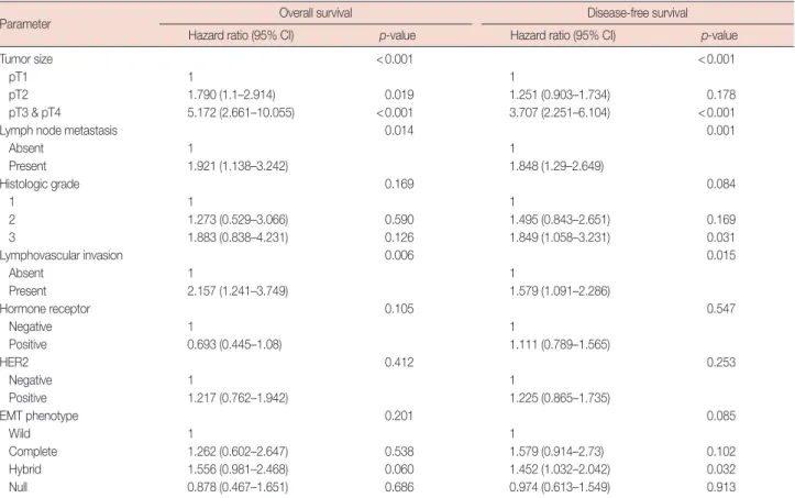
OS and DFS based on univariate analysis. In multivariate analysis, advanced pT stage (p<0.001), the presence of lymph node metastasis (p=0.014), and the presence of lymphovas- cular invasion (p=0.006) were independent prognostic fac- tors for poor OS in patients with IBC. For DFS, advanced pT
Table 2. Multivariate analysis of overall survival and disease-free survival
Parameter Overall survival Disease-free survival
Hazard ratio (95% CI) p-value Hazard ratio (95% CI) p-value
Tumor size <0.001 <0.001
pT1 1 1
pT2 1.790 (1.1–2.914) 0.019 1.251 (0.903–1.734) 0.178
pT3 & pT4 5.172 (2.661–10.055) <0.001 3.707 (2.251–6.104) <0.001
Lymph node metastasis 0.014 0.001
Absent 1 1
Present 1.921 (1.138–3.242) 1.848 (1.29–2.649)
Histologic grade 0.169 0.084
1 1 1
2 1.273 (0.529–3.066) 0.590 1.495 (0.843–2.651) 0.169
3 1.883 (0.838–4.231) 0.126 1.849 (1.058–3.231) 0.031
Lymphovascular invasion 0.006 0.015
Absent 1 1
Present 2.157 (1.241–3.749) 1.579 (1.091–2.286)
Hormone receptor 0.105 0.547
Negative 1 1
Positive 0.693 (0.445–1.08) 1.111 (0.789–1.565)
HER2 0.412 0.253
Negative 1 1
Positive 1.217 (0.762–1.942) 1.225 (0.865–1.735)
EMT phenotype 0.201 0.085
Wild 1 1
Complete 1.262 (0.602–2.647) 0.538 1.579 (0.914–2.73) 0.102
Hybrid 1.556 (0.981–2.468) 0.060 1.452 (1.032–2.042) 0.032
Null 0.878 (0.467–1.651) 0.686 0.974 (0.613–1.549) 0.913
CI=confidence interval; pT=pathologic tumor stage according to the 7th American Joint Committee on Cancer Staging System; pN=pathologic lymph node stage according to the 7th American Joint Committee on Cancer Staging System; HER2=human epidermal growth factor receptor 2; EMT=epithelial-mesenchy- mal transition.
stage (p=0.004), the presence of lymph node metastasis (p=0.001), histologic grade 3 (p=0.031), and the presence of lymphovascular invasion (p=0.015) were independent poor prognostic factors. Among EMT phenotypes, patients with the hybrid phenotype exhibited a 1.45-fold (95% CI, 1.03–
2.04; p=0.032) higher risk of disease recurrence than those with the wild-type phenotype (Table 2).
DISCUSSION
We investigated the aberrant expression of the EMT-related proteins E-cadherin and fibronectin in the cancer cells of 1,495 patients with IBC using immunohistochemistry. The EMT phenotype defined by the combined expression pattern of E-cadherin and fibronectin displayed significant correla- tions with clinicopathological factors indicating aggressive bi- ological behavior, including advanced pT and pN stages, high histologic grade, the presence of lymphovascular invasion, and triple negativity. In addition, patients with complete and hybrid EMT phenotypes exhibited poorer OS and DFS than those with the wild-type phenotype, and the hybrid EMT
phenotype was an independent prognostic factor in patients with IBC.
EMT is a dynamic and reversible process induced by a vari- ety of signaling pathways including Wnt, tumor necrosis fac- tor α/nuclear factor κB, Notch, MAPK/PI3K, and transform- ing growth factor β pathways [17]. These EMT signaling path- ways are also known to be involved in the generation of breast cancer stem cells (BCSCs) [18,19]. Sarrió et al. [20] reported an association of EMT with the basal-like phenotype of breast cancer, and they suggested that EMT is related to high aggres- siveness and the metastatic spread of basal-like breast cancer.
Prat et al. [21] recently identified a molecular subtype of triple- negative breast cancer, the claudin-low subtype, in which markers linked to EMT and BCSCs are expressed concurrent- ly. Breast cancer cells expressing these overlapping molecular features (EMT, BCSC, and claudin-low) are expected to have migratory potential, metastatic growth, and chemoresistance [21-23]. There have been reports demonstrating that residual breast cancer cells remaining after conventional therapy dis- played both BCSC and mesenchymal features [24,25]. These basic and fundamental findings support our results that the
EMT phenotype was significantly correlated with advanced stage, lymphovascular invasion, high histologic grade, triple- negativity, and poor clinical outcome.
The EMT phenomenon represents an interaction of com- plex EMT-related markers that are affected by each other [2].
Although EMT is characterized by a lack of epithelial features and attainment of the mesenchymal features of epithelial cells, a wide spectrum of EMT phenotypes have been reported.
Sung et al. [8], proposed four specific types of EMT, including wild-type (epithelial), complete (mesenchymal), and interme- diate (hybrid and null) phenotypes, based on the combined expression patterns of epithelial (E-cadherin) and mesenchy- mal (fibronectin, smooth muscle actin, and vimentin) mark- ers. They found that the EMT phenotype has significant prog- nostic value in esophageal squamous cell carcinoma. In their study, OS and DFS were worst in the complete EMT type group, better in the incomplete type group, and best in the wild type group. In addition, the EMT phenotype displayed significant associations with tumor size, histological differen- tiation and invasion depth. Ryu et al. [10] investigated the ex- pression status of five EMT-related markers (E-cadherin, vi- mentin, snail1, ZEB1, and β-catenin) and CD44 in gastric cancers to determine the roles of EMT-related proteins in gas- tric cancer progression. Although the loss of E-cadherin ex- pression and aberrant expression of vimentin were associated with poor patient survival, altered expression of snail1, ZEB1, CD44, and β-catenin did not have a significant effect on pa- tient survival. However, when they selected four markers, E- cadherin, snail1, vimentin, and CD44, tumors with altered expression of three or more proteins displayed highly aggres- sive clinical features and less favorable outcomes than those with altered expression of two or less proteins. Their results suggest that combined analysis of the expression of EMT-re- lated proteins may provide more information about the bio- logical behavior of tumors than the alteration of any single EMT-related marker.
In our previous study [9], we used three markers (E-cadherin, vimentin, and fibronectin) to define the EMT phenotype of small intestinal adenocarcinomas because none of the patients exhibited positivity for smooth muscle actin in their tumor cells, and the results obtained were similar to those reported by Sung et al. [8]. The complete EMT phenotype displayed significant correlations with undifferentiated histology and poor survival in patients with small intestinal adenocarcino- ma and a trend toward an association with advanced pT clas- sification. In this study, we also observed the expression of other mesenchymal markers including vimentin and smooth muscle actin in these patients. Unlike previous studies [8,9], vimentin and smooth muscle actin did not exhibit significant
prognostic value in breast cancer in our study (data not shown).
Therefore, only E-cadherin and fibronectin were used to de- fine EMT phenotypes in this study because we intended to stratify IBC according to EMT phenotypes with prognostic significance. These previous studies and our present research suggest that EMT phenotype defined by specific epithelial and mesenchymal markers may provide useful information for predicting patient outcome regardless of the location (esopha- gus, small intestine, and breast) and histology (squamous cell carcinoma and adenocarcinoma) of the tumors.
Logullo et al. [26] studied the concomitant expression of EMT-related markers (E-cadherin, β-catenin, Snail, trans- forming growth factor β1 [TGF-β1], TGFβ type II receptor [TBRII], and the HGF receptor [c-met]) in both ductal carci- noma in situ (DCIS, n=95) and invasive ductal carcinoma (IDC, n=55) using a TMA. Excluding c-met and TGF-β1, EMT markers were not associated with differences in positivi- ty rates between DCIS and IDC. In addition, none of the EMT markers was correlated with patient survival. Choi et al.
[27] recently reported that the expression of EMT markers (vimentin, smooth muscle actin, osteonectin, N-cadherin, E- cadherin, and β-catenin) and CD146 was significantly higher in invasive carcinoma than in DCIS of the basal-like subtype.
They suggested an important role of EMT in the progression from in situ to invasive basal-like breast cancer. To the best of our knowledge, this is the first study to perform survival anal- ysis according to the combined expression patterns of EMT- related proteins rather than the expression of a single EMT- related marker in patients with IBC.
There are several limitations in our study. First, the EMT process occurs primarily in the infiltrative tumor border [28];
however, our study was performed using TMAs. While con- structing TMA blocks, we attempted to remove tumor cores from the representative tumor area while avoiding areas tu- mor necrosis and central fibrous scars. Although some tumor cores may correspond to the peripheral portion of the tumor, most of tumor cores do not exactly reflect the infiltrative mar- gin of the tumor. Second, unlike other studies that used three or more epithelial and mesenchymal markers to define EMT phenotypes [8-10], only one epithelial marker and one mes- enchymal marker were selected for EMT phenotype analysis in the present study. In addition to these epithelial and mesen- chymal markers, there are many EMT-related markers, in- cluding transcription factors, which are more biologically sig- nificant in terms of the activation of signaling pathways.
Therefore, additional research using other EMT-related mark- ers is necessary to validate clinical significance of EMT phe- notypes in cancer samples.
In conclusion, we investigated the expression patterns of
EMT-related epithelial (E-cadherin) and mesenchymal (fibro- nectin) markers in 1,495 patients with IBC. The EMT pheno- types defined by the combination of expression patterns for both proteins exhibited significant associations with clinico- pathological factors, and they could further stratify patients with IBC into subgroups with prognostic significance.
CONFLICT OF INTEREST
The authors declare that they have no competing interests.
REFERENCES
1. Kim Z, Min SY, Yoon CS, Lee HJ, Lee JS, Youn HJ, et al. The basic facts of Korean breast cancer in 2011: results of a nationwide survey and breast cancer registry database. J Breast Cancer 2014;17:99-106.
2. Thiery JP, Acloque H, Huang RY, Nieto MA. Epithelial-mesenchymal transitions in development and disease. Cell 2009;139:871-90.
3. Roussos ET, Keckesova Z, Haley JD, Epstein DM, Weinberg RA, Condeelis JS. AACR special conference on epithelial-mesenchymal transition and cancer progression and treatment. Cancer Res 2010;70:7360-4.
4. Foroni C, Broggini M, Generali D, Damia G. Epithelial-mesenchymal transition and breast cancer: role, molecular mechanisms and clinical impact. Cancer Treat Rev 2012;38:689-97.
5. Zeisberg M, Neilson EG. Biomarkers for epithelial-mesenchymal tran- sitions. J Clin Invest 2009;119:1429-37.
6. Ioachim E, Charchanti A, Briasoulis E, Karavasilis V, Tsanou H, Arvanitis DL, et al. Immunohistochemical expression of extracellular matrix components tenascin, fibronectin, collagen type IV and laminin in breast cancer: their prognostic value and role in tumour invasion and progression. Eur J Cancer 2002;38:2362-70.
7. Christensen L. The distribution of fibronectin, laminin and tetranectin in human breast cancer with special attention to the extracellular ma- trix. APMIS Suppl 1992;26:1-39.
8. Sung CO, Park CK, Kim SH. Classification of epithelial-mesenchymal transition phenotypes in esophageal squamous cell carcinoma is strongly associated with patient prognosis. Mod Pathol 2011;24:1060-8.
9. Kim A, Bae YK, Gu MJ, Kim JY, Jang KY, Bae HI, et al. Epithelial-mes- enchymal transition phenotype is associated with patient survival in small intestinal adenocarcinoma. Pathology 2013;45:567-73.
10. Ryu HS, Park do J, Kim HH, Kim WH, Lee HS. Combination of epi- thelial-mesenchymal transition and cancer stem cell-like phenotypes has independent prognostic value in gastric cancer. Hum Pathol 2012;
43:520-8.
11. Kim A, Shin HC, Bae YK, Kim MK, Kang SH, Lee SJ, et al. Multiplica- tion of chromosome 17 centromere is associated with prognosis in pa- tients with invasive breast cancers exhibiting normal HER2 and TO- P2A status. J Breast Cancer 2012;15:24-33.
12. Bae YK, Kim A, Kim MK, Choi JE, Kang SH, Lee SJ. Fibronectin ex- pression in carcinoma cells correlates with tumor aggressiveness and poor clinical outcome in patients with invasive breast cancer. Hum Pathol 2013;44:2028-37.
13. Elston CW, Ellis IO. Pathological prognostic factors in breast cancer. I.
The value of histological grade in breast cancer: experience from a large study with long-term follow-up. Histopathology 1991;19:403-10.
14. Hammond ME, Hayes DF, Dowsett M, Allred DC, Hagerty KL, Badve S, et al. American Society of Clinical Oncology/College of American Pa- thologists guideline recommendations for immunohistochemical test- ing of estrogen and progesterone receptors in breast cancer (unabridged version). Arch Pathol Lab Med 2010;134:e48-72.
15. Wolff AC, Hammond ME, Hicks DG, Dowsett M, McShane LM, Allison KH, et al. Recommendations for human epidermal growth factor receptor 2 testing in breast cancer: American Society of Clinical Oncol- ogy/College of American Pathologists clinical practice guideline up- date. J Clin Oncol 2013;31:3997-4013.
16. Choi JE, Kang SH, Lee SJ, Bae YK. Androgen receptor expression pre- dicts decreased survival in early stage triple-negative breast cancer. Ann Surg Oncol 2015;22:82-9.
17. Huber MA, Kraut N, Beug H. Molecular requirements for epithelial- mesenchymal transition during tumor progression. Curr Opin Cell Biol 2005;17:548-58.
18. Morel AP, Lièvre M, Thomas C, Hinkal G, Ansieau S, Puisieux A. Gen- eration of breast cancer stem cells through epithelial-mesenchymal transition. PLoS One 2008;3:e2888.
19. Mani SA, Guo W, Liao MJ, Eaton EN, Ayyanan A, Zhou AY, et al. The epithelial-mesenchymal transition generates cells with properties of stem cells. Cell 2008;133:704-15.
20. Sarrió D, Rodriguez-Pinilla SM, Hardisson D, Cano A, Moreno-Bueno G, Palacios J. Epithelial-mesenchymal transition in breast cancer relates to the basal-like phenotype. Cancer Res 2008;68:989-97.
21. Prat A, Parker JS, Karginova O, Fan C, Livasy C, Herschkowitz JI, et al.
Phenotypic and molecular characterization of the claudin-low intrinsic subtype of breast cancer. Breast Cancer Res 2010;12:R68.
22. Chuthapisith S, Eremin J, El-Sheemey M, Eremin O. Breast cancer che- moresistance: emerging importance of cancer stem cells. Surg Oncol 2010;19:27-32.
23. Işeri OD, Kars MD, Arpaci F, Atalay C, Pak I, Gündüz U. Drug resistant MCF-7 cells exhibit epithelial-mesenchymal transition gene expression pattern. Biomed Pharmacother 2011;65:40-5.
24. Creighton CJ, Chang JC, Rosen JM. Epithelial-mesenchymal transition (EMT) in tumor-initiating cells and its clinical implications in breast cancer. J Mammary Gland Biol Neoplasia 2010;15:253-60.
25. Creighton CJ, Li X, Landis M, Dixon JM, Neumeister VM, Sjolund A, et al. Residual breast cancers after conventional therapy display mesen- chymal as well as tumor-initiating features. Proc Natl Acad Sci U S A 2009;106:13820-5.
26. Logullo AF, Nonogaki S, Pasini FS, Osório CA, Soares FA, Brentani MM. Concomitant expression of epithelial-mesenchymal transition biomarkers in breast ductal carcinoma: association with progression.
Oncol Rep 2010;23:313-20.
27. Choi Y, Lee HJ, Jang MH, Gwak JM, Lee KS, Kim EJ, et al. Epithelial- mesenchymal transition increases during the progression of in situ to invasive basal-like breast cancer. Hum Pathol 2013;44:2581-9.
28. Iwatsuki M, Mimori K, Yokobori T, Ishi H, Beppu T, Nakamori S, et al.
Epithelial-mesenchymal transition in cancer development and its clini- cal significance. Cancer Sci 2010;101:293-9.