Euptelea pleiosperma 에탄올 추출물의 항비만 활성
박정애1, 진경숙1, 권현주1,2, 김병우1,2*
1동의대학교블루바이오소재개발및실용화지원센터
2동의대학교생명응용학과
Received: September 1, 2015 / Revised: November 5, 2015 / Accepted: December 1, 2015
서 론
체내에지방조직이과다하게축적된상태를의미하는비 만은장시간에걸쳐에너지소비량에비해영양소를과다섭
취할경우발생하는에너지불균형이그원인이다
[8, 10].
에너지불균형을일으키는주요요인으로는과도한식이섭취
,
신체활동부족,
잘못된생활습관,
내분비기능이상,
유전적인 소인 등이 있으며
adipokine
의 생산과 분비를 통하여adipogenesis
과정으로분화가일어나비정상적인지방세포의크기증가
(hypertrophy)
및수의증가(hyperplasia)
로지 방축적이일어나게된다[2, 3].
세계보건기구
(World Health Organization, WHO)
에따르 면전세계적으로비만인구는1980
년이래두배이상증가 하여2014
년에는18
세이상성인약19
억명(39%)
이상이과체중이며이중
6
억명(13%)
이비만인것으로나타났다[20].
우리나라또한서구식식습관과생활방식이증가하면서전
체인구의
25%
가량이비만으로알려져있으며한국성인의과체중과비만율은빠른속도로증가하고있어
20
세이상남 성의과체중비율은36%
에이른다.
비만은심미적인관점에서뿐만아니라고혈압
,
당뇨,
고지 혈증,
관절염,
심혈관계질환,
암등수많은질환의직·
간접 적원인이되므로그예방과치료가매우중요하다.
비만의 치료를위해운동,
식이요법,
약물투여,
외과적수술등이 수행되고있으며이중식이요법은비만의예방과치료에가 장근본적이며중요한방법중하나이다[4, 6, 16].
시판되고있는대표적인비만치료제로는
sibutramine
과orlistat
이대표적이며이중orlistat
은췌장및위장에서분비되는지방분해효소인
lipase
의활성을억제하여섭취한지방중약
30%
의흡수를차단함으로서체중의감소에도움을준다
[13].
이에최근유용비만소재개발의많은연구가천연유래
lipase
활성저해제의개발에집중되고있다[12, 21].
또한비만은지방전구세포의지방세포로의분화및지방
Anti-Obesity Activity of Euptelea Pleiosperma Ethanol Extract
Jung Ae Park
1, Kyong-Suk Jin
1, Hyun Ju Kwon
1,2, and Byung Woo Kim
1,2*
1
Blue-Bio Industry Regional Innovation Center,
2Department of Life Science and Biotechnology, College of Natural Science and Human Ecology, Dong-Eui University, Busan 47340, Republic of Korea
Previously, Euptelea pleiosperma was identified as one of the useful sources containing anti-oxidative and anti-inflammatory activities for the first time in our research group. In this study, anti-obesity effect of E. pleiosperma ethanol extract (EPEE) was evaluated by using a pancreatic lipase enzyme inhibition assay and a cell culture model system. EPEE suppressed effectively pancreatic lipase enzyme activity dose dependently. Furthermore, EPEE significantly suppressed adipocyte differentiation, lipid accumulation, triglyceride contents, and triggered lipolysis activity on 3T3-L1 preadipocytes in a dose-dependent manner with- out cytotoxicity. Anti-adipogenic effect of EPEE was modulated by cytidine-cytidine-adenosine-adenosine-thymidine (CCAAT)/
enhancer binding proteins α (C/EBPα), C/EBPβ and peroxisome proliferator-activated receptor γ (PPARγ) gene and protein expressions. Taken together, these results provide the important new insight that E. pleiosperma possesses anti-obesity activi- ties such as pancreatic lipase inhibition, anti-adipogenic, and lipolysis effects. It might be utilized as promising sources in the fields of nutraceuticals. The identification of active compounds that confer anti-obesity activity of EPEE might be needed.
Keywords: Euptelea pleiosperma, anti-obesity activity, lipase inhibition activity, anti-adipogenic and lipolysis effects
*Corresponding author
Tel: +82-51-890-2900, Fax: +82-505-182-6951 E-mail: [email protected]
© 2015, The Korean Society for Microbiology and Biotechnology
생성과정에의하여지방세포의세포내중성지방
(triglyceride, TG)
의축적으로발생하므로이러한지방생성기전을조절하는것은비만억제의효과적인방법중하나이다
[1, 5].
지방세포의분화는세포형태및유전자발현의다양한변 화가동반되는복합적인과정이다
.
지방세포는mesenchymal
precursor
에서지방전구세포를거쳐지방세포로분화되며,
분화과정동안형태및생화학적변화를통해체내지방을 축적하면서크기가증가되는데이러한과정에는지방조직세 포에특이적으로발현되는유전자들의조절부위에작용하는 전사인자들이관여한다
[2, 19].
대표적인세포실험계인
3T3-L1
지방전구세포에insulin, dexamethasone(DEX), 3-isobutyl-1-methylxanthine(IBMX)
과 같은자극을유발하면cytidine-cytidine-adenosine-adenosine- thymidine(CCAAT)/enhancer binding protein
의종류인C/
EBP
β, C/EBP
δ로부터초기분화가시작되며이는상호작용또는단독으로
peroxisome proliferator-activated receptor
γ(PPAR
γ)
및C/EBP
α의발현을조절하게된다. C/EPB
α는PPAR
γ와분화초기에발현이유도되어분화후기가되면다양한
adipogenic
유전자들의발현을유도하여,
분화된지방 세포에서발현 양이 현저하게 증가되는양상을 나타낸다[15, 18].
한편전세계적으로비만치료제의개발을위한많은연 구가수행되고있으나시판되고있는비만치료제들의부작 용과내성등의문제가발생하면서사용기준이강화되고있 어우수한효능과함께안전성을보유한성분의개발이필
요하다
[12, 13].
이에특히천연유래소재로부터독성및부작용이없는항비만활성보유소재를발굴하기위한많은
노력이집중되고있다
[21].
Euptelea pleiosperma
는Eupteleaceae
과에속하는낙엽수 이며영춘목으로도불린다.
동아시아지역,
주로중국,
인도,
미 얀마및히말라야일대에분포한다.
잎은식용가능하며,
꽃 과가지의껍질은약용으로알려져있으나그구체적인효 능에대해서는알려진바가없다.
본연구자들은선행연구를통해
E. pleiosperma
에탄올추출물의항산화및항염증효과에대해서처음으로밝힌바있다
[11].
이에본연구에서는천연에서유래한생리활성보유신소재개발의일환으 로
E. pleiosperma 95%
에탄올추출물(EPEE)
이보유한항 비만활성을췌장리파아제효소활성저해,
지방세포분화 억제,
지방세포분화관련전사인자발현조절,
지방세포 분해등을중심으로분석하였다.
재료 및 방법
E. pleiosperma 에탄올 추출물의 준비
본연구에서사용한
E. pleiosperma 95%
에탄올추출물(EPEE)
은한국생명공학연구원,
해외생물소재허브센터에서구입
(
분양번호FBM123-004)
하였다.
건조및분쇄한시료를95%
에탄올을용매로하여45
oC
에서3
일간초음파추출을수행하고추출이끝난시료를
filter
로여과하여고형물을없앤후감압농축
(N-1000SW, EYELA, Japan)
및동결건조(FDU2100, EYELA, Japan)
한시료를구입하여사용전까 지4
oC
에보관하였다.
EPEE의 췌장 lipase 효소 활성 저해능 분석
EPEE
의항비만활성을세포실험계에서분석하기에앞서EPEE
의lipase
효소활성저해능보유유무를다음과같이측정하였다
.
먼저1.5 ml tube
에0.25 M Tris(pH 7.7), 250 mM CaCl
2, 5 mM 4-nitrophenyl dodecanoate(PNPD)
로구성된효 소액과기질을넣고잘섞어준후37
oC
에서5
분간예열하고, 0.25 M Tris(pH 7.7)
에녹인lipase
와시료를넣어37
oC
에서10
분간 반응시킨후20% sodium dodecyl sulfate(SDS)
를 첨가하여반응을종료하였다.
반응액을4
oC, 15,000 rpm
에 서20
분간원심분리하여,
분리한상층액을96-well tissue culture plate
에 분주하고multi-plate reader
를 이용하여412 nm
에서흡광도를측정한후, 10
분간반응시킨시료의흡광도로부터
0
분반응시료의흡광도를뺀값을control
대 비백분율로나타내었다.
측정값은3
회반복실험의평균값 으로나타내었다.
세포 배양 및 시료 처리
EPEE
의항비만활성을세포실험계에서분석하기위해항 비만활성분석에사용되는대표적인cell model system
인3T3-L1
지방전구세포를American Type Culture Collection (ATCC, VA, USA)
으로부터 구입하여10% fetal bovine serum(FBS)
및penicillin/streptomycin
이 포함된DMEM
배지에서배양하였다. 0.5
μM IBMX, 1
μM DEX, 10
μg/ml
의insulin(
이하MDI)
를처리하여adipogenesis
를유도하고EPEE
에의한지방세포분화및세포내지방축적저해능,
지방세포분화관련인자의유전자및단백질발현조절등 의항비만활성을분석하였다
[17].
세포 독성 유무 분석
항비만활성분석수행전시료가세포생존율에미치는영 향을확인함과동시에세포독성을유발하지않는시료의처 리농도를결정하기위해
water soluble tetrazolium(WST) assay
를수행하였다. 1
×10
5cell
을24-well tissue culture
plate
에분주하여24
시간동안부착시킨후시료를농도별로처리하여
72
시간동안배양하였다.
시료처리후WST
시약이든배지로교체하여한시간동안반응시킨후
multi-
plate reader
를이용하여450 nm
에서흡광도를측정하였다.
측정값은
3
회반복실험의평균값으로나타내었다.
Oil Red O staining을 통한 지방세포 분화 및 TG 생성 저해능 분석
3T3-L1
지방전구세포의지방세포로의분화는상기의MDI
를 처리하여 유도하였다
. 12-well tissue culture plate
에well
당2
×10
5개의세포를분주하고2
일후10% FBS
가든 배지로교체하였다. 2
일경과후MDI
가든배지로교체하 면서시료를농도별로처리하고2
일간격으로총4
회insulin
과시료를처리하였다.
마지막시료를처리하고2
일경과후 위상차현미경을이용하여지방세포분화정도및시료에의 한분화억제정도를200
배배율로관찰하여촬영한후,
지방세포 분화 억제능 및
TG
생성 저해능을Oil Red O
staining
을통해분석하였다.
지방세포분화및시료처리가완료된세포를
1
×phosphate buffered saline(PBS)
로씻어 준 다음10% formalin
으로 고정하고Oil Red O staining solution
을처리한후30
분간염색하였다.
염색완료후100%
isopropanol
을사용하여염색된지방을추출하고multi-plate reader
를이용하여500 nm
에서흡광도를측정하였다.
Reverse transcription-polymerase chain reaction (RT-PCR)을 통한 지방세포 분화 관련 유전자 발현 조절능 분석EPEE
가지방세포분화에중요한역할을담당하는유전자의 발현에 미치는 영향을 알아보기 위해
6-well tissue
culture plate
에3
×10
5개의세포를분주하고지방생성억 제능분석과동일한방법으로시료를처리한후RNA
를분리하여각유전자의발현을
RT-PCR
로분석하였다.
먼저시료 처리가 완료된 배양 세포의
total RNA
를TRIzol
(Invitrogen, CA, USA)
을사용하여추출한후NanoVue plus spectrophotometer(GE healthcare, WI, USA)
를 이용하여 정량하고SuperScript
TMFirst-Strand Synthesis System (Invitrogen)
을이용하여cDNA
를합성한후PCR
을수행하 였다.
유전자발현분석의internal control
로는housekeeping
gene
인glyceraldehydes-3-phosphate dehydrogenase (GAPDH)
를사용하였으며실험에사용한대상유전자의염기서열은Table 1
에제시한바와같다.
Western blot hybridization을 통한 지방세포 분화 관련 단백질 발현 조절능 분석
상기의
RT-PCR
분석에서와같은방법으로실험을수행한후시료처리가끝난세포에서단백질을분리하여지방생 성관련단백질의발현변화를
Western blot hybridization
으 로분석하였다.
먼저시료처리가 끝난배양세포에서cell lysate
를추출하여Bradford assay
로단백질 농도를결정 한 후50
μg
의 단백질을10% SDS-polyacrylamide gel electrophoresis
로 전기영동하고nitrocellulose membrane
에blotting
한후대상단백질의일차항체와hybridization
하였다.
실험에사용한C/EBP
α와C/EBP
β의일차 항체는Cell Signaling Technology(Beverly, MA, USA)
로부터 구 입하였고, PPAR
γ 및actin
의 일차 항체와horse radish peroxidase
가 부착된anti-goat, anti-rabbit, anti-mouse
등 의이차항체는Santa Cruz Biotechnology Inc.(Santa Cruz, CA, USA)
로부터구입하여사용하였다. Membrane
수세후 이차항체로한시간동안반응시키고chemiluminescence detection system(FluoChem
®FC2, AlphaInnotech, USA)
을 이용하여각단백질의발현을분석하였다.
지방세포내 중성지방 제거량 측정(lipolysis assay)
EPEE
가보유한지방세포내중성지방제거능(lipolysis
activity)
은 아래와 같이 분석하였다. Confluence
상태의3T3-L1
지방전구세포를2
일간배양한다음MDI
를첨가한DMEM
배지로2
일간배양하고10
μg/ml
이든DMEM
배지 에4
일간추가배양하였다.
이러한과정을통해지방세포분화가완료된
3T3-L1 cell
에서시료를농도별로처리한다음48
시간후중성지방분해에의해배지에방출된glycerol
양 을glycerol-3-phosphateoxidase(GPO)-TRINDER kit(Sigma, St. Louis, MO, USA)
를사용하여540 nm
에서흡광도를측Table 1. Primer sequences used for RT-PCR.
Gene name Sequence
C/EBPα Sense
Antisense
5'-GTG TGC ACG TCT ATG CTA AAC CA-3' 5'-GCC GTT AGT GAA GAG TCT CAG TTT G-3'
C/EBPβ Sense
Antisense
5'-GTT TCG GGA GTT GAT GCA ATC-3' 5'-AAC AAC CCC GCA GGA ACA T-3'
PPARγ Sense
Antisense
5'-CGC TGA TGC ACT GCC TAT GA-3' 5'-TGC GAG TGG TCT TCC ATC AC-3'
GAPDH Sense
Antisense
5'-GGG AGT CAA CGG ATT TGG TCG TAT-3'
5'-AGC CTT CTC CAT GGT GGT GAA GAC-3'
정하였다
.
측정값은3
회반복실험의평균값으로나타내었다.
데이터 처리 및 통계 분석모든실험결과는최소
3
회이상의반복실험을통하여얻 은 데이터를 평균(mean)
± 표준편차(standard deviation, SD)
로나타내었고,
필요시대표적인그림이나데이터를제 시하였다.
각데이터의통계분석은SPSS 20.0 software
를 이용한unpaired Student’s t-test
를통해p
값이0.05
미만( p < 0.05)
인경우유의성이있는것으로판단하였다.
결과 및 고찰
EPEE의 췌장 lipase 효소 활성 억제능 분석
지방분해효소인
lipase
는주로췌장에서분비되어TG
를지방산과글리세롤로가수분해하는효소이다
.
최근비만의치료를 위한 다양한 해법 중 천연에서 찾은 췌장
lipase
inhibitor
의중요성에관심이높아지고있으며췌장lipase
의 활성저해능은시료가보유한항비만활성을예측하기에매 우유용한시험법이다[7].
이에본연구에서는EPEE
의췌장lipase
효소활성억제능보유유무및그정도를알아보기위해농도별처리에따른
lipase
의활성변화를분석하였다.
그결과
0.05, 0.1, 0.15, 0.2, 0.25, 0.5, 1 mg/ml
의시료 처 리에의해시료비처리대조군(100%)
대비각각63.5, 46.3, 38.9, 33.9, 32.0, 26.4, 12.8%
의lipase
효소 활성을보였으 며효소활성을50%
저해하는농도(inhibitory concentration, IC
50)
는0.089 mg/ml
로나타났다(Fig. 1).
이를통해EPEE
가췌장
lipase
의활성을농도의존적으로유의적으로억제시키는 것을 확인하였으며 이러한 결과는
EPEE
가lipase
inhibitor
로작용하여항비만활성을보유할가능성을시사하였다
.
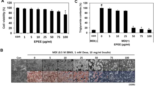
EPEE가 3T3-L1 preadipocyte의 세포 생존율에 미치는 영향
세포실험계에서 지방생성억제능의평가를위해먼저
EPEE
가3T3-L1 preadipocyte
의세포생존율에미치는영 향을WST assay
를이용하여분석하였다.
그결과1
−100
μg/
ml
의시료처리에서농도증가에따른약한세포증식억제 를보였으나세포독성은유발하지않음을확인하였다(Fig.
2A).
EPEE가 MDI로 유도한 3T3-L1 preadipocyte의 adipogenesis에 미치는 영향
EPEE
의항비만활성보유유무를알아보기위해MDI
로분화를유도한
3T3-L1 preadipocyte
의adipogenesis
에미치 는시료의영향을분석하였다.
그결과Fig. 2B
와2C
에서제 시된바와같이처리농도의증가에따라지방세포분화가억제되는것으로나타났고특히
50
μg/ml
이상의처리농도에서강한억제활성을보여
Oil Red O staining
결과염색 된지방의양이급격히감소되었다.
지방생성의억제정도 를정량적으로평가하기위해염색된지방을추출하여TG
생성량을측정한결과시료처리범위에서농도의존적으로 감소됨을보여50
μg/ml
의처리에의한억제능이80.3%
로 나타났다.
EPEE가 adipogenesis 관련 유전자 및 단백질 발현에 미 치는 영향
지방전구세포가지방세포로분화되는
adipogenesis
의과 정에는초기,
중기,
후기의각단계별로중요한분화조절인 자가관여한다. MDI
처리초기에는가장먼저c-fos, c-jun, c-myc
등의유전자발현이증가하며이와함께C/EBP
β와 δ의발현또한유도된다
. C/EBP
β와 δ는지방전구세포가MDI
등의분화인자에노출되었을때분화초기에가장먼저작 용하는전사 인자로서
MDI
의경우C/EBP
β는DEX
에, C/
EBP
δ는IBMX
에의해활성이유도되는것으로알려져있다
.
이러한C/EBP
β와δ의활성은분화중기및후기에서작 용하는PPAR
γ와C/EBP
α의발현매개인자로서,
활성화된PPAR
γ와C/EBP
α는단독혹은상호작용을통해지방세포특이적유전자의발현
(adipocyte specific gene expression)
을유도하여지방세포분화및지방형성을완성한다
[1, 9,
14].
따라서이와같은지방세포분화에관여하는주요인자들의발현조절유무는소재가보유한지방생성억제능 및그작용기전을판단하는주요지표중하나이다
.
본연구에서는
EPEE
가보유한지방생성억제능의작용 기전을알아보기위하여EPEE
가adipogenesis
에관여하는 주요핵심조절자인C/EBP
α, C/EBP
β,
그리고PPAR
γ의유 전자및단백질발현에미치는영향을분석하였다.
그결과Fig. 1. Lipase enzyme inhibition activity of EPEE. The effects
of EPEE on pancreatic lipase activity. Values are represented as
the mean ± SD (n = 3) *p < 0.01 vs vehicle control (0).
Fig. 3
의결과에서와같이MDI
처리에의해지방세포분화 가일어난대조군에서는세인자의유전자및단백질발현 이유의적으로 증가되었으며시료의처리에의해농도의존 적인감소를보여50
μg/ml
의처리에서는모든 인자의유 전자및단백질발현이미분화대조군과유사한정도의낮은발현을보였다
.
이러한결과를통해EPEE
가보유한지방세포분화억제능이
adipogenesis
에관여하는핵심인자 의유전자및단백질발현저해를통해나타나는것으로판 단된다.
EPEE의 중성지방 제거능(lipolysis activity)
EPEE
가지방세포분화를억제할뿐만아니라생성된 지Fig. 2. Effect of EPEE on 3T3-L1 cell proliferation (A), morphological change and lipid accumulation (B), and TG contents (C).
(A) Cells were treated with the indicated concentrations of EPEE for 72 h and viability was determined by WST assay. Data are expressed as the mean ± SD of triplicate experiments. (B) Differentiation of confluent 3T3-L1 preadipocytes was initiated with MDI treatment and maintained in DMEM containing 5% FBS in presence and absence of EPEE. After day 8, cells were fixed and stained with Oil red O.
The morphological change and lipid droplet accumulation were visualized using by inverted microscopy ( ×200). (C) TG contents were determined by Oil red O staining after treatment of EPEE. TG contents were measured at 500 nm by multi-plate reader. Data are expressed as the mean ± SD of triplicate experiments. *, # Significantly different from the undifferentiated cell control (Con) and untreated cell control (0), respectively (p < 0.05).
Fig. 3. Effect of EPEE on adipogenesis related gene and protein expressions. (A) Modulation of adipogenic transcription factors by
EPEE was evaluated by RT-PCR. GAPDH was used as an internal control. (B) Modulation of adipogenesis related protein expressions
by EPEE was evaluated by Western blot analysis. Actin was used as an internal control. The data are representative of three independent
experiments.
방의제거에도효과적인지를판단하기위해지방세포 내 중성지방제거능을
lipolysis activity assay
를통해분석하였다
. MDI
로분화시킨 지방세포에EPEE
를후처리한 결과세포내축적되어있던중성지방의분해를통해배지로 방출된글리세롤의 양이농도의존적으로증가되는것으로 나타났다
(Fig. 4A).
배지를제거한후Oil Red O staining
을수행하고현미경 관찰
(Fig. 4B)
및세포 내에남아있는TG
의양을정량분석(Fig. 4C)
한결과배지에방출된글리세롤양의증가와비례적으로세포내에남아있는
TG
의양이감소되는것으로나타나
EPEE
가지방세포내에 축적되어있는중성지방을농도의존적으로제거하는것을확인 하였다
.
이상의결과를통해
EPEE
가췌장lipase
효소활성억제능
,
지방세포분화억제능,
지방세포내중성지방제거능을 통한항비만활성을보유함을확인하였다.
이러한결과는E.
pleiosperma
추출물의항비만활성을처음으로밝혀낸것이며추후계속적인연구를통해활성물질의규명이필요할 것으로판단된다
.
요 약
선행연구에서
Euptelea pleiosperma
가항산화능과항염증 활성을나타내는유용한소재임을처음으로밝혔다.
본연구 에서는E. pleiosperma
에탄올추출물(EPEE)
의항비만활 성을췌장리파아제효소활성억제능및세포실험모델계를 이용하여분석하였다.
먼저EPEE
는농도의존적으로lipase
효소활성을유의적으로억제시켰으며, 3T3-L1 preadipocyte
에서지방세포분화
,
세포내지방축적, TG
함량등을독성없이농도의존적으로억제하였으며지방세포내중성지방을 유의적으로분해시키는것으로나타났다
.
이러한EPEE
의지방세포 분화 억제능은 핵심 작용 인자인
C/EBP
α, C/
EBP
β,
그리고PPAR
γ의유전자및단백질발현조절에서기 인함을확인하였다.
이러한결과는E. pleiosperma
가보유 한췌장lipase
활성저해능,
지방세포분화억제능,
지방세 포내지방분해능을통한항비만활성을처음으로밝혀낸 것이며추후계속적인연구를통해활성물질의규명이필 요할것으로판단된다.
Fig. 4. Stimulatory effect of EPEE on glycerol release in MDI-induced 3T3-L1 adipocytes (A), morphological change and lipid
accumulation (B), and TG contents (C). (A) Amount of released glycerol in culture media was measured after EPEE treatment. Glycerol
contents were measured 540 nm by multi-plate reader. (B) Differentiation of confluent 3T3-L1 preadipocytes was initiated with MDI treat-
ment and maintained in DMEM containing 5% FBS. After day 8, MDI-induced adipocytes were treated with EPEE for 48 h. Cells were
fixed and stained with Oil red O. The morphological change and lipid droplet accumulation was visualized using by inverted microscopy
( ×200). (C) TG contents were determined by Oil red O staining after treatment of EPEE. TG contents were measured at 500 nm by multi-
plate reader. Data are expressed as the mean ± SD of triplicate experiments. *, # Significantly different from the undifferentiated cell control
(Con) and untreated cell control (0), respectively (p < 0.05).
Acknowledgments
This work was supported by Blue-Bio Industry Regional Innova- tion Center (RIC08-06-07) at Dong-Eui University as a RIC pro- gram under Ministry of Trade, Industry and Energy (MOTIE) and Busan city.
References