大 韓 不 妊 學 會 誌 : 第 28 卷 第 3 號 2 001 Kor. J. Fertil. Steril., Vol. 28, No. 3, 2001, 9
배양액 내의 마그네슘 이온이 생쥐 초기 배아 발생에 미치는 영향
삼성제일병원 생식생물학 및 불임 연구실1, 성신여자대학교 생물학과2
최수진1,2・전진현1・박용석1・배인하2
Effect of Magnesium Ion in the Culture Medium on the Development of Preimplantation Mouse Embryos In Vitro
Soo-Jin Choi1,2, Jin Hyun Jun1, Yong-Seog Park1, In-Ha Bae2
1Laboratory of Reproductive Biology and Infertility, Samsung Cheil Hospital &
Women's Healthcare Center, Seoul 100-380; 2Department of Biology, College of Natural Sciences, Sungshin Women's University, Seoul 136-742, Korea
Objective: The present study was undertaken to examine the effects of magnesium ion in the culture medium on the development of mouse fertilized oocytes either before or after pronuclear formation, and to investigate whether the effect of magnesium ion is related with the redistributional change of mitochondria.
Methods : Fertilized oocytes obtained from the oviducts of mice at 15 hr after hCG injection before pronuclear formation (pre-PN) or 21 hr after hCG injection after pronuclear formation (post-PN) were used. The embryos were cultured for 3 days with basic T6 medium-magnesium free and various concentrations of magnesium ion, 0.0, 0.5, 1.0, 2.0, 4.0 or 8.0 mM, respectively. After culture, the developmental stages of embryos and the number of nuclei were evaluated. To observe the effects of magnesium ion on the mitochondrial distribution, fertilized oocytes were collected at 21 hr after hCG injection and cultured for 6 hr with various concentration of magnesium ion. As a control, fertilized oocytes with pronuclei at 27 hr after hCG injection were used.
Results: The concentration of magnesium ion to accelerate the in vitro development of mouse fertilized oocytes appeared to be at 2.0 mM for the pre-PN and the post-PN stage embryos. In the mitochondrial redistribution patterns, the embryos cultured in 2.0 mM concentration of magnesium ion showed the highest percentage (22.6%) of distinct perinuclear clustering pattern comparing to other experimental group.
Conclusion: The effect of magnesium ion may be related to the cytoplasmic redistribution of mitochondria. This relationship seems to connect the developmental competence of preimplantation mouse embryos in vitro. These results can suggest that higher concentration of magnesium ion (2.0 mM) than those of conventional culture medium (0.2~1.2 mM) is more suitable for in vitro culture of preimplantation mouse embryos.
Key Words: Magnesium ion, Culture medium, Preimplantation mouse embryos, Developmental competence, Mitochondrial distribution
- 200 - 포유류 초기 배아의 체외 배양이 시작된 이래 배 아의 발생률을 향상시키기 위한 많은 노력이 있었으 며 생체 내 환경에 가장 가까운 배양액과 배양 환경 에 대한 연구는 계속되어 왔다.1~10 그러나 포유류 배 아의 체외 배양 (in vitro culture ) 시 그 발생률은 체내 에 비해 낮은 것으로 알려져 있다. 특히, 체외 배양 시 착상 전 배아는 종에 따라서 특정 시기에 난할 중지 현상이 나타나기도 한다.
생체의 생리적인 작용에 필수적인 칼슘 이온과 마 그네슘 이온은 여러 세포 유형에서 다양한 세포 기 능 (cellular function)을 조절한다고 보고되었다.11,12 세 포 분열을 진행하는 과정에는 세포질 내 칼슘 이온 의 진동 현상 (calcium oscillation)이 나타나며, 이는 세포 분열을 조절하는데 매우 중요한 요소로 작용 한다.13 또한, 세포 내 칼슘 이온 농도의 조절 이상 (abnorma l control)은 단백질 생성, DNA 복제, 미토콘 드리아의 기능과 세포간의 communication에 악영향 을 가져올 수 있다고 보고되었다.14,15 칼슘 이온이 부 족한 배양액에서는 DNA 합성과 세포의 증식이 억 제되며, 마그네슘 이온이 부족한 배양액에서는 uri- dine의 세포질 내로의 흡수와 이용률이 매우 감소되 었다.16,17 Fisher는18 칼슘 이온이 없는 배양 조건에 서 고농도의 마그네슘 이온이 생쥐 포배기 배아에서 최적 자극 (optimal stimulation)을 유도함을 관찰하여 마그네슘 이온이 칼슘 이온과 대체될 수 있음을 제 시하였다. 또한, 다양한 칼슘과 마그네슘 농도의 배 양액에서 병아리 배아 세포 (Chic k embryo cell)의 DNA 합성에 관한 연구를 통해, 마그네슘이 DNA 합성 조절의 일차적 작용체 (effector)이고 칼슘은 이 차적 조절체 (modifier)로19 세포 증식에 마그네슘이 중추적 역할을 한다고 보고되었다.20,21
최근에 햄스터 배아를 이용한 실험에서 Lane 등 은22 1-세포기 햄스터를 이용한 실험에서 0.5 mM~
4.0 mM 의 다양한 마그네슘 농도에서 배양하였을 때 2.0 mM에서 가장 높은 발생률을 나타내었다고 보고 하였다. 이 실험에서 배양액 내 마그네슘 농도가 증 가되면 배양된 2-세포기 배아의 세포 내 칼슘 농도 증가가 억제되었다. 이는 여분의 배양액 내 마그네 슘 농도가 배양된 배아 내의 칼슘 항상성 (homeos- tasis)의 조절 요인으로 작용한다는 것을 시사한다.
세포 내 칼슘 농도의 항상성은 세포막의 유지, gap
junction communication, 대사 과정과 같은 세포의 생 리학적인 기능 조절에 기본이다. 체외 배양 시 햄스 터 1-세포기 배아의 발생 능력 약화는 칼슘 항상성 (homeostasis)의 이상과 관계 있으며,22 마그네슘은 이 러한 세포 내로의 칼슘 이온의 이동을 조절함으로 써 항상성에 중요한 역할을 한다고 보고되었다.23 배 양 환경에서 배아의 세포 내 이온 항상성이 배아의 생존 능력과 발생 과정에 작용하는 기전은 현재까지 명확하게 알려져 있지 않다.
이중 미토콘드리아는 살아있는 세포의 에너지 대 사에서 중요한 역할을 하며, 초기 배아의 발생에는 많은 양의 ATP가 필요한 것으로 알려져 있다. 이러 한 미토콘드리아의 세포 내 분포는 에너지가 요구 되는 부분에 밀집되는 경향이 있으며, 미토콘드리아 의 배치와 분포는 세포 증식과 분화 과정 등 세포 기능의 변화에 따라 다양하게 나타난다.24~27
생쥐 배아의 체외 발생 중지 현상에 대한 연구에 서 미토콘드리아는 배아의 초기 난할 과정에서 특정 부위로 이동하고 그 이동이 발생 중지 현상과 관계 있다고 보고되었으며,28 Barnett과 Bavister는29 햄스터 1-세포기 배아에서 미토콘드리아의 전핵 주위에 응 축 현상이 배아의 발생 능력과 관련이 있음을 제시 하였다.
이에 본 연구에서는 배양액 내의 마그네슘 이온이 생쥐 초기 배아의 발생에 미치는 영향을 알아보기 위해 전핵 형성 전/후 수정란을 각각 다른 농도의 마그네슘 이온이 첨가된 배양액에서 체외 배양하여 발생률과 세포 수를 관찰하였다. 또한, 이러한 배양 액의 마그네슘 이온 농도의 변화가 배아 내 미토콘 드리아의 재배열 현상과 관계가 있는지 알아보았다.
연구 대상 및 방법
1. 실험 동물 및 수정란의 회수 1) 과배란 유도
본 실험에서는 사료와 물을 충분히 공급하면서 14 시간 조명, 10시간 소등 환경에서 자란 생후 5~6 주령의 암컷 생쥐 (C57BL/6 × CBA F1)와 생후 12 주령 이상의 생식력 있는 수컷을 사용하였다.
생쥐 암컷의 복강에 5 IU (international unit)의 pre- gnant mare's serum gonadotropin (PMSG, Sigma)을 투
여하고 48시간 후 human chorionic gonadotropin (hCG, Sigma)을 주사하여 과배란을 유도한 후 수컷 생쥐와 각각 교미시키고 다음날 아침 질전 (vaginal plug)을 확인하여 교미 여부를 판정하였다.
2) 전핵 형성 전 수정란 (pre-PN) 수집 질전이 확인된 암컷을 hCG 주사 후 15시간째 경 추골 파열 방법으로 도살한 후 양쪽 난관을 채취하 였다. 채취 난관을 0.4% bovine serum albumin (BSA, Gibco)이 첨가된 본 연구의 기본 배양액인 modified Tyrode's solution-Magnesium free (T6-Mg free 배양액) 을 이용하여 수란관 팽창 부위로부터 수정란을 수 집하였다. 필요에 따라 0.1% hyaluronidase (Sigma)를 사용하여 수정란의 mucin 성분과 난구 세포 (cumu- lus cell)를 제거하였다. 수집된 배아는 기본 배양액 으로 3회 세척하였다. 전핵 (pronucleus)이 관찰되지 않은 수정란만을 선별하여 실험에 사용하였다.
3) 전핵 형성 후 수정란 (post-PN) 수집 질전이 확인된 암컷을 hCG 주사 후 21시간째 기 본 배양액을 이용하여 위와 같은 방법으로 수정란을 수집하였다. 전핵이 관찰되는 수정란만을 선별하여 실험에 사용하였다.
2. 배양 방법
수집된 수정란은 microdroplet 방법으로 배양하였
다. 배양 접시 (Falcon 3002)에 마그네슘 이온이 0.0, 0.5, 1.0, 2.0, 4.0, 8.0 mM 농도로 첨가된 각각의 배양
액으로30,31 20 ìl의 배양액 소적을 만들고, 그 위에
멸균된 mineral oil (Sigma)로 덮은 후 배아를 키우는 방법을 이용하였다. 수집된 배아는 각 농도의 마그 네슘 배양액으로 3회 이상 세척한 후 배양 소적에 분배하였다. 이때 배아들은 37℃ 온도에서 5% CO2
와 95% 공기, 100% 습도가 유지되는 배양기에서 배양하였다. 배아의 발생 정도는 hCG 주사 후 96 시간에 관찰하여 2~8 세포기, 상실기 (morula), 포배 기 (blastocyst) 그리고 퇴화 배아 (degenerated embryo) 로 구분하였다 (Figure 1).
3. 배아의 염색 및 할구 수 계수
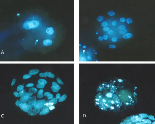
체외 배양한 각 배아를 hCG 주사 후 96시간째 관 찰하여 상실기 혹은 포배기 배아를 선별하여 염색하 였다. 이 배아들을 1% glutaraldehyde에 고정하고, 10 ìg/ml Hoechst 33342 (bisbenzimide solution, Sigma)이 포함된 phosphate buffered saline (PBS)에서 염색하였 다.32 염색된 배아는 0.4% polyvinyl-pyrrolidine (PVP, Sigma)-PBS로 세척 후 mounting 하고 형광 현미경 (Nikon, Japan)하에서 (400×) 관찰하여 배아의 할구 수를 측정하였다 (Figure 2).
A B
Figure 1. Microphotographs of mouse embryos collected from oviduct 21 hr after hCG injection followed by culture for 75 hr. Magnification, ×200. Bar indicates 50 ìm. For details, see Table 2. A, 0.0 mM Mg2 concentration, 1.78 mM Ca2+ concentration; B, 2.0 mM Mg2+ concentration, 1.78 mM Ca2+ concentration
- 202 - 4. 미토콘드리아의 관찰
배아 내의 미토콘드리아의 분포 상태를 관찰하기 위해 미토콘드리아의 transmembrane electrical poten- tial에 반응하는 rhodamine 123 (Rh123, Molecular pro- bes R-302, Eugene, USA)을 이용하였다. Rh123 stock solution은 methanol로 10 mg/ml로 희석하여 -20℃에 저장하였다.
마그네슘 이온이 미토콘드리아의 재배열에 미치 는 영향을 관찰하기 위해 hCG 주사 후 21시간에 전 핵이 형성된 수정란을 회수하여 0.0, 2.0, 8.0 mM 마 그네슘 이온 농도의 배양액에 6시간 동안 체외 배 양한 실험군과 hCG 주사 후 27시간에 수란관에서 수집한 대조군 (control)을 각각 염색하여 비교, 분석 하였다.
Rh123 stock을 기본 배양액으로 (1 ìg/ml; final con- centration 10 ìg/ml) 희석하여 실험군과 대조군의 수 정란을 배양 환경 (culture condition)에서 15분 동안 염색하고 각 배양액에서 완전히 세척하였다. 염색된 배아는 slide에 옮겨 cover slip을 덮은 후 B-2A filter 를 사용하여 형광 현미경 (Nikon, Japan)하에서 (200×) 관찰하였다.10,29,33
미토콘드리아의 세포 내 재배열 양상은 전핵 주 위 응축 양상 (distinct perinuclear clustering), 전핵 주 위 밀집 양상 (perinuclear clustering), 세포질 내 균등 양상 (dispersed throughout the cytoplasm)으로 구분하 였다 (Figure 3).
5. 통계처리
각 실험값들의 통계적 유의성은 Student's t-test를
A B
C D
Figure 2. Fluorescence micrographs of mouse embryo stained with Hoechst 33342 (bisbenzimide solution). Magnifi- cation, ×400. For details, see Table 1 & 2. A, 4-cell embryo; B, Morula; C, Early Blastocyst; D, Degenerated embryo
이용하였다.
결 과
1. 배양액 내 마그네슘 이온이 초기 배아 발생에 미치는 영향
전핵이 형성되기 전 초기 1-세포기 배아를 배양 했을 때 마그네슘 0.0 mM 농도군의 상실기 혹은 포 배기 배아 발생률은 25.7% 이였으며, 2.0, 4.0, 8.0 mM 농도군에서의 발생률은 각각 62.0%, 55.6%, 47.1%로 유의하게 높게 나타났다 (p<0.01). 그 중에서 2.0 mM 농도의 배양액에서 가장 높은 발생률을 나타내었다.
Hoechst 33342 염색을 이용한 세포 내 할구 수 관찰 에서도 전핵 형성 전 수정란의 할구 수는 마그네슘 2.0, 4.0, 8.0 mM 농도군에서 25.2, 25.2, 22.5로 마그 네슘 0.0 mM 농도군 (17.4) 보다 유의하게 많게 나 타났으며 (p<0.01), 2.0과 4.0 mM 농도군에서 가장 많은 평균 할구수가 관찰되었다 (Table 1).
또한, 전핵이 형성된 후기 1-세포기 배아를 배양 했을 때는 마그네슘 농도가 0.0 mM (25.1%)인 배양 액 보다 2.0과 4.0 mM 일 때 71.3%, 64.4%로 높은 발생률을 나타냈으며, 2.0 mM 일 때 가장 높게 나 타났다 (p<0.01). 전핵이 형성된 수정란의 할구 수는 마그네슘 1.0, 2.0, 4.0 mM 농도일 때 각각 20.2, 21.0, 18.8개로 마그네슘 0.0 mM 농도군 (15.5) 보다 유의 하게 많았으며 (p<0.05), 2.0 mM 농도군에서 가장 많은 평균 할구 수가 관찰되었다 (Table 2, p<0.01).
2. 배양액 내 마그네슘 이온에 따른 미토콘드리 아의 재배열 양상
발생이 진행됨에 따라 나타나는 전핵 주위의 미토 콘드리아 응축 양상을 나타낸 비율은 hCG 주사 후 27시간째에 수집한 대조군에서 64.4%로 가장 높은 비율을 나타내었으며, 이는 각각의 마그네슘 이온 농도의 배양액에서 6시간 체외 배양이 이루어진 실 험군들과 유의한 차이를 나타내었다 (p<0.01).
실험군들 중에서는 2.0 mM 마그네슘 이온 농도 군에서 미토콘드리아의 전핵 주위 응축 양상을 보이 는 수정란의 비율이 22.6%로 0.0, 8.0 mM 배양액의 4.3%, 6.0% 보다 유의하게 높게 관찰되었다 (Table 3, p<0.05).
A
B
C
Figure 3. Fluorescence micrographs of the mitocho- ndria in the one-cell embryo stained with Rhodamine 123. Magnification, ×400. For details, see Table 3. A, distinct perinuclear clustering; B, perinuclear clustering;
C, dispersed throughout the cytoplasm
- 204 -
Table 1. Effect of various magnesium concentrations in the culture medium on the in vitro development of pre -PN stage embryos
Developmental stage reached after 3 days of culture (%) Magnesium
concentration (mM)
Total no. of embryos
Morula (Mo) Blastocyst (Bl) Mo. + Bl.
Cell number of Mo. and Bl.
0.0 66 25.7±3.9 0.0±0.0 25.7±3.9 17.4±1.2
0.5 62 33.6±3.7 8.1±6.5 41.7±6.2 19.8±1.5
1.0 64 34.8±3.2 5.2±3.4 39.9±3.3* 18.9±1.0
2.0 71 48.6±6.5 13.4±3.1 62.0±5.5** 25.2±1.1**
4.0 67 41.7±6.8 8.7±3.4 55.6±5.2** 25.2±0.9**
8.0 68 47.1±4.8 0.0±0.0 47.1±4.8** 22.5±1.0**
The concentration of calcium ion was adjusted to 1.78 mM. The results were obtained by pooling of seven replicates (Student's t -test). P values significantly differ from the 0.0 mM concentration group (**p<0.01, *p<0.05). Embryos were flushed at 15 hr after hCG injection and those of PN formed embryos at 26~28 hr after hCG were further culture in the present study. Data are mean±SEM.
Table 2. Effect of various magnesium concentrations in the culture medium on the in vitro development of post-PN stage embryos
Developmental stage reached after 3 days of culture (%) Magnesium
concentration (mM)
Total no. of embryos
Morula (Mo) Blastocyst (Bl) Mo. + Bl.
Cell number of Mo. and Bl.
0.0 77 23.7±5.1 1.4±1.3 25.1±5.9 15.5±1.0
0.5 74 42.1±9.3 0.0±0.0 42.1±9.3 15.3±0.6
1.0 79 39.6±6.2 7.7±6.9 47.3±10.3 20.2±1.4*
2.0 74 60.7±7.7 10.5±3.2 71.3±10.2** 21.0±1.2**
4.0 76 59.0±3.5 5.5±2.6 64.4±5.9** 18.8±0.9*
8.0 77 32.1±8.8 0.0±0.0 32.1±8.8 16.3±0.9
The concentration of calcium ion was adjusted to 1.78 mM. The results were obtained by pooling of five replicates (Student's t -test). P values significantly differ from the 0.0 mM concentration group (**p<0.01, *p<0.05). Embryos were flushed at 21 hr after hCG injection and those of PN formed embryos were further culture in the present study. Data are mean±SEM.
Table 3. Effect of various magnesiu m concentrations on mitochondrial distribution of one-cell embryos in vitro culture Pattern of mitochondrial distribution (%)
Magnesium concentration
Total No. of
embryos Distinct Perinuclear clustering
Perinuclear clustering
Dispersed throughout the c ytoplasm
Control 67 64.4±4.4a 26.5±3.0 9.2±4.1
0.0 74 4.3±1.9b 28.6±4.3 67.0±3.1
2.0 79 22.6±4.7c 35.6±4.2 41.7±3.0
8.0 77 6.0±2.7d 39.9±5.2 54.1±4.9
The concentration of calcium ion was adjusted to 1.78 mM. The results were obtained by pooling of seven replicates (student's t-test). P values significantly differ from the control group (ab, ac, ad p<0.01); P values significantly differ from the 2.0 mM concentration group (bc, p<0.01, cd p<0.05). Control, all embryos were flushed at 27 hr after hCG injection. The other group, all embryos were flushed at 21 hr after hCG injection and 6 hr further culture were done in vitro system. Data are mean±SEM.
고 찰
마그네슘 농도에 따른 발생률은 전핵이 형성되기 전 초기 1-세포기 배아를 배양했을 때 마그네슘 2.0, 4.0, 8.0 mM 농도 첨가 배양액에서 0.0 mM 일 때 보 다 유의하게 높게 나타났으며 (p<0.01), 2.0 mM 일 때 가장 높은 발생률을 보였다. 세포 내 할구 수 관 찰 실험에서도 전핵 형성 전 수정란은 2.0, 4.0, 8.0 mM 마그네슘 첨가 배양액에서 많은 수가 관찰되었 다 (Table 1). 또한 전핵이 형성된 후기 1-세포기 배 아를 배양했을 때는 마그네슘 농도가 2.0과 4.0 mM 일 때 높은 발생률을 나타냈으며, 고농도 마그네슘 첨가군 (8.0 mM)에서는 오히려 수정란의 발생률이 떨어지는 결과가 나타났다. 전핵이 형성된 수정란 배양 시 할구 수는 1.0, 2.0, 4.0 mM에서 많게 나타 났으며 2.0 mM에서 21.0으로 가장 많은 수가 관찰 되었다 (Table 2).
이는 2-세포기 햄스터 배아를 포배기까지 배양한 Mckiernan 등과4 햄스터 수정란을 배양한 Bavister와 Golden의34 칼슘 이온과 마그네슘 이온 비율에 의한 농도 차이는 발생률에 영향을 미치지 않는다는 보고 와 다른 양상을 보이나 McKiernan는 0.5 mM~0.125 mM , Bavister는 0.1 mM ~0.8 mM 의 저농도 마그네슘 배양액에서만 실시되었다. 반면 1-세포기 햄스터를 이용한 Lane 등의22 실험에서 0.5 mM ~4.0 mM 의 마 그네슘 농도에서 배양하였을 때 2.0 mM에서 가장 높은 발생률을 나타내었다고 보고하였다. Barnett와 Bavister가35 햄스터로 실험한 결과에서 난자 활성화 후 3시간째 수정란을 회수하여 체외 배양 시 상실기 혹은 포배기 배아의 발생률은 16%로 난자 활성화 후 9시간째 회수된 수정란의 발생률 64% 와 매우 큰 차이를 나타내었다.
본 연구에서는 전핵 형성되기 전 수정란보다 전핵 이 형성된 수정란의 발생률이 각 농도별 비교하여 전반적으로 높게 나타났으며, 8.0 mM의 고농도 마 그네슘이 첨가된 군에서는 오히려 전핵이 형성된 수 정란의 발생률이 떨어지는 결과가 나타났다. 이는 과도한 농도의 마그네슘 이온 역시 배아 발생을 저 해하는 것으로 보여진다. 1-세포기의 전핵 형성이 이루어지는 시간에 칼슘 이온 농도 변화에 민감하
며 갑작스런 세포 내 칼슘 이온 농도 증가 현상은 배아 발생에 이롭지 않는 것으로 판단된다.34,36 이때 마그네슘 이온은 칼슘 이온의 농도 증가를 막아주 는 buffe r 역할을 함으로써 배아 발생률을 증가시키 는 것으로 추정된다.
Myocardia l cell 등 다른 세포 유형에서 배양액 내 마그네슘 이온이 세포 내 칼슘 이온 농도 증가를 완 화시킨다는 논문이 많이 발표되고 있으며,23,37 myo- cardia l cell 내 칼슘 농도를 낮추는데 효과적인 마그 네슘 농도는 2.0 mM 로 관찰되었다. 이는 본 실험에 서와 비슷한 결과를 보이고 있어 마그네슘 이온의 buffering 작용으로 인한 것이라고 추정된다.
인간의 배아에서 미토콘드리아는 수정 전에는 세 포질 (cytoplasm) 내에 퍼져 있는 형태이고 수정란에 서 전핵 주위로 응축된다고 보고되었다.27 Van-Bler- kom과 Runner는24 생쥐 난자를 이용한 연구에서 미 토콘드리아의 전핵 주위로 응축되는 현상은 spindle 형성과 소멸, 염색체 이동에 필요한 에너지 생산과 활용 때문이라고 주장하였으며, Muggleton-Harris 와 Brown은26 생쥐의 '2-cell block'은 에너지 상태와 미 토콘드리아의 재배열 때문이며 미토콘드리아의 위 치 변화는 체외 배양 시 배아 발생 능력과 관계 있 다고 보고하였다.
본 연구에서 미토콘드리아 재배열 양상을 관찰한 결과 마그네슘 농도 2.0 mM 일 때 미토콘드리아의 전핵 주위 응축 양상을 보이는 배아가 22.6% 로 실험 군 중에서 가장 높게 나타났으며, 이는 체내 배양이 이루어진 대조군에서 가장 높게 나타난 미토콘드리 아 분포 양상이다 (Table 3).
미토콘드리아 재배열 파괴 현상은 spindle 형성 또 는 소멸과 염색체 이동에 필요한 에너지 생산과 활 용의 조절 기능을 약화시키며 미토콘드리아의 대사 작용의 변화를 일으켜 초기 발생에 영향을 미친다고 생각되어진다.
미토콘드리아가 핵 주위에 응축되어 있는 형태가 체내 (in vivo) 상태에서 가장 높은 비율을 차지하는 것으로 보아 hCG 주사 후 27시간째인 수정란에 가 장 적합한 미토콘드리아의 배열 방식이라고 생각된 다. 그러나 체외 배양 후 (6시간) 세포질 내에 고르 게 퍼져 있는 형태 (dispersed throughout the cytoplasm) 가 많고 또 배아 발생이 잘 되지 않는 점으로 보아
- 206 - 미토콘드리아는 세포질에 퍼져 있는 형태에서 전핵 주위에 밀집되는 형태 (perinuclear clustering)를 거쳐 응축되는 형태 (distinct perinuclear clustering)로 변하 고 있는 것으로 추정된다. 미토콘드리아의 응축 양 상은 마그네슘 2.0 mM 농도 첨가군에서 가장 높게 나타났으며 상실기 및 포배기 배아의 체외 발생률이 가장 높은 점으로 보아, 마그네슘 이온 농도에 따 라 미토콘드리아 분포 조절이 달라지며 발생률에 차 이가 있음을 알 수 있었다.
이상의 결과로 보아 생쥐 배아 발생에는 기존의 연구들에서 보고된 0.2 mM~1.2 mM 마그네슘 이온 농도와 달리 더 높은 마그네슘 이온 농도가 요구되 어진다고 생각된다.
참 고 문 헌
1. Borland RM, Hazra S, Biggers JD, Lechene CP.
The elemental Composition of the environments of the gametes and preimplantation embryo during the initiation of pregnancy. Biol Reprod 1977; 16: 147- 57.
2. Bavister BD, Leibfried ML, Lieberman G. Deve- lopment of preimplantation embryos of the golden hamster in a defined culture medium. Biol Reprod 1983; 28: 235-47.
3. Spindle A. In vitro development of one-cell em- bryos from outbred mice: Influence of culture me- dium composition. In Vitro Cell Dev Biol 1990; 25:
151-6.
4. McKiernan SH, Bavister BD. Environmental vari- ables influenceing in vitro development of hamster 2-cell embryos to the blastocyst stage. Biol Reprod 1990; 43: 404-13.
5. Lawitts JA, Biggers JD. Optimization of mouse embryo culture media using simplex methods. J Reprod Fert 1991a; 91: 543-56.
6. Lawitts JA, Biggers JD. Overcoming the 2-cell block by modifying standard components in a mo- use embryo culture medium. Biol Reprod 1991b;
45: 245-51.
7. Lawitts JA, Biggers JD. Joint effects of sodium
chloride, glutamine, and glucose in mouse preim- plantation embryo culture media. Mol Reprod Dev 1992; 31: 189-94.
8. Biggers JD, Lawitts JA, Lechene CP. The protec- tive action of betaine on the deleterious effects of NaCl on preimplantation mouse embryos in vitro.
Mol Reprod Dev 1993; 34: 380-90.
9. Erbach GT, Lawitts JA, Papaioannou VE, Biggers JD. Differential growth of the mouse preimplan- tation embryo in chemically defined media. Biol Reprod 1994; 50: 1027-33.
10. Barnett DK, Bavister BD. What is the relationship between the metabolism of preimplantation embryos and their developmental competence? Mol Reprod Dev 1996a; 43: 105-33.
11. Carvalho AP, Sanui H, Pace N. Calcium and ma - gnesium binding properties of cell membrane mate- rials. J Cell Comp Physiol 1963; 62: 311-8.
12. Yamamoto Y, Chen G, Miwa K, Suzuki H. Per- meability and Mg2+ blockade of histamine-operated cation channel in endotherial cells of rat intrapul- monary artery. J Physiol 1992; 450: 395-408.
13. Bos-Mikich A, Whittingham DG, Jones KT. Meio- tic and mitotic Ca2+ oscillations affect cell composi- tion in resulting blastocysts. Dev Biol 1997; 182:
172-9.
14. McCormack JG, Halestrap AP, Denton RM. Role of calcium ions in regulation of mammalian intra - mitochondrial metabolism. Physiol Rev 1990; 70:
391-425.
15. Lazrak A, Peracchia C. Gap junction gating sen- sitivity to physiological internal calcium regardless of pH in Novikoff hepatoma cells. Biophys J 1993;
65: 2002-12.
16. Bowen-Pope DF, Rubin H. Magnesium and cal- cium effects on uptake of hexose and uridine by chick embryo fibroblasts. Proc Natl Acad Sci USA 1977; 74: 1585-9.
17. Bowen-Pope DF, Vidair C, Sanui H, Rubin AH.
Separate roles for calcium and magnesium in their synergistic effect on uridine uptake by cultured cells:
significance for growth control. Proc Natl Acad Sci USA 1979; 76: 1308-12.
18. Fisher SB. The role divalent cations in the metabo- lic response of mouse blastocysts to serum. J Em- bryol Exp Morph 1980; 58: 217-29.
19. Rubin H, Koide T. Mutual potentiation by magne- siu m and calcium of growth in animal cells. Proc Nat Acad Sci USA 1976; 73: 168-72.
20. Rubin H. Central role for magnesium in coordinate control of metabolism and growth in animal cells.
Proc Nat Acad Sci USA 1975; 72: 3551-5.
21. Rubin AH, Berbie Chu. Reversible regulation by magnesium of chick embryo fibroblast proliferation.
J Cell Physiol 1978; 94: 13-20.
22. Lane M, Boatman DE, Albrecht RM, Bavister BD.
Intracellular divalent cation homeostasis and deve- lopmental competence in the hamster preimplanta- tio n embryo. Mol Reprod Dev 1998; 50: 443-50.
23. Altura BM, Altura BT, Carella A, Turlapaty PD.
Ca2+ coupling in vascular smooth muscle: Mg2+ and buffer effects on contractility and membrane Ca2+
movements. Can J Physiol Pharmacol 1982; 60:
459-82.
24. Van-Blerkom J, Runner M. Mitochondrial reorga- nization during resumption of arrested meiosis in the mouse oocyte. Am J Anat 1984; 171: 335-55.
25. Batten BE, Albertini DF, Ducibella T. Patterns of organelle distribution in mouse embryos during pre - implantation development. Am J Anat 1987; 178:
204-13.
26. Muggleton-Harris AL, Brown JJG. Cytoplasmic factors influence mitochondrial reorganization and resumption of cleavage during culture of early mo - use embryos. Hum Reprod 1988; 3: 1020-8.
27. Noto V, Campo R, Roziers P, Swinnen K, Vercruy- ssen M, Gordts S. Mitochondrial distribution after fast embryo freezing. Hum Reprod 1993; 8: 2115-8.
28. Tokura T, Noda Y, Goto Y, Mori T. Sequential observation of mitochondrial distribution in mouse
oocyte and embryos. J Assisted Reprod Gen 1993;
10: 417-26.
29. Barnett DK, Bavister BD. Inhibitory effect of glu- cose and phosphate on the second cleavage division of hamster embryos: is it linked to metabolism?
Hum Reprod 1996b; 11: 177-83.
30. Bae IH, Fooke RH. Maturation and rabbit follicular oocytes in a defined medium of varied osmolality. J Reprod Fert 1980; 59: 11-3.
31. Quinn P, Warnes GM, Kerin JF, Kirby C. Culture factors in relation to the success of human in vitro fertilization and embryo transfer. Fertil Steril 1984;
41: 202-9.
32. Koong MK, Jun JH, Song SJ, Lee HJ, Song IO, Kang IS. A second look at the embryo toxicity of hydrosalpingeal fluid: an in-vitro assessment in a murine model. Hum Reprod 1998; 13: 2852-6.
33. Barnett DK, Clayton MK, Kimura J, Bavister BD.
Glucose and phosphate toxicity in hamster preim- plantation embryos involves disruption of cellular organization, including distrivution of active mito- chondria. Mol Reprod Dev 1997; 48: 227-37.
34. Bavister BD, Golden M. Alteration of extracellular cation concentrations and ratios in culture medium does not affect first cleavage division of hamster zygotes in vitro nor overcome the 'two-cell block'.
Repro Fertil Dev 1989; 1: 231-6.
35. Barnett DK, Bavister BD. Hypotaurine requirement for in vitro development of golden hamster one-cell embryos into morulae and blastocysts, and produc- tion of term offspring from in vitro-fertilized ova.
Biol Reprod 1992; 47: 297-304.
36. Lane M, Bavister BD. Calcium homeostasis in early hamster preimplantation embryos. Biol Reprod 1998;
59: 1000-7.
37. Tzivoni D, Keren A, Cohen AM. Magnesium the- rapy for torsade de pointes. Am J Cardiol 1984; 53:
528-31.