대 한 생 식 의 학 회 지 : 제 37 권 제 1 호 2010
인간 배아줄기세포의 생식세포로의 분화 및 효소에 의해 분리된 단일줄기세포 배양조건
미즈메디병원 불임의학연구소 지희준*·최순영·정다연
Differentiation of Human Embryonic Stem Cells into Germ Cell and Culture Condition for Single Embryonic Stem Cells Dissociated by Enzyme
Hee Jun Chi
*, Soon Young Choi, Da Yeon Chung
Mizmedi Hospital ART Research Center
Objective: The present study was carried out to induce differentiation of human embryonic stem cells (hESCs) into germ cells and
to establish a culture condition for single hESCs dissociated by enzyme.Methods: Embryonic body (EB) was formed by hanging drop culture for 3 days from hESCs colony. The EBs were cultured in
the medium supplemented with retionic acid (RA) or/and bone morphogenetic protein-4 (BMP4) for 14 days to differentiate into germ cells. Germ cell specific markers, c-kit and VASA were used for immunohistochemistry of EB. Human ESCs colonies were dissociated into single cells by Collagenase, Tryple and Accutase, and then colony formation rate of the single cells was examined.Rho-associated kinase inhibitor (ROCK inhibitor, Y27632) was added into the culture medium of single cells to reduce the apoptotic damage during the dissociation.
Results: Single cells dissociated with Tryple or Accutase showed higher colony formation rates compared to the cells dissociated
with Collagenase. Seeding of 5×10³ cells/well (4 well dish) was efficient to obtain high colony formation rate compared to other concentrations of seeding cell. Addition of Y27632 significantly increased the colony formation rate of the single cells dissociated by Tryple. Immunohistochemistry of EB with c-kit and VASA markers showed a weak fluorescence signals compared to the signals from the testicular tissue.Conclusion: Dissociation with Tryple was useful to obtain healthy single cells and addition of Y27632 was beneficial for survival
and colony formation of the single cells. Unlike other studies, we just observed a dim fluorescence staining of the germ cell markers, probably caused by the short-term culture for the differentiation of EB compared to other studies.[Korean. J. Reprod. Med. 2010; 37(1): 13-23.]
Key Words: HESCs, Germ cells, Tryple, Retionic acid, Y27632
배아줄기세포 (ES cell)는 생식세포 (germ cell)를 포함한 신체를 구성하는 모든 형태의 세포로 분화 되는 잠재력을 가진 세포로 알려져 있다. 이러한 특성을 이용하여 배아줄기세포로부터 신경세포,
1심근세포,
2조혈세포,
3인슐린분비세포
4등 다양한 체세포로의 분화와 관련한 연구들이 진행되었다.
또한 배아줄기세포를 체외에서 장시간 배양할 경 우 이들이 자발적으로 생식세포 특이적 유전자의 발현을 나타내었으며
5,6난포와 유사한 구조를 갖는 세포로 분화됨으로써 생식세포 분화 연구에 대한 촉매제가 되었다.
7주관책임자: 지희준, 우) 157-723 서울시 강서구 내발산동 701-4, 미즈메디병원 불임의학연구소
Tel: (02) 2007-1840, Fax: (02) 2007-1852 e-mail: [email protected]
이처럼 배아줄기세포의 자발적인 생식세포로의 분화가능성이 제시되었지만 생식세포는 체세포와 는 다른 독특한 분화과정과 발달경로를 가지고 있 어서 특별한 배양조건과 분화방법이 요구되었고
8이와 관련한 다양한 연구들이 진행되었는데 생쥐 의 배아줄기세포를 생쥐의 난소과립막세포 (ovarian granulosa cells)와 공동 배양함으로써 형태적으로 난자와 유사한 세포로 분화시켰으며
9생쥐의 EB (embryonic body)를 testicular cell conditioned medium 에서 배양한 경우에도, 난소유사구조 (ovarian-like structure)로 분화되었다.
10또한 체외에서 원시생식 세포 (primordial germ cell, PGC)의 증식과 특성화 에 중요한 역할을 하는 것으로 알려진 retionic acid
(RA)
11,12와 bone morphogenetic protein-4 (BMP4)
producing M15 cells을 이용하여 생식세포로의 분 화를 유도하였다.
13최근에는 영장류인 cynomolgus monkey embryonic body (EB)의 일부 세포괴에서 초 기 생식세포인 gonocyte의 특이발현유전자인 VASA 의 발현을 확인하였으며
15인간 배아줄기세포로부 터 형성된 EB를 BMP4, BMP7, BMP8b 등을 이용하 거나,
14돼지 난소섬유아세포 (mitotically inactivated porcine ovarian fibroblasts)와 공동 배양하였을 경우 에도 초기 생식세포로의 분화성공이 보고되었다.
16생식세포의 분화는 다양한 특이유전자들의 발현 을 통해 확인하였는데 이들 분화된 세포들은 난자 특이유전자인 Figalpha, GDF-9, and ZP1-3 뿐만 아니 라 정소 특이유전자인 SCP3를 발현하였다.
9이외 에도 다양한 특이유전자인 VASA, c-kit, Mvh, Stella, Dazl, Piwil 2, Pdrd 1, Rex 14, Rnf 17, Bmp8b, Stra-8, Haprin, LH-R, Sycp1, Sycp3, EMA-1, FE-J1, Fragilis, Acrosin, acetylated alpha-tubulin) 등의 발현을 관찰한 연구들이 보고되었다.
5,6,11,12,14,15,17최근에는 이러한 유전자발현을 역으로 이용하여 배아줄기세포로부 터 분화된 생식세포에서 VASA의 발현을 시각적으 로 관찰할 수 있는 형광표지유전자 (GFP, LacZ)를 생쥐 배아줄기세포 내에 삽입한 knock-in ES cell line을 구축하여 germ cell 분화과정을 연구한 연구 결과가 보고되었으며
13Kee 등은 최근에 본 연구와
같은 목적으로 인간 배아줄기세포를 대상으로 외 래유전자인 VASA-GFP를 삽입한 knock-in ES cell을 만들어 분화유도 후 초기 생식세포를 분리한 다음 이들을 유전자조절을 통해 다시 반수체의 정자세 포로 분화시키는데 성공을 하였다.
18이처럼 외래유전자를 삽입 또는 특정 세포만을 선별하기 위해서는 배아줄기세포의 효과적인 단일 세포로의 분리방법이 필요한데 이를 위해 Trypsin
19,20
외에 recombinant-trypsin (Tryple)
21과 혼합효소 인 Accutase 등이 사용되었으며
22분리 후 배양액에 ROCK (Rho-associated kinase) inhibitor (Y-27632)를 첨가하여 단일세포 분리에 의한 apoptosis를 줄여 생존성을 향상시키는 연구가 진행되었다.
22,23한편 Novak 등은 생쥐 배아줄기세포에서 생식세 포로 분화를 시켰을 경우, 분화된 유사생식세포들 이 반수체가 되기 위한 감수분열이라는 마지막 장 벽을 넘어서지 못한다는 결과를 보고하였으나
17같 은 해 Nayernia 등은 생쥐의 배아줄기세포로부터 spermatogonial stem cells (SSCs)을 구축한 뒤 이들 세포가 체외에서 meiosis를 통해 반수체 (haploid) 생식세포로의 분화를 확인하였으며 이들 세포를 생쥐 난자에 미세주입하여 수정 및 산자 생산에 성 공함으로써 배아줄기세포로부터 분화된 생식세포 가 정상적인 수정과 발달능력을 갖추었다는 상반 된 결과를 보고하였다.
24이처럼 배아줄기세포로부터 생식세포로의 분화 연구를 통하여 많은 지식과 경험이 축적되면서 생 쥐에서는 인위적으로 분화된 생식세포를 이용하여 배반포 형성
25및 산자의 생산
24까지 성공을 거두었 고 인간의 배아줄기세포로부터는 반수체 생식세포 로의 분화에 성공함으로써
18수정능력이 있는 생식 세포 분화의 가능성도 열어놓았다고 할 수 있다.
이에 본 연구는 형광표지유전자 (GFP, LacZ)와 생식
세포 발현유전자 (VASA)를 결합한 외래유전자를
인간 배아줄기세포 내에 삽입하여 새로운 기능을
부여한 knock-in hECs cell line을 구축하여 생식세포
로 분화를 유도함으로써 생식세포 발생에 대한 근
본적인 기전을 규명하고 이해하고자 하는 궁극적
인 목표를 두고 그 기초단계 연구로서 배아줄기세 포의 생식세포로의 분화유도 조건과 유전자삽입을 위한 단일세포배양 조건의 확립을 위해 본 연구를 수행하였다.
연구대상 및 방법
본 연구는 본원의 배아연구기관 IRB 심의를 통 과하여 2008년 6월부터 2009년 12월까지 1.5년간 연구를 수행하였던 연구로서 사용된 재료 및 방법 은 다음과 같다.
1. 인간 배아줄기세포 (human embryonic stem cells) 배양 및 유지
미분화 상태의 인간 배아줄기세포주 (hESCs)인 Miz-4는 세포성장 억제 물질인 마이토마이신 C를 처리한 mouse embryonic fibroblast (MEF) 세포 위에 서 공배양을 하였다. 이때 hESCs는 기초배양액인 DMEM/F12 (50:50, Gibco BRL, Gaithersberg, MD)에 20% (v/v) serum replacement (Gibco), 1 mM L-glutamine (Gibco), 1% nonessential amino acids (Gibco), 100 mM β-mercaptoethanol (Gibco), 4 ng/mL bFGF (Sigma, St.
Louis, MO)를 첨가하여 조성한 배양액을 사용하였 다. 배양액은 매일 갈아주며, 7일 간격으로 새로운 지지세포에 계대배양하였다.
2. hESCs colony의 single cell 분리, 배양 및 colony formation 유도
hESCs의 특성을 손상하지 않고 single cell로 분 리한 다음 다시 colony 를 재구성하는 배양조건을 확립하기 위해서 기존에 널리 사용되던 Collagenase (Sigma, Type IV, 1 mg/ml)를 비롯하여 최근에 상품화 된 Tryple (Gibco, 1 ml/well) 또는 Accutase (Chemicon, Temecula, CA, 1 ml/well) 등의 효소들을 이용하여 가장 적합한 효소를 선택하고자 하였다. 일정농도 의 효소를 처리한 뒤 각 효소의 최적의 반응시간을 할애한 후 분리된 세포들을 회수하여 세척한 뒤 hESCs의 배양과 동일한 조건으로 배양을 하였다.
또한 hESCs의 특성 상 single cell 배양이 쉽지 않기 에 일정농도의 single cell이 함께 배양되어야 상호 작용에 의해 colony를 형성하는데 이로울 것이라 는 가정하에 single cell seeding 농도를 달리하여 적 절한 single cell seeding 농도를 구하고자 하였다. 한 편 배아줄기세포의 특성 상 single cell로 분리하여 배양하였을 경우, 세포활착력이 떨어져 배양접시 또는 지지세포 위에 부착되지 못한 세포들이 결국 apoptosis로 빠져 colony formation 능력이 현저하게 감소하게 되는데 세포활착력 향상효과가 있다고 알려진 Rho-associated kinase inhibitor (ROCK inhibitor, Y27632, Calbiochem, La Jolla, CA)를 10 μM 농도로 배양액에 첨가하여 colony formation rate의 향상효과 를 조사하였다. Colony formation은 hESCs의 대표적 인 전분화능 marker인 alkaline phosphatase (AP)로 염색을 하여 염색이 된 colony의 수를 측정하였다.
3. hESCs의 germ cell로의 분화유도
hESCs의 germ cell로의 분화유도를 위해 hESCs colony를 Collagenase (0.25 mg/ml)를 20분간 처리 하여 떼어낸 뒤 hanging drop에서 3일간 배양하여 embryonic body (EB)를 형성하였다. 이렇게 형성된 EB를 Bone morphogenetic protein-4 (BMP4) 100 nM 또는 Retionic acid (RA, Sigma) 각각 100 nM 농도로 첨가된 배양액과 함께 EB의 3차원적 구조를 지속 적으로 유지하도록 ultra-low attachment surface dish (Corning, Wilkes Barre, PA)에서 배양함으로써 생식 세포로의 분화를 유도하였다.
4. EB조직의 germ cell로의 분화확인
일정기간 EB를 분화유도한 뒤 이를 파라핀을
이용한 조직절편을 한 뒤 초기 germ cell에서 발현
되는 유전자인 c-kit (primordial germ cell marker)과
VASA (gonocyte marker) 등을 germ cell specific marker
로 이용하여 면역형광염색방법으로 germ cell로의
분화가 이루어졌는지를 확인하였다. 분화유도한 EB
를 4% paraformaldehyde 용액에서 1시간 동안 고정
시킨 후에 파라핀 블럭을 만들었다. 파라핀으로 고
정된 샘플은 5 μm 두께로 잘라서 슬라이드에 올린 후에 5% normal goat serum과 1% BSA로 blocking을 시킨 다음 일정농도로 희석된 marker를 (chicken anti-human VASA 1/100, c-kit 1/100) 이용하여 면역 형광염색을 실시하였다. 대조군으로 사용된 정소 조직은 폐쇄성 무정자증으로 예상되는 환자의 현 미경적 정소조직검사 이후 폐기 처리되는 남은 조 직 파편 일부를 본 연구에 사용하였다.
5. 통계분석
연구결과에 대한 통계분석은 Student t-test를 이 용하여 실시하였으며 p<0.05인 경우 통계학적 유의 성이 있는 것으로 판정하였다.
결
과1. 효율적인 단일세포 분리방법
특정유전자를 삽입하기 위해서는 hESCs의 단일 세포배양이 필수적이기에 hESCs의 효율적인 단일 세포 분리방법을 구축하고자 하였다. hESCs colony 를 single cell로 만들기 위해서 크기가 유사한 150 개의 hESCs colony (colony 1개당 약 1~1.2×10
3개 의 세포로 구성)를 각각 50개씩 Collagenase, Tryple 또는 Accutase 등의 효소로 처리하여 single cell로 분리한 다음 이들의 single cell 회수율을 조사하였 고 분리된 single cell들을 각 well당 1×10
3개의 농 도로 seeding하여 7일간 배양한 후 이들의 colony formation을 비교하였다 (Table 1). hESCs colony를
single cell로 분리하는데 Collagenase를 사용하였을 경우, 60분의 긴 시간 동안 처리를 하여도 single cell로 분리되는 효율성이 상대적으로 낮고 회수 율도 낮았다. 또한 분리된 single cell을 배양하여 colony formation을 유도하였지만 단 한차례도 성공 하지 못하였다. 반면 Tryple과 Accutase를 이용하였 을 경우 상대적으로 짧은 처리시간인 5분 이내에 쉽게 single cell로 분리가 용이하였으며 이들을 체외 배양하였을 경우 colony formation이 관찰되었다. 이 러한 결과를 통해 ES cell을 single cell로 분리한 뒤 이를 다시 colony로 재구성하는데 있어서는 Tryple 또는 Accutase를 사용하는 것이 시간적인 면에서 유리할 뿐만 아니라 처리과정 중에 세포에 대해 상대적으로 손상을 덜 주는 방법이라는 것을 확인 하였다.
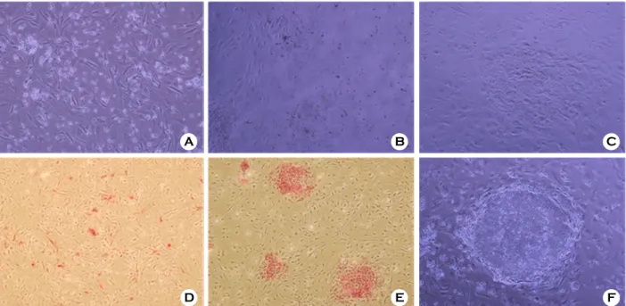
효소를 이용하여 hESCs을 single cell로 만든 다음 이들을 배양하여 colony를 형성하는 과정을 Figure 1에 제시하였다. Single cell에서 처음으로 colony를 형성할 때는 세포 수가 적고 치밀하지 못한 엉성 한 colony 형태를 나타내지만 (Figure 1-E) 이러한 colony를 계대배양하였을 때 이들 colony의 모양이 정상적인 상태로 다시 치밀화 된다는 것을 (Figure 1-F) 확인하였고 이들의 colony 수는 alkaline phosphatase (AP) staining을 통해 염색된 colony를 계수하여 측정하였다.
2. 분리된 단일세포의 적정한 seeding 농도 Tryple로 분리된 single cell을 농도를 달리하여 Table 1. Colony formation of single ES cells dissociated with enzymes
Enzymes Exposure time Cell recovery Concentration of
seeding cells No. of colonies (%) (7 day culture) Collagenase 60 min 2.2±1.3×10
41×10
3/ml/well Failure (0%)
aTryple 5 min 5.4±0.7×10
41×10
3/ml/well 4±2.5 (0.4%)
bAccutase 5 min 3.0±2.2×10
41×10
3/ml/well 5.8±3.0 (0.58%)
bValues are mean ± S.D.
a,bNumbers in columns with different superscripts are significantly different (p<0.05)
Hee Jun Chi. Differentiation of Human Embryonic Stem Cells into Germ Cell and Culture Condition for Single Embryonic Stem Cells Dissociated by Enzyme. Korean J Reprod Med 2010.
feeder cell 위에 seeding할 때 세포의 농도에 따른 colony formation rate를 비교하였다 (Table 2). 분리된 single cell을 feeder cell이 깔린 4 well-dish의 각 well 당 1×10
3, 5×10
3, 1×10
4개/1 ml로 농도를 달리하여 seeding한 결과, 1×10
3개의 적은 수의 single cell을 seeding하여도 colony가 형성되지만 형성된 colony
가 지속적인 성장을 보여주지는 못하였다. 반면 세포 수를 well 당 5×10
3개 seeding한 경우 colony formation의 유의한 증가를 나타냈으며 지속적인 계 대배양을 할 경우에도 건강한 colony를 유지한다는 것을 확인하였다. 1×10
4개의 세포를 seeding한 경 우에는 건강한 colony를 얻을 수 있었지만 colony formation rate는 5×10
3개를 seeding하는 것 보다 비 교적 낮은 효율성을 나타내었다.
3. ROCK inhibitor (Y27632)의 첨가가 단일세 포 colony formation에 미치는 영향
Tryple과 Accutase를 사용하여 단일세포로 분리한 후 이들을 well 당 5×10
3개의 농도로 seeding하여 colony formation을 유도할 때 체외배양액에 첨가한 Y27632의 효과를 조사하여 Table 3에 나타내었다.
hESCs colony를 single cell로 분리하는데 Tryple을 사용한 군에서 보다 높은 colony formation rate를 나타내었고 Y27632를 첨가하였을 경우 양쪽 군 모 Table 2. Colony formation rate according to the
concentrations of seeding single cells Concentrations of
seeding cells No. of colonies (%) (7 day culture) 1×10
3/ml/well 3.6±3.5 (0.36%)
a5×10
3/ml/well 37.3±28.7 (0.74%)
b1×10
4/ml/well 47.0±18.5 (0.47%)
a,bValues are mean ± S.D.
a,bNumbers in columns with different superscripts are significantly different (p<0.05)
Hee Jun Chi. Differentiation of Human Embryonic Stem Cells into Germ Cell and Culture Condition for Single Embryonic Stem Cells Dissociated by Enzyme.
Korean J Reprod Med 2010.
A B C
D E F
Figure 1. Colony formation of single hESCs dissociated by Tryple. (A) single hESCs seeded on MEF feeder layer (magnification ×200), (B) early colony formation on day 7 after seeding (×200), (C) colony formation on day 14 after seeding (×200), (D) alkaline phosphatase (AP) staining of early colony on day 7 (×100), (E) AP staining of colony on day 14 (×100), (F) colony formation after two passages of subculture (×200).
Hee Jun Chi. Differentiation of Human Embryonic Stem Cells into Germ Cell and Culture Condition for Single Embryonic Stem Cells Dissociated by Enzyme. Korean J Reprod Med 2010.
두에서 colony formation이 증가하였으나 Tryple군에 서 보다 유의하게 높은 colony formation rate를 나타 내었다. 따라서 Y27632를 배양액에 첨가하는 것이 분리된 세포의 지지세포 위에 활착력을 향상시키 고 세포를 분리하는 과정에서 발생하는 apoptotic damage를 감소시켜 colony formation rate를 증가시 킨 다는 것을 확인하였다.
4. hESCs의 생식세포로의 분화유도
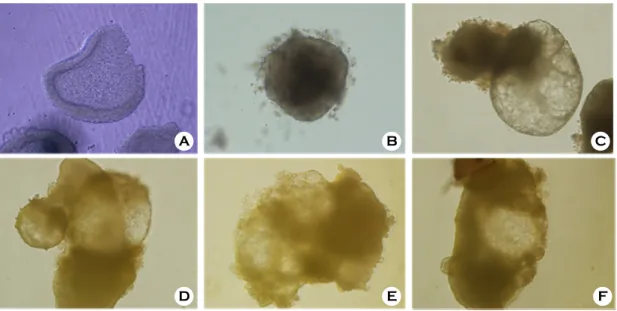
hESCs를 생식세포로 분화유도하는 물질로 알 려진 RA와 BMP4를 이용하여 생식세포로 분화시 키고자 하였다. 분화유도를 하기 위해서 계대배양 직전의 hESCs colony 중 그 크기가 크면서 분화 가 이루어지지 않은 건강한 colony만을 선별하여 Collagenase에 20분간 노출시켰다가 피펫팅으로 colony를 물리적인 손상 없이 배양접시 바닥에서 떼어낸 후 세척을 하여 분화용 배양액이 담긴 배 양접시로 옮겨 EB의 형성을 유도하였다 (Figure 2).
RA와 BMP-4를 이용한 분화유도는 low attachment dish를 이용하여 가능한 한 바닥에 부착되지 않는 3차원적 구조를 유지한 상태로 배양을 하였다. 이 러한 배양조건에서 EB가 cyst 형태로 변화하며 성 장이 이루어지는 현상을 나타내었으나 BMP-4를 첨가한 조건에서 배양된 EB는 상대적으로 cyst 형 성이 떨어지고 이미 형성된 EB도 부서지면서 작아 지는 현상을 나타내었다 (Figure 2-E). 그리고 두 가 지 물질을 함께 첨가하여 분화유도를 하였을 경우 (Figure 2-F), RA 단독 첨가한 실험군과 (Figure 2-D)
차이를 나타내지 않아 두 분화유도제의 공동상승 작용 (synergy)은 발견할 수 없었다. 이러한 결과를 관찰한 이후, 분화유도에 RA를 주로 활용하였다.
5. 생식세포로 분화유도 후 생식세포 특이유전 자의 발현
형성된 EB를 RA가 첨가된 배양조건하에서 14일 간 배양하여 생식세포로의 분화를 유도한 후 이들 을 조직절편하여 슬라이드에 도포한 뒤 생식세포 로 분화하였는지 여부를 초기 생식세포에서 발현 하는 유전자인 c-kit과 VASA 유전자에 대한 형광 표지인자를 이용하여 생식세포로의 분화여부를 조 사하였다 (Figure 3). 대조군으로 사용된 정소조직은 생식세포 특이유전자인 c-kit과 VASA의 발현에 대 한 명확한 표지인자의 형광염색을 나타낸 반면 분 화유도된 EB 절편으로부터는 이들 유전자의 표지 인자에 대한 매우 희미한 형광염색을 나타냄으로 써 생식세포로의 분화에 성공하였다고 확신을 할 수는 없었다.
고
찰일반 조직이나 세포괴에서 single cell을 분리하는 데 많이 사용되는 Collagenase를 hESCs colony를 single cell로 분리하는데 사용하였을 경우, 다른 체 세포와는 달리 60분 동안 처리를 하여도 single cell 로 분리되는 효율성이 낮아 처리 후 single cell의 회수율도 낮았다. 또한 분리된 single cell에서 colony Table 3. Effect of ROCK inhibitor (Y27632) on colony formation of single hESCs
Enzymes ± Y27632 Concentration of seeding cells No. of colonies (%) (7 day culture) Tryple 5×10
3/ml/well 5.67±1.15 (0.11%)
a,bTryple + Y27632 5×10
3/ml/well 106.67±5.03 (2.12%)
cAccutase 5×10
3/ml/well 2.67±0.58 (0.05%)
aAccutase + Y27632 5×10
3/ml/well 17.67±2.08 (0.34%)
bValues are mean ± S.D.
a,b,cNumbers in columns with different superscripts are significantly different (p<0.05)
Hee Jun Chi. Differentiation of Human Embryonic Stem Cells into Germ Cell and Culture Condition for Single Embryonic Stem Cells Dissociated by Enzyme. Korean J Reprod Med 2010.
Figure 2. Formation of EB and differentiation of EB to germ cells (magnification ×200). (A) detached hESCs colony, (B) EB formation after 3 day culture, (C) EB differentiated in the basic medium DMEM F-12 for 14 days, (D) EB differentiated in the presence of retionic acid (RA), (E) EB differentiated in the presence of BMP4, (F) EB differentiated in the presence of RA and BMP4.
Hee Jun Chi. Differentiation of Human Embryonic Stem Cells into Germ Cell and Culture Condition for Single Embryonic Stem Cells Dissociated by Enzyme. Korean J Reprod Med 2010.
A
D
B
E
C
F
A
C D
B
Figure 3. Immunohistochemistry of EB with germ cell specific markers (magnification ×200). (A) testicular tissue stained with DAPI and c-kit marker, (B) testicular tissue stained with DAPI and VASA marker, (C) EB stained with c-kit marker, (D) EB stained with VASA marker.
Hee Jun Chi. Differentiation of Human Embryonic Stem Cells into Germ Cell and Culture Condition for Single Embryonic Stem Cells Dissociated by Enzyme. Korean J Reprod Med 2010.
formation이 이루어지지 않은 것은 Collagenase의 처리과정에 의해 hESCs의 세포표면 또는 기능적인 부분에 손상을 받아 지지세포 위에 활착이나 증식 이 이루어지지 않은 것으로 생각된다. Tryple이나 Accutase 등은 Collagenase보다 세포손상력이 강한 Trypsin을 포함하고 있지만 처리시간이 5분이라는 매우 짧은 시간 때문에 오히려 세포에 손상을 주지 않은 것으로 사료된다.
Tryple과 Accutase를 이용하여 분리한 군들 간에 colony formation rate에서 차이를 나타내지는 않았 다. 그러나 단일세포 분리에 따른 apoptosis를 감소 시키는 Y27632를 첨가하여 배양하였을 때 Tryple 과 Accutase를 사용한 두 그룹 모두에서 Y27632 를 첨가하지 않은 그룹들에 비해 유의한 colony formation의 증가가 나타났으며 이러한 Y27632의 분리된 single cell의 생존성에 대한 긍정적인 효과 는 다른 연구자들의 연구결과에서도 보고되었다.
22,23
Li 등은 이러한 Y27632의 효과가 분리된 cell
의 활착력을 높여주어 다른 세포에 비해 anokis (detachment induced apoptosis)에 민감한 hESCs의 생 존성을 높여주었다고 설명하고 있다.
22본 연구에서 도 Y27632의 유익한 효과를 apoptosis와 연결시켜 확인하려고 하였으나 FACS 장비를 활용하는데 필 요한 충분한 세포를 확보하는 현실적인 어려움이 있어서 이와 관련한 실험은 다음 연구에 수행할 계 획이다. Y27632의 긍정적인 효과는 특히 Tryple을 이용한 군에서 두드러지게 나타나 Accutase를 이용 한 군에 비해 유의하게 높은 colony formation rate 를 나타내었다. 이러한 결과는 같은 조건하에서는 Tryple이 Accutase 보다는 single cell로 분리하는데 보다 세포에 손상을 덜 준다는 것을 의미하는 것이 며 Tryple이 single cell로 분리하는데 3~5분 정도 소요되는데 비해
21Accutase의 경우 이보다 2배에 가까운 8분을 필요로 하였다는 이전의 연구결과들 이
22이를 뒷받침한다고 할 수 있다.
분리된 single cell 농도를 달리하여 feeder cell 위 에 seeding한 결과, 농도에 따라 colony formation rate는 차이를 나타내지 않았다. 그러나 계대배양을
위해 장기간 형성된 colony를 배양한 결과 낮은 농도보다는 일정 수 이상의 높은 농도의 세포를 (well 당 5×10
3개 ) 뿌리는 것이 보다 건강한 colony 를 형성하는데 도움이 된다는 것을 확인하였다. 이 러한 결과는 배아줄기세포 특성 상 단일세포로 생 존 및 증식이 어렵다는 점을 확인시켜 주었으며 일정농도의 세포들을 함께 배양하는 것이 single cell간의 상호작용뿐만 아니라 paracrine signals 등이 이들 single cell의 생존과 colony formation에 도움을 준다는 주장과
26일치한다고 할 수 있다. Colony formation rate (%)는 seeding한 세포 수와 형성된 colony 수의 비율로 나타내었으며 colony의 형태를 쉽게 구별하고 이들이 전분화능을 유지하고 있는지 여부를 확인하기 위하여 가장 널리 사용되는 stem cell marker인 alkaline phosphatase (AP)로 염색하여 colony 수를 측정하였다. 본 연구에서는 우선적으로 효소적으로 분리된 단일세포의 colony formation에 중점을 두었기에 몇 차례의 계대배양 후의 전분화 능 유지여부에 대한 실험은 시행하지 못하였다. 이 와 관련한 실험은 보다 안정된 단일세포 분리 및 배양방법이 구축된 후에 다양한 전분화능 표지인자 를 사용하여 확인할 예정이다. 그리고 효소를 이용 하여 분리된 single cell로부터 형성된 colony는 물리 적으로 작은 세포괴로 나뉘어 새로 형성된 colony 에 비해 얇고 엉성한 형태를 나타내었다가 몇 차례 의 계대배양을 통해 정상적인 형태의 colony의 형 태를 갖추게 되는데 이러한 현상은 이들 세포가 새로운 변화에 대해 적응하는 시간이 필요한 것으 로 생각되며 이러한 형태적인 특성은 Ellerstrom 등의 연구
21에서도 관찰되었다.
Retionic acid를 이용하여 14일 동안 분화유도 후
생식세포 특이유전자에 대한 표지인자 (c-kit, VASA)
를 이용하여 면역조직학적 검사를 수행한 결과 대
조군인 정소조직은 유전자의 발현이 선명하게 확
인되었으나 EB 절편으로부터는 매우 희미한 germ
cell marker의 형광염색만이 확인 가능하였기에 생
식세포로의 분화에 성공하였다고 확신을 할 수는
없었다. 그러나 Richards 등도 RA를 이용하여 15일
동안 hESCs를 germ cell로 분화유도하였을 때 본 연구결과처럼 매우 희미한 유전자 (VASA) 발현의 형광염색을 관찰하였으나 30일 분화유도 시에는 강한 유전자발현을 관찰하였다. 따라서 본 연구에 서도 분화유도 배양기간을 길게 하였다면 좀 더 확실한 유전자발현을 관찰할 수 있었으리라 생각 된다. 생쥐의 EB로부터 분화유도를 한 경우에는 7 일 후면 생식세포의 특이유전자를 발현하는 것이 관찰되었지만
11인간의 EB는 이보다 더 긴 30일 정도의 분화유도기간을 필요로 하는 것으로 종간 에 차이가 있는 것으로 생각된다.
16한편 자료를 제시하지는 않았지만 본 연구에서도 Lacham-Kaplan 등의 연구방법과 유사하게 갓 태어 난 생쥐의 난소와 정소조직을 배양한 conditioned medium을 이용하여 hESCs의 생식세포 분화를 유 도하였지만 EB가 conditioned medium 내에서는 3 차원적 구조를 유지하지 못하고 culture dish 바닥에 부착되어 single layer와 유사한 형태로 바뀌기에 파 라핀절편을 이용한 immunohistochemistry를 수행할 수가 없어서 이들 배양액에 대한 분화효과를 제대 로 확인할 수가 없었다. 이에 대한 연구는 향후 실 험디자인을 다시 세워 수행할 예정이다.
이상의 연구결과로 hESCs colony를 Tryple로 단 일세포로 분리하여 Y27632를 첨가한 배양액에서 배양하고 well당 약 5×10
3개의 단일세포를 seeding 하는 것이 보다 건강하고 많은 colony를 얻을 수 있다는 것을 확인하였다. 한편 생식세포 분화유도 제로 RA를 이용하였을 때 희미하지만 생식세포 특이유전자의 발현을 확인하였으며 분화유도기간 을 연장할 경우 좀 더 뚜렷한 생식세포 특이유전자 의 발현을 관찰할 수 있으리라 생각된다. 아직은 초기단계의 연구라서 더 많은 지속적인 연구가 필 요하다.
참 고 문 헌
1. Ben-Hur T, Idelson M, Khaner H, Pera M, Reinhartz E, Itzik A, et al. Transplantation of human embryonic stem cell-
derived neural progenitors improves behavioral deficit in Parkinsonian rats. Stem Cells 2004; 22: 1246-55.
2.Mummery C, Ward D, van den Brink CE, Bird SD, Doevendans PA, Opthof T, et al. Differentiation of human embryonic stem cells to cardiomyocytes: role of coculture with visceral endoderm-like cells. Circulation 2003; 107: 2733-4.
3. Kaufman DS, Hanson ET, Lewis RL, Auerbach R, Thomson JA. Hematopoietic colony-forming cells derived from human embryonic stem cells. Proc Natl Acad Sci USA 2001; 98:
10716-21.
4. Assady S, Maor G, Amit M, Itskovitz-Eldor J, Skorecki KL, Tzukerman M. Insulin production by human embryonic stem cells. Diabetes 2001; 50: 1691-7.
5. Chen HF, Kuo HC, Chien CL, Shun CT, Yao YL, Ip PL, et al.
Derivation, characterization and differentiation of human embryonic stem cells: comparing serum-containing versus serum-free media and evidence of germ cell differentiation.
Hum Reprod 2007; 22(2): 567-77.
6. Clark AT, Bodnar MS, Fox M, Rodriquez RT, Abeyta MJ, Firpo MT, et al. Spontaneous differentiation of germ cells from human embryonic stem cells in vitro. Hum Mol Genet.
2004; 13(7): 727-39.
7. Hubner K, Fuhrmann G, Christenson LK, Kehler J, Reinbold R, De La Fuente R, et al. Derivation of oocytes from mouse embryonic stem cells. Science 2003; 300(5623): 1251-6.
8. Kiatpongsan S. From embryonic stem cells to functioning germ cells: science, clinical and ethical perspectives. J Med Assoc Thai 2007; 90(10): 2233-7.
9. Qing T, Shi Y, Qin H, Ye X, Wei W, Liu H, et al. Induction of oocyte-like cells from mouse embryonic stem cells by co-culture with ovarian granulosa cells. Differentiation 2007;
75(10): 902-11.
10.Lacham-Kaplan O, Chy H, Trounson A. Testicular cell conditioned medium supports differentiation of embryonic stem cells into ovarian structures containing oocytes. Stem Cells 2006; 24: 266-73.
11.Kerkis A, Fonseca SA, Serafim RC, Lavagnolli TM, Abdelmassih S, Abdelmassih R, et al. In vitro differentiation of male mouse embryonic stem cells into both presumptive sperm cells and oocytes. Cloning Stem Cells 2007; 9(4): 535 -48.
12. Silva C, Wood JR, Salvador L, Zhang Z, Kostetskii I, Williams CJ, et al. Expression profile of male germ cell-associated
genes in mouse embryonic stem cell cultures treated with all-trans retinoic acid and testosterone. Mol Reprod Dev 2009; 76(1): 11-21.
13. Toyooka Y, Tsunekawa N, Akasu R, Noce T. Embryonic stem cells can form germ cells in vitro. Proc Natl Acad Sci U S A 2003; 100(20): 11457-62.
14.Kee K, Gonsalves JM, Clark AT, Reijo Pera RA. Bone morphogenetic proteins induce germ cell differentiation from human embryonic stem cells. Stem Cells Dev 2006; 15(6):
831-7.
15. Teramura T, Takehara T, Kawata N, Fujinami N, Mitani T, Takenoshita M, et al. Primate embryonic stem cells proceed to early gametogenesis in vitro. Cloning Stem Cells 2007;
9(2): 144-56.
16. Richards M, Fong CY, Bongso A. Comparative evaluation of different in vitro systems that stimulate germ cell differentiation in human embryonic stem cells. Fertil Steril 2010; 93(3): 986 -94.
17. Novak I, Lightfoot DA, Wang H, Eriksson A, Mahdy E, Hoog C. Mouse embryonic stem cells form follicle-like ovarian structures but do not progress through meiosis. Stem Cells 2006; 24(8): 1931-6.
18. Kee K, Angeles VT, Flores M, Nguyen HN, Reijo Pera RA.
Human DAZL, DAZ and BOULE genes modulate primordial germ-cell and haploid gamete formation. Nature 2009; 462;
222-5.
19. Hasegawa K, Fujioka T, Nakamura Y, Nakatsuji N, Suemori H. A method for the selection of human embryonic stem cell
sublines with high replating efficiency after single-cell dissociation. Stem Cells 2006; 24: 2649-60.
20. Nicholas CR, Gaur M, Wang S, Reijo Pera RA, Leavitt AD. A method for single-cell sorting and expansion of generally modified human embryonic stem cells. Stem Cells Dev 2007;
16: 109-17.
21.Ellerstrom C, Strehl R, Noaksson K, Hyllner J, Semb H.
Facilitated expansion of human embryonic stem cells by single-cell enzymatic dissociation. Stem Cells 2007; 25: 1690 -6.
22. Li X, Krawetz R, Liu S, Meng G, Rancourt DE. ROCK inhibitor improves survival of cryopreserved serum/feeder- free single human embryonic stem cells. Hum Reprod 2009;
24(3): 580-9.
23. Watanabe K, Ueno M, Kamiya D, Nishiyama A, Matsumura M, Wataya T, et al. A ROCK inhibitor permits survival of dissociated human embryonic stem cells. Nature Biotechnology 2007; 25(6): 681-6.
24. Nayernia K, Nolte J, Michelmann HW, Lee JH, Rathsack K, Drusenheimer N, et al. In vitro-differentiated embryonic stem cells give rise to male gametes that can generate offspring mice. Dev Cell 2006; 11(1): 125-32.
25. Geijsen N, Horoschak M, Kim K, Gribnau J, Eggan K, Daley GQ. Derivation of embryonic germ cells and male gametes from embryonic stem cells. Nature 2004; 427(6970): 148-54.
26. Pyle AD, Lock LF, Donovan PJ. Neurotropins mediate human embryonic stem cell survival. Nat Biotechnol 2006; 24: 344 -50.
= 국문초록 =