Abstract
Purpose: Computer-aided design/computer assisted manufacture (CAD/CAM) guides for surgery are finding widespread use as a tool for implant dentistry. This study sought to evaluate the accuracy and the precision of stereolithographic guide surgery system.
Materials and Methods: Twenty epoxy resin mandibles with artificial silicone gum were fabricated, and each was fitted with an individual computed tomography (CT) guide. Stereolithographic guides were created using CAD/CAM technology and planning software. All of the stereolithographic guides had 2 holes for the right second premolar and molar. After implant was placement, the model was positioned for CT scan to compare the implant position against that which had been planned implant. The linear and angular deviations between the planning data and surgical results were evaluated.
Results: The average differences between the planned and actual implant position in mesiodistal and buccolingual direction and angles of implants showed a considerable reduction in CAD/CAM group versus the conventional group.
Conclusion: This system showed promising accuracy with respect to the planned implant position within the limitations of this study.
Key Words: computer-aided design/computer assisted manufacture surgical guide, computer-aided implantology, dental implant, stereolithography, surgical guide
Stereolithographic Surgical Guide를 이용한 임플란트 수술법의 정확도 평가: In Vitro Study
박수정, 안수진, 서진호, 이성복
경희대학교 치과대학 강동경희대학교치과병원 생체재료보철과학교실
Evaluation of the Accuracy of Implant Surgery with Stereolithographic Surgical Guide: In Vitro Study
Su Jung Park, Su Jin Ahn, Jin Ho Seo, Richard Leesungbok
Department of Biomaterial-Prosthodontics, Kyung Hee University Dental Hospital at Gangdong, School of Dentistry, Kyung Hee University, Seoul, Korea
ISSN 1229-5418 Implantology 2015; 19(1): 10~15
Reprint requests: Richard Leesungbok
Department of Biomaterial-Prosthodontics, Kyung Hee University Dental Hospital at Gangdong, School of Dentistry, Kyung Hee University, 892, Dongnam-ro, Gangdong-gu, Seoul 134-727, Korea
Tel: 82-2-440-7518, Fax: 82-2-440-7549 E-mail: [email protected]
Received for publication: March 20, 2015 Accepted for publication: March 24, 2015
교신저자: 이성복
(134-727) 서울시 강동구 동남로 892
경희대학교 치과대학 강동경희대학교치과병원 생체재료보철과학교실 Tel: 82-2-440-7518, Fax: 82-2-440-7549
E-mail: [email protected] 원고접수일: 2015년 3월 20일 게재확정일: 2015년 3월 24일
Copyright © 2015. The Korean Academy of Oral & Maxillofacial Implantology
This is an Open Access article distributed under the terms of the Creative Commons Attribution Non-Commercial License (http://creativecommons.org/licenses/by-nc/3.0/) which permits
하고 수술까지 이어지도록 하는 이러한 과정은 수술 전 에 임플란트가 식립될 부위의 골을 평가·예상할 수 있 으며, 수술 부위를 재현하여 임플란트가 최적의 위치에 식립될 수 있도록 한다. 또한 flap을 거상하지 않고 최소 침습적인 수술을 할 수 있으므로 수술 시간을 단축시키 고 술 전, 술 후 환자의 불편감을 감소시킨다6-8. 또한 수 술 전 단계에서 보철물의 결과를 예상할 수 있으므로 고 정성 임시보철을 수술 전에 제작해놓고 수술 후 바로 장 착하도록 하여 즉시부하를 가능하게 한다. 이러한 CAD/
CAM을 이용한 stereolithographic surgical guide는 임플 란트 식립 위치의 정확성을 증진시키고 술자에 따른 편 차를 줄이고 있다.
이는 기존의 전통적인 surgical guide와 비교하여 추가 적인 시간과 비용, 노력이 요구되므로, 그 유용성에 대한 검토가 필요하다. 이번 실험에서는 치아상실 부위가 서 로 떨어져있는 경우 삽입로를 일정하게 유지하기 어렵다 는 점을 고려하여, 유치악 모델에서 임플란트 브릿지의 식립을 재현하여 서로 떨어진 치아 상실 부위에 임플란 트를 식립하는 경우를 재현하였다. 이 연구를 통해 CAD/
CAM stereolithographic stent의 정확성을 검증하고, 이 를 사용하였을 때와 사용하지 않았을 때의 임플란트 식 립 위치의 오차를 비교함으로써 stereolithographic sur- gical guide의 유용성을 검토하고자 하였다.
II
연구재료 및 방법우측 하악 제2소구치, 제1대구치, 제2대구치를 치아 상
임플란트 식립의 방법과 기술이 계속적으로 발 전하고 있지만, 이의 성공은 골에 대한 임플란 트 골유착 정도와 임플란트 위치에 영향을 받 는다1. 해부학적 중요구조물을 손상시키지 않으며 수술 중 예기치 못한 상황을 최소화하고 임플란트가 좋은 예 후를 갖도록 하기 위해서는 임플란트 고정체를 정확하게 위치시키는 것이 매우 중요하다. 이를 위해 술자는 수술 전 식립 위치를 적절히 계획함과 동시에 환자의 구강에 서 정확히 재현해야 한다.
의학 분야에서 computer-aided design/computer- assisted manufacture (CAD/CAM) 시스템은 일찍이 이용 되어 왔다. 수술 전 computed tomography (CT)를 통해 획 득한 3차원(three-dimensional, 3D) 이미지를 통하여 수 술 과정을 예상하고 계획하며, 이를 prototype model로 재현하여 수술 과정을 시뮬레이션하고 최소 침습적인 수 술을 할 수 있도록 하였다2,3. 20여 년 전부터 치의학에서 도 CT를 통한 3D 이미지는 임플란트 시술의 술 전 진단에 많이 이용되고 있다. 증가된 3D 이미지의 해상도는 임플 란트 식립이 계획된 위치의 골을 술 전에 평가하여 정확 한 수술 계획을 세울 수 있게 하였으며, 환자의 CT 사진 을 분석해 신경관의 위치를 측정한 후, 신경관 위치를 고 려한 임플란트 fixture 선정, 드릴링 자세 및 깊이 계산 등 계획을 수립하여 임플란트를 식립할 수 있게 하였다4,5.
이에 더하여 최근에는 컴퓨터의 3D 이미지상에서 임플 란트의 위치를 미리 계획할 수 있도록 하는 CAD program 이 앞다투어 개발되었다. 치아 구조 및 턱뼈, 그리고 식립 계획 정보를 바탕으로 임플란트의 식립 위치를 계획하며 술자는 CAD 기술을 사용한 3D 결과를 컴퓨터 모니터로 쉽게 볼 수 있다. 이는 계획한 위치대로 임플란트 식립을
Original Article
실 부위로 하는 20개의 하악 유치악 모델(M.Tech Korea, Seoul, Korea)을 제작하였다. 무치악 부위의 골질은 상부 2 mm가 D1의 피질골의 골질과 유사하도록, 하부가 D3의 해면골의 골질과 유사한 강도를 갖도록 에폭시(epoxy) 레진으로 제작하였다. 치은 조직을 재현하기 위해 상부 는 3 mm의 silicone (Henkel, Dusseldorf, Germany)으로 덮이도록 하였고 임상적인 순서에 따라서 모델을 CT scanning (patient CT scanning)하였다. 이 모델을 3D scanner (Trios; 3Shape, Copenhagen, Denmark)로 scanning한 뒤 CT scanning 이미지와 정합하였다.
1. Planning procedure
CT scan 이미지는 i-CAT cone-beam CT (Imaging Science International, Hatfield, PA, USA)를 통하여 얻 었으며, 0.2 mm 사이즈의 voxel로 40초간 각 모델마다 시행하였다. 90 kVp, 23 Ma, 314의 axial cut으로 이미지 를 얻었다. NeoBiotech software (NeoBiotech Korea, Seoul, Korea)를 이용하여 planning 실시를 위한 정합을 마치고, 컴퓨터상에서 임플란트 planning kit를 선정하 여 인접치아의 치축과 대합치와의 교합, 신경관과의 거 리와 임플란트와 치아 간 거리, 임플란트 간 거리를 고려 하여 #45, #47의 임플란트 식립 부위와 보철의 위치를 planning하였다. 가상 kit를 사용하여 channel을 형성하
고, 직경 4.0 mm, 길이 10.0 mm의 임플란트 고정체에 맞는 surgical drill에 맞도록 확대하였다(Fig. 1). 이에 따 른 #45, #47 식립을 위한 stereolithographic surgical guide (NeoBiotech Korea)를 제작하였다.
2. Validation of the technique
기제작된 stereolithographic surgical guide를 모델에 장착하고, fit checker (GC Co., Tokyo, Japan)를 사용하 여 조기접촉을 제거한 뒤 완전히 적합되도록 하였다. 치 과용 모의실습장비(DSE Plus; Sirona, Charlotte, NC, USA)에 장착 후 제조사의 지시에 따라 직경 4.0 mm, 길 이 10.0 mm의 임플란트 고정체(IS II; NeoBiotech Korea)를 #45, #47 치아상실 부위에 식립하였다. 임플란 트 고정체의 식립을 마친 뒤 같은 장비로 같은 조건하에 식립한 모델을 CT로 재촬영하였다. 대조군은 stereo- lithographic surgical guide를 사용하지 않고 술자의 임 의적인 판단으로 #45, #47을 식립하였으며, 두 그룹 모 두 한 명의 술자(S.J.P)가 시행하였다. Planning된 고정 체의 위치와 식립된 고정체의 위치의 비교를 위하여, 각 고정체의 기준점을 설정하여 두 기준점의 거리를 측정하 였다. 임플란트의 계획된 위치와 식립된 위치 간의 임플 란트 고정체 neck의 중심 사이의 거리, apex의 중심 사이 의 거리, 장축 간의 각도의 차이를 측정하였으며 이를
Fig. 1.
Presurgical scan (A) and postsurgical images (B, C) were performed.Su Jung Park et al. : Evaluation of the Accuracy of Implant Surgery with Stereolithographic Surgical Guide: In Vitro Study. Implantology 2015
A B C
Fig. 2에 나타내었다. SPSS version 15.5 (SPSS Inc., Chicago, IL, USA)를 사용하여 각 그룹 간 통계적 유의 차이를 검토하였고, 두 그룹 간 비교이므로 사후검정은 실행하지 않았다.
III
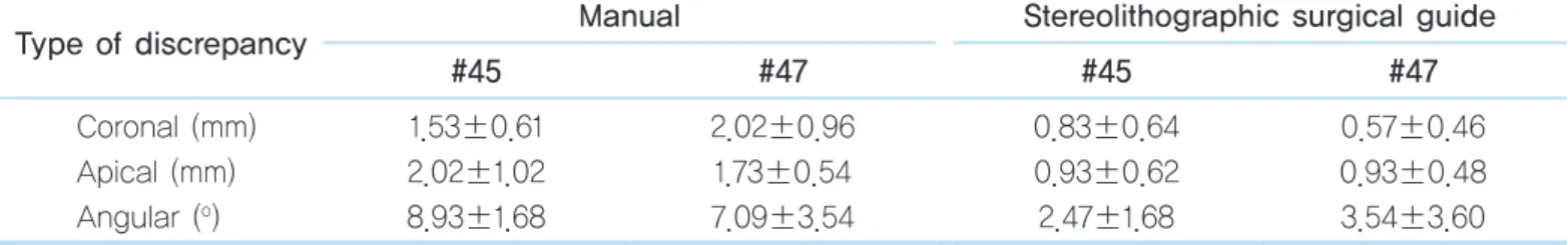
연구결과결과는 Table 1과 같다. CAD로 계획한 임플란트의 가
상 식립 위치를 기준으로 하였을 때 술자에 의존하여 식 립한 그룹이 stereolithographic guide를 이용하여 식립 한 그룹에 비하여 모든 측정치에서 큰 변위량을 가졌으 며, 특히 각도에서 큰 측정치를 가졌다(Fig. 3). 전후방의 식립 위치에 따른 유의차는 없었으며 두 그룹을 비교하 였을 때 #45 고정체의 식립 각도, #47의 고정체의 coro- nal deviation과 apical deviation의 유의확률이 0.05 이하 였다.
Fig. 2.
Measurement of deviations between placed and planned implants.Su Jung Park et al. : Evaluation of the Accuracy of Implant Surgery with Stereolithographic Surgical Guide: In Vitro Study. Implantology 2015
Fig. 3.
Mean distance and standard deviation between the planned implants and placed implants.Su Jung Park et al. : Evaluation of the Accuracy of Implant Surgery with Stereolithographic Surgical Guide: In Vitro Study. Implantology 2015
y
x z
A
B Coronal difference (sum)
Apical (sum)
difference
Table 1.
Discrepancies between planned and actual implant positions (n=10)Type of discrepancy Manual Stereolithographic surgical guide
#45 #47 #45 #47
Coronal (mm) Apical (mm) Angular (o)
1.53±0.61 2.02±1.02 8.93±1.68
2.02±0.96 1.73±0.54 7.09±3.54
0.83±0.64 0.93±0.62 2.47±1.68
0.57±0.46 0.93±0.48 3.54±3.60 Values are presented as mean±standard deviation.
Su Jung Park et al. : Evaluation of the Accuracy of Implant Surgery with Stereolithographic Surgical Guide: In Vitro Study. Implantology 2015 Coronal
(mm) 14 12 10 8 6 4 2 0
Meandiscrepancy
Apical (mm)
Angular ( )
#45 #47
Manual
Stereolithographic surgical guide
Coronal (mm)
Apical (mm)
Angular ( )
Original Article
IV
총괄 및 고찰치의학의 임플란트는 보다 더 비침습적인 시술과 시간 의 단축, 보다 심미적인 방향을 향해 진보하고 있다.
CAD/CAM surgery system은 제한된 골이 있거나 해부학 적으로 주의해야 할 구조물이 임플란트를 식립할 위치에 근접하게 있을 때 종종 flap을 거상하지 않고 고정체를 위치시키는 수술에 추천된다4,9,10. Flapless 임플란트 수 술에서 CT data를 이용한 surgical stent는 술 전 모델을 통하여 정확한 위치로의 임플란트 식립을 가능하게 한 다. 때문에 임플란트 식립에 이용하는 guide surgery system의 정확성을 검증하는 것은 매우 중요하다. 이 연 구는 임플란트의 위치에 있어서 계획된 위치와 stereo- lithographic surgical guide를 따라 실제로 식립한 임플 란트 고정체의 차이를 모형상에서 측정한 것이다. 술 전 과 술 후 이미지를 같은 방법으로 얻었기 때문에 오차를 최소화할 수 있도록 하였다.
Stereolithographic surgical guide는 컴퓨터상에서 계 획된 수술 계획을 구강 내로 옮길 수 있는 정보를 가지고 있으며, 환자에게 장착시킨 상태로 수술이 진행된다. 이 번에 사용한 surgical guide에는 금속 재질의 작은 부품 (sleeve)이 식립 드릴의 사이즈에 맞도록 고정되어 있고, 이 sleeve는 드릴의 방향과 임플란트의 식립 방향을 guide시켜주는 역할을 한다. 또한 임플란트 식립 kit에 vertical stop이 있어 식립 깊이가 계획된 위치보다 깊어 지지 않도록 해주었으며, 실제로 결과에서 planning된 위치와 비교하여 식립 깊이의 오차가 매우 작았다.
이번 실험의 stereolithographic surgical guide는 부분 무치악에 이용하는 치아지지형으로, 잔존치상에서 위치 하여 안정성을 얻게 하는 장치이다. 수술 전 골과 인접 치아 등 해부학적 구조물이 광범위하게 평가되므로 flap
을 거상하지 않아도 안정성 있는 수술이 가능하다11-13. 본 실험의 결과에서는, 수술 가이드 없이 술자의 경험 에 의존하여 식립한 고정체에 비하여 stereolithographic surgical guide를 사용한 그룹이 계획한 임플란트 위치에 대해 모든 측정치에서 작은 변위량을 가졌다(Fig. 3). 특 히 변위량이 큰 측정치는 두 그룹 모두 고정체의 식립각 도였으며 악궁의 전, 후방부에 위치한 소구치, 대구치의 위치에 따른 유의차는 없었다. 본 실험에서 사용한 임플 란트 수술 kit (Neoguide; NeoBiotech Korea)는 선택한 고정체에 따른 vertical stop을 가지고 있기 때문에 식립 깊이의 차이를 내포하는 고정체의 coronal, apical devi- ation이 작고 다른 수치에 비하여 식립 각도의 차이 (angular deviation)가 큰 결과가 나왔다고 추측해 볼 수 있다. 또한 stereolithographic surgical guide를 사용하였 음에도 일정한 변위가 존재하는 것은 surgical guide를 제작하는 중에 있는 여러 가지 오차와 함께 sleeve 내면 과 임플란트 드릴, 고정체 사이의 공간이 존재하기 때문 이라고 예상해 볼 수 있다. Stereolithographic surgical guide가 제작되기까지 다양한 과정을 거치는데, 환자의 CT 정보와 구강 내 scan 정보를 정합하고, 환자의 CT data를 3D data와 이미지로 변환하는 과정, 이러한 정보 를 각 장비에 전송하고 변환하는 과정, 컴퓨터상에서 디 자인한 surgical guide를 실제 사용할 stereolithographic guide로 제작하는 과정 등을 거치며 실제 구내 정보와 오 차가 생길 수 있다. 이러한 CAD/CAM surgery implant system을 제조하는 회사들은 data 정합과 stereolitho- graphic surgical guide 제작의 정확도를 향상시키기 위 한 여러 가지 오차 수정과정을 거치고 있지만 이러한 오 차는 발생할 수 밖에 없으며, 술자는 보다 정확한 수술을 위해 사전에 꼭 surgical guide를 구내에 위치시켜서 안 정적인 적합을 확인해 보아야 한다. 또한 잘 맞지 않는 경우, 현재 통용되는 시스템 자체의 과정과 한계에 대한 숙지하에, stereolithographic surgical guide를 제조하는
회사와 지속적인 커뮤니케이션을 하며 시스템 process- ing 과정에서 발생하는 각 과정의 오차를 수정한다면 보 다 적합도 있는 stereolithographic surgical guide를 제공 받을 수 있을 것이다.
본 실험은 하악 유치악 모형에서 술자에 의존하여 식 립한 경우와 비교하여 stereolithographic surgical guide 의 유용성을 검증하였다. 그 결과, 사용한 그룹의 모든 측정치가 사용하지 않은 그룹에 비하여 정확하게 식립된 것으로 측정되었으며, 특히 소구치 부위의 각도와 대구 치 부위의 깊이에서 사용 여부에 대한 유의한 차이가 있 었다. Stereolithographic surgical guide를 사용하였을 때에도 planning data와 비교하여 오차가 존재하지만, 이를 줄여줄 수 있다는 점에서 stereolithiographic sur- gical guide의 사용은 의미를 가질 수 있겠다. 또한 술자 의 시술 경험에 크게 좌우되지 않는, 보다 안정적인 결과 를 가져옴으로써 환자의 임플란트 시술에 대해 보다 나 은 예후를 가져올 수 있을 것이다.
V
결 론하악 부분 무치악 환자의 상실 부위에서, stereolithio- graphic surgical guide를 이용하는 것은 술자의 경험에 의존하여 식립하는 것과 비교하였을 때 임플란트 식립 위치의 정확성을 증가시킨다.
References
1. Soares MM, Harari ND, Cardoso ES, et al. An in vitro model to evaluate the accuracy of guided surgery systems. Int J Oral Maxillofac Implants.
2012; 27: 824-831.
2. Tahmaseb A, Wismeijer D, Coucke W, et al. Computer technology applications in surgical implant dentistry: a systematic review. Int J Oral Maxillofac Implants. 2014; 29(Suppl): 25-42.
3. Cassetta M, Stefanelli LV, Giansanti M, et al. Accuracy of implant placement with a stereolithographic surgical template. Int J Oral Maxillofac Implants. 2012; 27: 655-663.
4. Valente F, Schiroli G, Sbrenna A. Accuracy of computer-aided oral implant surgery: a clinical and radiographic study. Int J Oral Maxillofac Implants. 2009; 24: 234-242.
5. Cassetta M, Giansanti M, Di Mambro A, et al. Accuracy of positioning of implants inserted using a mucosa-supported stereolithographic surgical guide in the edentulous maxilla and mandible. Int J Oral Maxillofac Implants. 2014; 29: 1071-1078.
6. Ozan O, Turkyilmaz I, Ersoy AE, et al. Clinical accuracy of 3 different types of computed tomography-derived stereolithographic surgical guides in implant placement. J Oral Maxillofac Surg. 2009; 67: 394-401.
7. Jacobs R, Adriansens A, Verstreken K, et al. Predictability of a three- dimensional planning system for oral implant surgery. Dentomaxillofac Radiol. 1999; 28: 105-111.
8. Nokar S, Moslehifard E, Bahman T, et al. Accuracy of implant placement using a CAD/CAM surgical guide: an in vitro study. Int J Oral Maxillofac Implants. 2011; 26: 520-526.
9. Ersoy AE, Turkyilmaz I, Ozan O, et al. Reliability of implant placement with stereolithographic surgical guides generated from computed tomography: clinical data from 94 implants. J Periodontol. 2008; 79:
1339-1345.
10. Testori T, Robiony M, Parenti A, et al. Evaluation of accuracy and precision of a new guided surgery system: a multicenter clinical study.
Int J Periodontics Restorative Dent. 2014; 34(Suppl 3): S59-S69.
11. Pettersson A, Komiyama A, Hultin M, et al. Accuracy of virtually planned and template guided implant surgery on edentate patients. Clin Implant Dent Relat Res. 2012; 14: 527-537.
12. Van Assche N, van Steenberghe D, Guerrero ME, et al. Accuracy of implant placement based on pre-surgical planning of three-dimensional cone-beam images: a pilot study. J Clin Periodontol. 2007; 34: 816-821.
13. Arisan V, Karabuda ZC, Ozdemir T. Accuracy of two stereolithographic guide systems for computer-aided implant placement: a computed tomography-based clinical comparative study. J Periodontol. 2010; 81:
43-51.