ISSN: 2233-601X (Print) ISSN: 2093-6516 (Online)
− 123 −
Received: May 19, 2016, Revised: August 5, 2016, Accepted: August 12, 2016, Published online: April 5, 2017
Corresponding author: Young Mog Shim, Department of Thoracic and Cardiovascular Surgery, Samsung Medical Center, Sungkyunkwan University School of Medicine, 81 Irwon-ro, Gangnam-gu, Seoul 06351, Korea
(Tel) 82-2-3410-1677 (Fax) 82-2-3410-0489 (E-mail) [email protected]
© The Korean Society for Thoracic and Cardiovascular Surgery. 2017. All right reserved.
This is an open access article distributed under the terms of the Creative Commons Attribution Non-Commercial License (http://creativecommons.org/
licenses/by-nc/4.0) which permits unrestricted non-commercial use, distribution, and reproduction in any medium, provided the original work is properly cited.
Squamous Cell Carcinoma Arising from the Pleural Cavity After Pneumonectomy for Chronic Empyema
Yeong Jeong Jeon, M.D., Sumin Shin, M.D., Young Mog Shim, M.D., Ph.D.
Department of Thoracic and Cardiovascular Surgery, Samsung Medical Center, Sungkyunkwan University School of Medicine
Malignant tumors associated with chronic empyema have been reported in the literature, and a majority of these tumors are lymphomas. Epithelial tumors originating from the post-pneumonectomy space in patients with chronic empyema are extremely rare. Here, we present the cases of 2 patients with squamous cell car- cinoma arising from the pleural cavity after pneumonectomy for chronic empyema.
Key words: 1. Squamous cell carcinoma 2. Empyema
3. Pneumonectomy
Case reports
1) Case 1
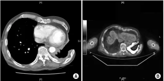
A 74-year-old man underwent left pleuropneumo- nectomy for tuberculous empyema in 1997. He devel- oped a bronchopleural fistula (BPF) and underwent an Eloesser operation following BPF closure with an omental flap in 1998. The patient was then doing well. However, in 2015, he was referred to Samsung Medical Center because of increased wound discharge and pain. Chest computed tomography (CT) demon- strated pleural thickening and irregularity, along with bony destruction in the left 10th rib, at the Eloesser aperture (Fig. 1A). A F-18 fluorodeoxyglucose posi- tron emission tomography (FDG-PET) revealed hyper- metabolic pleural thickening with invasion into the left 10th rib (maximum standardized uptake value [SUV]=21.0) (Fig. 1B). He underwent incisional biop- sy for tissue confirmation. Th e biopsied specimen was examined microscopically; the results led to a diagnosis of squamous cell carcinoma. The patient then received definitive radiotherapy with a dose of
60 Gy in 20 fractions, and the tumor regressed markedly. He is currently in a good general condition and is being followed up as an outpatient.
2) Case 2
A 53-year-old woman underwent right pneumo- nectomy because of a destroyed tuberculous lung with pleurisy in 1990. She was diagnosed with post- pneumonectomy empyema without BPF in 2006 and underwent repeated irrigation and debridement, fol- lowed by the Clagett procedure. She visited Samsung Medical Center complaining of right shoulder and chest pain in October 2010. Chest CT revealed extra- thoracic extension of enhancing soft-tissue lesions originating from the right pneumonectomy space.
Percutaneous needle biopsy was performed, and the pathologic findings included a fibrinous exudate and inflammatory cells. The patient was treated with anti- biotics; however, her pain was progressive, and she received a follow-up chest CT scan in March 2011.
The chest CT revealed progression of the extra- thoracic extension of the enhancing soft-tissue lesion,
Korean J Thorac Cardiovasc Surg 2017;50:123-125 □ CASE REPORT □
https://doi.org/10.5090/kjtcs.2017.50.2.123
Yeong Jeong Jeon, et al
− 124 −
Fig. 1. (A) Chest computed tomog- raphy demonstrated pleural thick- ening and irregularity, along with bony destruction in the left 10th rib, at the Eloesser aperture. (B) An F-18 fluorodeoxyglucose positron emission tomography scan revealed hypermetabolic pleural thickening with invasion into the left 10th rib (maximum standardized uptake val- ue=21.0).
Fig. 2. (A) Chest computed tomography revealed progression of the extrathoracic extension of the enhancing soft-tissue lesion, including rib invasion (black arrow) and particularly involving the neural foramen and the epidural space at the level of the thoracic spine (T7 ver- tebral body) (white arrow), indicating an empyema-associated malignancy. (B) An F-18 fluorodeoxyglucose positron emission tomography scan showed a hypermetabolic malignant mass in the right pneumonectomy space (maximum SUV=35.3), right chest wall (maximum SUV=9.9), right seventh rib, and T7 (maximum SUV=26.1). SUV, standardized uptake value.
including rib invasion and particularly involving the neural foramen and the epidural space at the level of the thoracic spine (T7 vertebral body), indicating an empyema-associated malignancy (Fig. 2A). An F-18 FDG-PET scan showed a hypermetabolic malignant mass in the right pneumonectomy space, right chest wall, righ t seventh rib, and T7 spine (SUV=35.3) (Fig.
2B). Pathologic diagnosis of the squamous cell carci- noma was confirmed by the percutaneous needle bi- opsy of the soft-tissue lesions. The patient received palliative radiotherapy with a total dose of 42 Gy in 12 fractions. She developed bladder dysfunction in addition to back and leg pain. A spine CT showed an
increase in the size of the malignant mass involving the T6, T7, and T8 vertebral bodies. The patient’s status declined, and she eventually died.
Discussion
Chronic empyema, due to either tuberculosis or bacterial causes, can result in the development of a malignancy [1]. The most common tumor is non- Hodgkin lymphoma; however, thus far, only a few cases of epithelial tumors have been reported [2-4].
In the literature, only 6 cases of squamous cell carci- noma arising from the pleura have been reported [5].
Squamous Cell Carcinoma Arising from Pleural Cavity After Pneumonectomy for Chronic Empyema
− 125 − At our medical center, 72 patients experienced post-pneumonectomy empyema between 1998 and 2015. Nineteen of these 72 patients underwent a pneumonectomy because of an infective disease re- sulting from tuberculosis, aspergillosis, or chronic empyema due to BPF. Two of these patients devel- oped malignancies in the pleural cavity.
The pathogenesis of a malignant tumor arising in the post-pneumonectomy cavity associated with chronic empyema remains unknown. According to Deaton [6], squamous cell carcinoma may originate from the metaplasia of the pleural endothelium, metaplastic bronchial epithelium that has extended into the cavity, or metaplastic skin epithelium grow- ing into the cavity.
In the cases discussed her, 2 patients experienced tuberculous empyema for several years before under- going pneumonectomy. Chronic tuberculous empyema is known as a predisposing factor for developing a thoracic malignancy [1]. Both patients developed post- pneumonectomy empyema with or without BPF, and underwent a thoracotomy (Eloesser or Clagett). The tumors in these 2 cases were diagnosed after 17 years and 4 years, respectively. The chronic inflammatory state after post-pneumonectomy empyema is believed to have led to the development of the malignancy.
Radiographic findings in patients with chronic em- pyema, such as increased opacity in the thoracic cav- ity, soft-tissue bulging, nodular pleural thickening, and destruction of the bone near the empyema, sug- gest the possibility of a chronic empyema-associated malignancy [7]. Osteolytic changes with pleural thick- ening in the imaging findings were observed in the 2 cases reported, and were suggestive of a malignancy.
Squamous cell carcinoma was confirmed by biopsy.
The treatment of tumors arising from the post- pneumonectomy space should be determined on the basis of the patient’s condition. Most of the patients
who underwent pneumonectomy had poor pulmonary function; therefore, radiation therapy was recom- mended. The abovementioned 2 patients received ra- diotherapy because of poor pulmonary function.
These are rare case reports of squamous cell carci- noma arising from the pleural cavity after pneumo- nectomy for chronic empyema. We suggest that the possibility of a malignancy originating from the pleu- ral cavity be considered in patients who experience post-pneumonectomy empyema.
Conflict of interest
No potential conflict of interest relevant to th is ar- ticle was reported.
References
1. Tamura A, Hebisawa A, Sagara Y, et al. Thoracic malig- nancies in patients with chronic tuberculous empyema.
Kekkaku 2004;79:301-7.
2. Nakatsuka S, Yao M, Hoshida Y, Yamamoto S, Iuchi K, Aozasa K. Pyothorax-associated lymphoma: a review of 106 cases. J Clin Oncol 2002;20:4255-60.
3. Yun JS, Kang SK, Kim JH, Jung Y, Choi YD, Song SY. Diffuse large B-cell lymphoma arising from chronic tuberculous empyema. Korean J Thorac Cardiovasc Surg 2015;48:82-5.
4. Chung WJ, Lee SH, Kim KT, et al. High grade sarcoma aris- ing from the chest wall of a chronic tuberculous empye- ma: a case report. Korean J Thorac Cardiovasc Surg 2008;
41:795-8.
5. Franke M, Chung HD, Johnson FE. Squamous cell carcinoma arising from the pleura after pneumonectomy for squ- amous cell carcinoma of the lung. Am J Surg 2010;199:
e34-5.
6. Deaton WR Jr. Carcinoma arising in chronic empyema cav- ity: case report with review of the literature. Dis Chest 1962;42:563-6.
7. Minami M, Kawauchi N, Yoshikawa K, et al. Malignancy as- sociated with chronic empyema: radiologic assessment.
Radiology 1991;178:417-23.