c Korean Medical Association
This is an Open Access article distributed under the terms of the Creative Commons Attribution Non-Commercial License (http://creativecommons.
org/licenses/by-nc/3.0) which permits unrestricted non-commercial use, distribution, and reproduction in any medium, provided the original work is properly cited.
한국 성형외과 재건 미세수술의 조망
전 병 준·문 구 현* | 성균관대학교 의과대학 삼성서울병원 성형외과
Perspectives on reconstructive microsurgery in Korea
Byung-Joon Jeon, MD·Goo-Hyun Mun, MD*
Department of Plastic Surgery, Samsung Medical Center, Sungkyunkwan University School of Medicine, Seoul, Korea
*Corresponding author: Goo-Hyun Mun, E-mail: [email protected] Received May 11, 2011·Accepted May 24, 2011
W
ith the advancement of modern medicine, there have been increasing demands for reconstructive surgeries. The operative technique using free flaps makes it possible for reconstructive surgeons to restore various defects and deformities more precisely. Furthermore, functional problems, such as facial paralysis and lymphedema, can be managed with microsur- gical procedures. The need for the composite tissue allograft, including that of the face, has been noticed, and this transplantation surgery required complex microsurgical procedures. With the very high success rate of free flap and popularization of perforator flap, which provides improved outcomes, reconstructive microsurgeons now play major role in various reconstructive fields.Keywords: Reconstruction; Microsurgery; Free flap; Allograft
서 론
수 술 현미경과 미세한 혈관을 다루기 위해 특별히 고안 된 기구들을 이용한 미세수술은 산업화에 따른 각종 사고들로 인한 외상의 재건을 위해 빠르게 발전해 왔다. 한 국에서 미세수술은 1970년대 이르러 행해지기 시작하였으 며, 국문으로 발표된 최초의 피판 관련 논문은 1974년에[1], 그리고 유리피판의 경우 1978년 대한성형외과학회지에 게 재되었다[2]. 벼농사 문화와 젓가락 사용을 공통으로 가지고 있는 한국과 중국 그리고 일본은 비교적 섬세한 손 움직임을 필요로 하는 미세수술 분야에서 두각을 나타내어 왔고, 현재 국내 여러 대학병원과 미세재건센터에서 미세혈관문합술 또 는 신경문합술을 이용하여 어려운 결손을 성공적으로 재건
하고 있다. 이러한 여러 국내 술자들의 우수한 결과들은 국 제학술지에 지속적으로 게재되고 있으며, 성형외과 분야에 서 중에서 특히 미세수술 분야는 매우 높은 국제경쟁력을 가 지고 있다.
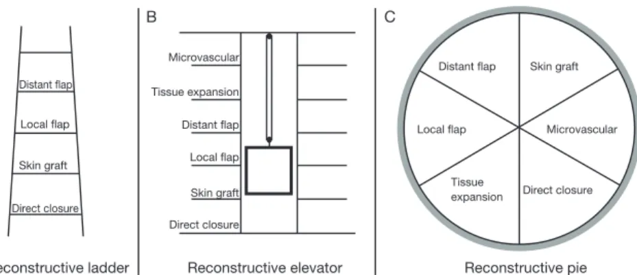
전통적으로 결손부가 있을 때 이를 해결하는 방법으로 단
계적인 ‘재건 사다리(reconstructive ladder)’ 개념이 있었
고 그 이후 미세수술, 조직확장술의 발전으로 ‘재건 엘리베
이터(reconstructive elevator)’ 개념으로 발전했다. 이는 일
차봉합 같이 비교적 쉽고 간단한 수술로부터 재건을 시도하
여, 단계적으로, 피부이식, 국소피판, 유리피판 등의 어렵고
복잡한 수술을 통한 재건이 고려되어야 한다는 개념(재건
사다리)에서 점차 엘리베이터와 같이 위계가 있긴 하지만
원하는 층의 단추를 누르면 멈추는, 즉 기능적, 미용적으로
최적의 결과를 얻기 위해 가장 적합한 방법을 사용하여야 한 다는 개념에 이르게 되었음을 뜻한다. 이러한 개념의 변화 는 근래 10년간 재건미세수술의 결과가 더욱 향상됨에 따라 모든 재건법을 수평에 놓고 술자의 경험이나 선호도, 환자의 선호도 등을 함께 고려하여 보다 자유롭게 재건 방법을 선택 하는 ‘재건 파이(reconstructive pie)’의 개념에 이르게 되 었다(Figure 1).
미세혈관문합(microvascular anastomosis)을 이용한 피 판의 원격 전이를 통해 과거에는 수 차례에 걸친 피부이식으 로도 얻기 어려웠던 피부를 포함한 충분한 연부조직을 단 한 번의 수술로 옮길 수 있게 되었다. 또한 외상으로 인한 광범 위한 손상이 생긴 경우나 방사선 조사를 받은 창상의 경우 창상과 주변 조직의 치유가 어려운데, 미세수술을 이용하여 손상 부위와 멀리 떨어져 있고, 쉽게 가려질 수 있는 부위를
공여부(donor site)로 피판을 거상하여 피복하면, 상처의 치 유를 돕고 재건에 필요한 충분 한 조직을 공급할 수 있다. 아 울러 근육, 뼈, 소화관의 일부도 이식할 수 있는데, 이를 통해 기 능적으로 개선된 재건이 가능 하고, 환자가 당뇨와 같이 혈액 순환의 장애를 초래하는 질병 을 갖고 있는 경우에도 혈관문 합을 통해 옮겨진 조직의 생존 율을 높이고 상처의 치유도 촉 진할 수 있게 되었다. 국내에서 시행된 유리피판의 이전에 따 른 성공률은 미세문합이 비교 적 활발히 시행되기 시작한 1970년대 말에서 1980년대 초 반까지 66.7- 75%로 비교적 낮게 보고되었으나[3-5], 동일 한 시기의 국제적인 보고에 따 르더라도 60 -75%로 국내의 성적이 나쁘지 않음을 알 수 있 다[6,7]. 이후 1990년대 중반을 넘어서면서 국내 학회지에 보고된 유리피판의 성공률은 97.3-100%로 매우 높아져 유 리피판의 성적이 크게 향상되었음을 알 수 있다[8-11].
비교적 최근에 소개되어 널리 이용되고 있는 천공지피판 (perforator flap)은 혈액공급을 받아 생존하는 조직인 피판 의 혈관경으로 근육을 관통하여 피부 및 피하지방에 혈류를 공급하는 천공지(perforator vessels)를 사용하는 피판이다 [12,13]. 과거에는 결손부위 피복을 위해 근육피부피판(mu- sculocutaneous flap) [14] 또는 근육피판(muscle flap)과 피부이식(skin graft) [15]을 이용한 재건이 널리 행해졌지 만 기능적인 목적이 아닌, 피복만을 위해 근육을 희생하는 것은 공여부의 손실이 과도하다는 인식이 널리 퍼지면서 천 공지피판을 이용한 재건이 각광을 받고 있다[16-18]. 이러 한 천공지피판은 결손부의 피복과 연부조직의 재건을 위해
Reconstructive ladder Reconstructive elevator Reconstructive pie C
B A
Distant flap Microvascular
Tissue expansion
Skin graft Direct closure Distant flap Local flap
Skin graft
Microvascular
Direct closure Tissue
expansion Local flap Distant flap
Local flap Skin graft Direct closure
Figure 1. Concept change from reconstructive ladder (A) through reconstructive elevator (B) to recon- structive pie.
Figure 2. Preoperative planning with computed tomography (CT) angiography. (A) Transverse view of rendered 3D CT angiography of the abdomen. (B) Coronal view of rendered 3D CT angio- graphy. (C) Evaluation of recipient vessels (in this case, internal mammary vessels are used as recipient vessels) can be done with CT angiography. P, perforating vessels.
A B C
필요한 피부와 피하지방만을 거상함으 로써 공여부의 이환율을 감소시키고, 피하지방의 두께 조절이 비교적 용이 하여 기존의 방법들을 많은 분야에서 대체하여 널리 사용되고 있다.
하지만 천공지피판은 혈관경의 변이 가 심해 피판을 거상하는데 어려움을 초래하기도 한다. 이러한 어려움을 해 소하기 위해 여러 가지 방법이 고안되 어 사용되고 있으며, 이 중에 공여부에 대한 컴퓨터단층혈관조영술(c o m- puted tomography angiography)이 활발히 시행되고 있다. 이는 술전에 미 리 적합한 천공지를 선택할 수 있을 뿐 만 아니라 혈관의 주행경로, 추가적인 문합을 위해 필요한 주변 혈관의 주행 과 굵기 등의 다양한 정보를 제공하여 구체적인 수술 전 계획 하에 신속한 수 술 진행을 도와준다(Figure 2) [19].
현재 가장 대표적인 재건방법 가운 데 하나로 기능적, 미용적으로 만족스 러운 재건을 위해 널리 이용되고 있는 미세수술을 이용한 유리피판술들을 살 펴보고, 앞으로 재건미세수술 분야가 나아갈 방향에 대해서도 조망해보기로 한다.
두경부 재건
두피부터 목에 이르기까지 광범위하 거나 복잡한 결손부를 재건할 때, 재건 에 필요한 국소조직이 부족하므로 유 리피판술의 역할이 매우 큰 곳이다. 미 세수술을 이용한 두피재건은 악성 흑 색종(malignant melanoma), 상피세 포암(squamous cell carcinoma), 혈
Figure 3. Burn scar contracture, scalp. (A) Preoperative view. (B) Flap design (thoracodorsalartery perforator flap). (C) Elevated flap. (D) Two months postoperative view.
Figure 4. Recurred nasopharyngeal cancer, left temporal region. (A) Defect after subtotal petrosectomy. (B) Chimeric pattern anterolateral thigh flap with vastus lateralis muscle. (C) Elevated muscle flap can be used for obliteration of dead space after ablative surgery. (D) Immediate posto-perative view.
A B
C
A
C
B
D D
관육종(angiosarcoma) 등의 발생 후 광범 절제술을 시행한 뒤 생긴 결손 부위를 채우기 위해 활용되고 있다[20-23]. 이 외에 사고 등을 통해 발생한 외상이나 화상 반흔 등이 발생 한 경우에도 미세수술을 이용한 피판은 결손부 재건을 위해
이용되며, 흉배동맥천공지피판(tho- racodorsal artery perforator flap) [24] 전외측대퇴피판(anterolateral thigh flap) [25] 심하복벽천공지피판 (deep inferior epigastric artery per- forator flap) [26]과 같은 천공지피판 이나, 더 큰 넓이의 조직이 필요한 경우 광배근(latissimus dorsi muscle)과 흉배동맥천공지피판을 키메라형으로 거상하여 결손을 피복하고 근육 부위 에 피부이식을 시행하는 방법을 사용 하기도 한다(Figure 3) [27].
뇌종양 등을 절제하고 나서 발생하는 두개저(skull base) 결손을 재건하는 경우에도 미세수술을 이용한 유리피판 의 이식은 널리 활용되고 있다. 이 경우 에도 각종 근육피판을 이용하거나[28], 전외측 대퇴피판을 이용하거나[29], 부 피가 필요한 경우 심하복벽천공지피판 을 사용하기도 하며[30], 추가적인 부피 를 얻기 위해 복직근(rectus abdomi- nis muscle)을 키메라형으로 함께 거 상하여 사용하기도 한다(Figure 4).
악성종양 절제 후 발생한 안와를 포 함하는 중안면과 상악의 결손을 재건 하는 것은 가장 어렵고 고려할 사항들 이 많은 재건으로 손꼽힌다. 이는 인접 한 구조물들이 뇌나 안구 등과 같이 중 요한 구조물이며, 이러한 구조물들이 밀도가 높게 모여있어 주의가 필요하 며, 일상생활에서 시선이 많이 머무는 부분으로 미용적으로 만족스러워야 함 과 아울러 기능적으로도 우수한 3차원적 재건이 요구되는 부분이기 때문이다[31]. 따라서 요구되는 기능에 따라 연부 조직 단독으로 또는 골조직과 연부조직을 함께 이식해 주어 야 하는 경우가 있다. 대표적으로 이용되는 재건 방법으로
Figure 5. Recurred maxillary cancer, right. (A) Defect after extended radical maxillectomywith orbital exenteration. (B) Elevated vertical rectus abdominis musculocutaneous flap and design. (C,D) Fortyone months postoperative view. (E) Intraoral view.
Figure 6. Retromolar trigone cancer, right. (A) Defect after mandibulectomy, right. (B) Flap design, right peroneal region. (C) Elevated fibular osteocutaeous flap. (D) Inset of fibular osteocutaneous flap was done. (E) Three months postoperative view. (F) Split thickness skin graft was done on the donor-site.
A B
C D E
A B C
D E F
는 연부조직 단독으로 사용하는 경우 요측전완피판(radial forearm flap) [32], 복직근피판[33], 광배근피판[34,35] 등 을 이용할 수 있고, 연부조직과 아울러 골조직이 함께 필요 한 경우, 요골을 포함한 요측전완피판[36], 비골피판[37], 장 골릉피판[38], 견갑골의 일부를 이용한 여러 가지 피판[34]
등을 활용하여 재건에 활용할 수 있다(Figure 5). 아울러 해 부학적 복잡성과 기능적 중요성 등의 이유로 중안면부 재건에 서도 동종이식을 이용한 재건이 활발히 연구되고 있다[39].
하악의 재건은 주로 악성 신생물의 제거에 따른 결손부위
를 채우기 위해 시행하게 된다. 혈관 없 이 뼈만 단독으로 이식하거나[40], 보형 물만 이용하여 재건할 경우 대체로 그 결과가 만족스럽지 못하다. 하악 재건 을 성공적으로 수행하기 위해 요구되는 공여부의 조건으로는 뼈의 혈행이 풍부 하고, 길이가 재건에 충분하며, 결손부 위의 형태와 유사하고, 최소한의 조작 으로 결손부위와 유사한 모양을 얻을 수 있고, 공여부의 이환율을 최소화하 며 아울러 두 개의 팀에 의한 수술이 가 능한 것 등이 제시되고 있다. 또한 연부조직을 충분히 제공할 수 있어 하악, 인두 및 혀 부위의 피부결손을 피복할 수 있는 것이어야 한다[41]. 이러한 특성들을 만족시키는 공여부로 는 장골릉(iliac crest)과 비골(fibula)을 포함하는 골-피부 피판(osteocutaneous flap)을 고려할 수 있다(Figure 6).
과거 구강의 재건을 위해 요측전완피판(radial forearm flap)이 널리 사용되었으나 현재 이와 함께 다른 피판들이 함께 사용되고 있다. 전외측대퇴피판(anterolateral thigh flap)은 충분한 피부를 얻을 수 있고, 피부판과 별도로 주변
Figure 7. Tongue cancer, right. (A) Defect after near total glossectomy. (B) Preoperative computed tomography angiography was done to select appropriate perforators. (C) Design according to the defect. (D) Elevated anterolateral thigh flap. (E) Inset of the flap. (F) Six weeks postoperative view.Figure 8. Esophageal cancer. (A) Jejunal free flap can be a successful option for esophageal reronstruction. (B) The esophagogram after reconstruction shows patent luminal structure.
A B C
F E
D
A B
근육을 포함하는 키메라형 피판의 거상이 가능하며, 외측대 퇴피부신경(lateral femoral cutaneous nerve)을 포함할 경우 감각 회복이 가능한 형태의 피판을 거상할 수 있어 구 강 재건에 널리 이용되고 있다(Figure 7) [42].
미세수술을 이용한 조직의 이전을 통해 양성 또는 악성 병변의 제거 후 발생한 하인두-식도의 결손을 성공적으로 재건할 수 있다. 이를 위해 대표적으로 사용되는 재건방법 으로 공장유리피판술(jejunal free tissue transfer)이나 요 측전완피판(radical forearm flap)을 관 모양으로 말아서 사용하는 방법 등이 대표적으로 이용되고 있다(Figure 8).
안면마비의 재건에서도 미세수술을 이용한 기능적 신경- 근 이식술(functional neuromuscular transfer)이 중추적 인 역할을 하고 있다. 안면 신경마비에 따라 나타나는 모든 증상을 기능적으로 재건하는 것은 현재로서는 불가능하나 환자의 사회생활에 큰 영향을 미칠수 있는 웃는 모습의 교정 을 위해, 웃을 때 구각(mouth corner)의 비대칭을 개선하 기 위한 다양한 동적 재건방법들이 고안되어왔다. 박근 (gracilis), 전방거근(serratus anterior muscle), 소흉근
(pectoralis minor) 등을 이용한 이단 계 재건법이 일찍이 제안되어 사용해 왔다[43,44]. 이 외에도 광배근의 일부 와 이를 지배하는 흉배신경(thoraco- dorsal nerve)분지를 포함한 단단계 유리피판술도 하안면부 소생(reani- mation)에서 효과적으로 이용되고 있다(Figure 9) [45].
체간 및 유방의 재건
악성종양의 근치적 절제 후나 외상 등으로 인해 발생한 흉벽 또는 복벽의 결손을 재건하기 위해 국소피판 또는 유리피판이 널리 활용되고 있다. 결손 의 위치와 정도에 따라 적절한 재건 방 법의 고려가 필요한데, 몸통의 경우 다 양한 근육을 포함하는 국소피판을 거 상할 수 있어 만족스러운 재건이 가능한 경우가 많다[46].
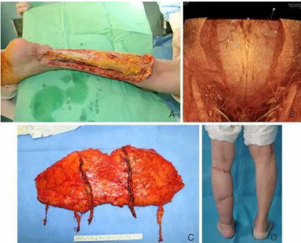
하지만 외상에 따른 광범위한 손상이 조직에 가해진 경우, 방 사선 조사 등이 시행된 경우 결손부 주변의 조직이 재건에 적 합하지 않을 수 있고 이 경우 유리피판을 이용할 경우 만족스 러운 재건이 가능할 수 있다. 흉벽 결손의 재건을 위해 대흉 근피부피판(pectoralis major musculocutaneous flap)이 나 광배근피부피판(latissimus dorsi musculocutaneous flap)이 유경피판의 형태로 널리 사용되고 있으나 전술한 바 와 같이 외상이나 방사선 조사 등으로 이용이 어려울 경우, 결 손부가 매우 광범위 한경우 등은 심하복벽천공지 유리피판과 같은 유리피판을 이용하면 넓은 면적과 충분한 연부조직을 제 공하여 만족스러운 재건이 가능하다(Figure 10) [47].
한국에서 유방암은 1996년 100,000명 당 16.7명에서 2006년 46.8명으로 세 배 가까이 증가하였으며, 호발 연령 이 40대가 40%, 50대가 25.7%로 사회활동이 활발한 연령 에서 발생하여 더욱 주목받고 있는 실정이다. 미국의 통계 에 따르면 유방암 환자의 25% 가량이 재건수술을 받는 것으 로 알려져 있으며 향후 우리나라에서도 유방암재건이 더욱
Figure 9. Facial nerve schwannoma, left. (A) Preoperative view. (B) Elevated latissimus dorsimuscle flap with thoracodorsal nerve. (C) Seven months postoperative view.
Figure 10. Fibromatosis, sternal region. (A) Defect after radical excision of mass. (B) Elevated deep inferior epigastric artery perforator flap. (C) Immediate postoperative view.
A
A B C
B C
증가할 것으로 예상된다.
유방암의 재건은 보형물을 이용하는 방법과 자가 직을 이 용하는 방법으로 크게 나누어 볼 수 있다. 보형물 단독 [48,49], 또는 광배근유경피판과 보형물을 이용한 경우[50], 확장광배근유경피판을 이용한 경우[51], 유경횡복직근피판 [52,53] 등 다양한 술식이 이용되어 왔으나 최근 미세혈관수 술을 이용한 횡복직근유리피판(free transverse rectus ab- dominis musculocutaneous flap) [54,55]이나 심하복벽 천공지유리피판(free deep inferior epigastric artery perforator flap) [56,57]으로 유방재건이 주목을 받고 있고 그 이용이 증가하고 있다. 이는 이 두 유리피판은 심하복벽 동맥(deep inferior epigastric artery)을 혈관경으로 하며
충분한 혈류를 피판에 공급해 유방재건 에 필요한 충분한 크기의 조직을 옮길 수 있게 해준다는 장점이 있다. 특히 심 하복벽천공지피판은 횡복직근피판과 달리 복직근을 포함시키지 않아 복근의 약화나 탈장을 초래할 가능성이 매우 낮 고, 통증이 적고 빠른 공여부 회복을 가 능하게 해주는 추가적 장점이 있다 (Figure 11).
상지 및 하지의 재건
하지의 결손은 다양한 원인에 의해 발생하며 뼈와 연부조직 및 피부를 포 함한 손상이 발생할 수 있다. 외상에 의 한 경우가 많으나 당뇨병성 족부병변, 만성창상, 악성종양 절제 및 말초혈관 질환도 주요한 요인들이라 할 수 있다.
과거에는 결손부위 피복을 위한 유리 피판으로 근육피부피판(musculocu- taneous flap) [14] 또는 근육피판과 피 부이식[15]을 이용한 재건이 널리 행해 졌지만 기능적인 목적이 아닌, 피복을 위한 목적으로 근육을 희생하는 것은 공 여부에 손실을 초래하는 정도가 크다는 인식이 널리 퍼지면서 천공지피판을 이용한 재건이 각광을 받고 있다[16-18]. 결손 부의 크기에 따라, 심하복벽천공지피판, 흉배동맥천공지피 판이나 전외측대퇴피판 순으로 재건을 고려할 수 있고 술자 의 선호에 따라 다양한 천공지피판의 이용이 가능하다 (Figure 12) [58-60].
발바닥의 경우 하지의 다른 피부와 구별되는 특징들을 가 지는데, 피부의 각질층이 두껍고, 충격을 흡수하는데 특화되 어 있다. 이처럼 특화된 부위를 재건하기 위한 방법으로 크 기와 위치에 따라 유경피판(pedicled flap)을 포함한 국소 피판[61-64], 유리근육피부피판(free musculocutaneous flap) [65-67], 근육피판에 더한 피부이식(free muscle flap
Figure 11. Invasive ductal carcinoma, right. (A) Defect after nipple sparing mastectomy anddesign of deep inferior epigastric artery perforator flap. (B) Elevated flap. (C,D) Seven months postoperative views.
A B
C D
with skin graft) [68,69] 등의 방법이 사용되고 있고, 역시 천공지피판의 도 입으로 전외측대퇴피판[70]과 흉배동 맥천공지피판을 이용한 발바닥 부위의 재건이 이루어지고 있다(Figure 13).
상지의 결손 역시 전술한 바와 같이 외 상을 비롯한 다양한 원인에 의해 발생 할 수 있으며, 수부에서 미세수술의 역 할은 매우 중요하다. 이 분야는 본 특집 호 다른 논문에서 심도 깊게 다뤄질 것 이다. 이러한 사지의 재건은 단순히 결 손 부위를 피복하는 것에 그치지 않고, 안면에서와 마찬가지로 수술이나 외상 등으로 인해 이차적으로 발생한 연부 조직 결손에 따른 함몰을 교정하여 윤 곽을 호전시키기에 이르기까지 미용적 으로도 개선하기 위한 노력이 지속되 고 있다.
자궁 경부암 수술 후 방사선 조사를 받거나 유방암 수술과 림프절 절제를 동시에 받고 방사선 치료를 함께 받은 환자들에게서 하지 또는 상지가 붓고 붓기가 지속되면서 피부의 색깔과 질 감마저 달라지는 림프부종이 발생하는 경우가 있다. 림프계에 관한 많은 지식 이 알려졌음에도 불구하고 림프부종의 원인이 무엇인지 아직 정확히 알지 못 하고 있으며 고식적인 치료법을 포함 하여 다양한 치료법들이 제시되고 있 으나 만족스러운 결과는 얻지 못하고 있는 실정이다.
미세수술을 이용한 치료법에는 림프 절-정맥연결법(lymphonodovenous anastomosis), 림프관-정맥연결법 (lymphovenous anastomosis), 그리 고 비교적 최근에 소개되어 이용되고
Figure 12. Paraffinoma, left calf. (A) Defect after radical excision. (B) Preoperative renderedCT angiography for selection of appropriate perforators. (C) Elevated deep inferior epigastric perforator flap with 2 deep inferior epigastric vessels and 2 superficial inferior epigastric veins. (D) Two years postoperative view.
Figure 13. Malignant melanoma, left sole. (A) 3x1.5 cm sized melanoma on the left sole. (B) Elevated thoracodorsal artery perforator flap. (C) Fifteen months postoperative view of donor-site. (D) Fifteen months postoperative view.
A B
C D
D C
A B
있는 림프관-세정맥연결법(lymphatico-venular anasto- mosis) 등이 있다. 이 중에 림프관-세정맥연결법은 0.3 - 0.5 mm 지름의 세정맥과 림프관을 연결하는 것으로 11- 0, 12- 0 나일론 봉합사와[71] 보다 특별하게 고안된 초미세수 술용 기구를 이용하여 시행하는 것이다(Figure 14). 이 방 법을 이용할 경우 기존의 림프관-정맥연결법과 비교하여 적 응증을 확대하여 적용할 수 있고, 숙련된 의사들이 시행할 경우 국소마취 하에서 복수의 팀이 수술에 참가하여 수술 시 간을 단축시킬 수도 있다. 국내 몇몇 림프부종 센터에서 림 프관-세정맥연결법(lymphaticovenular anastomosis)을 이용한 치료를 도입해 시행하고 있고 곧 그 결과가 보고 될 것 으로 기대한다.
안면이식
수 년 전 개봉되었던 영화를 떠올리지 않더라도 안면이식
은 오래 전부터 사람들의 상상력을 자 극하는 주요한 소재들 가운데 하나였 다. 안면이식에 앞서 1998년 프랑스의 Jean-Michel Dubernard에 의해 세계 최초의 수부 동종이식이 시행되었다 [72]. 기본적으로 수부에서 피부, 뼈, 근 육, 신경 등의 동종이식이 가능함을 확 인한 후 안면에서도 동일한 조직을 포 함한 이식편의 전이가 이루어 질 수 있 다는 생각에서 안면이식의 가능성에 대해 고려하던 중 2005년 11월 27일 프랑스 리용에서 세계 최초의 안면이 식이 성공적으로 시행되었다[73]. 대상 환자는 38세 여자 환자로 기능적으로 평가했을 때, 가벼운 접촉이나 열 또는 차가움에 대한 감각은 6개월 무렵부터 정상에 가까워졌고, 입술은 수술 후 10 개월 경에 완전히 다물어지게 되었으 며, 이후 표정도 점차 자연스러워졌다.
면역거부반응은 수술 후 18일, 214일 경에 발생하였으나 해결되었고 환자는 정신적, 미용적으로 만족스러워하고 있는 상태였다[74].
이후 중국, 미국, 스페인 등의 나라에서 총 8 예의 동종 안 면이식이 추가로 이루어져왔으며, 이식 거부 반응은 면역 억 제제를 규칙적으로 복용하지 않은 1 예에서, 사망은 2 예에 서 발생하였다. 사망의 원인은 1 예에서는 이식과 무관한 심 장 관련 합병증으로 발생하였고, 나머지 1 예의 원인은 알려 져 있지 않은 상태이다.
얼굴은 개인의 정체성을 결정하는데 중요한 요소로서, 안 면이식수술을 받을 환자의 적절한 선택이 중요하며, 심리적 인 지원이 반드시 필요하다. 또한 환자가 수술에 대한 열의 가 있어야 하고, 평생 면역억제제를 복용하여야 하는 이유를 이해하고 이에 순응해야 한다[75].
동종이식은 자가조직으로 얻기 어려운 넓은 피부가 필요 하고, 피판만으로는 3차원적으로 만족스러운 모양을 얻을 수 없으며, 기능적으로나 미용적으로 뛰어난 재건이 어려운
Figure 14. Lymphedema, left calf. (A) Preoperative view. (B) Three slit incisions were done.(C) Lymphaticovenular anasto-mosis. (D) Three months postoperative view.
A B
C D
현실적인 이유들로 인해 고안된 치료방법이다[76]. 또한 안 면추형이 초래하는 사회생활의 장애나 이에 따른 절망감으 로 생에 대한 의욕이 저하된 환자를 위해 반드시 필요한 치 료적 수단으로서 바라보는 것이 필요하다. 하지만 평생 면 역억제제를 복용해야 한다는 것과 그에 따르는 잠재적 부작 용을 감수하면서 치료를 진행하는 것에 대해서는 이견이 있 을 수 있다. 국내에서도 안면이식에 대한 준비가 조심스럽 게 이루어지고 있다. 성공적인 안면이식이 이루어지기 위해 서는 숙련된 미세수술 성형외과 의사들뿐 아니라 두개 안면 성형, 이식면역, 이식 관련 심리, 마취, 중환자 관리, 사회사 업, 치과, 재활, 언어치료, 안과, 이비인후과와 생명윤리와 관련된 전문가에 이르기까지 다양한 분야의 전문가로 구성 된 팀과 이를 조화롭게 이끌 수 있는 리더가 필요하다[77].
결 론
재건성형에 있어 이상적인 목표는 유사한 조직으로 결손 부위를 기능적, 미용적으로 재건하고 아울러 공여부의 결손 을 최소화하는 것이라고 할 수 있다. 이러한 이상적 재건에 대한 열망은 동종이식이라는 새로운 방법을 통해 한층 가까 이 다가설 수 있게 되었다. 향후 면역학적 문제를 해결하기 위한 지식과 기술이 발전할 것이며, 이에 따라 다양한 부위 의 동종이식술이 재건미세수술분야 성형외과의사에 의해 시행될 것으로 보인다.
지속적인 유리피판술의 발전과 새로운 수술 방법인 천공 지 피판의 도입 및 정련(refinement)을 통해 우리는 과거보 다 더 높은 수술 성공률과 괄목할 수준의 기능-미용적 재건 을 할 수 있게 되었다. 목적에 따라 자유로운 재건 방법의 선 택이 가능하다는 재건 파이(reconstructive pie)의 개념이 확산되면서, 앞으로 유리피판술은 재건성형외과 분야에서 더욱 널리 시행될 것이다. 또한 결과의 완성도는 더욱 높아 지고, 환자가 부담하여야 할 위험과 희생은 더욱 감소할 것 으로 예상된다.
따라서 의료계 전반에서 점증하는 미세 수술의 필요성과 중요성에 대한 환기가 필요하며, 대형병원과 재건센터에서 미세혈관수술을 이용한 재건을 담당하는 성형외과 의사들
의 중요한 역할에 대한 재조명이 필요한 시점이다. 미세수 술을 이용한 성공적인 재건술의 지속적인 발전을 통해, 환자 에게 더욱 만족스러운 수술 결과를 제공할 수 있게 될 뿐만 아니라, 환자의 행복을 증진시킬 수 있고, 삶의 질 향상에도 기여할 수 있게 될 것이다
핵심용어: 재건; 미세수술; 유리 피판; 동종이식
REFERENCES
21. Chung CN. Clinical observation on skin graft and flap. J Korean Soc Plast Reconstr Surg 1974;1:23-39.
22. Lim P, Cho MJ, Cho HS, Hur E, Lee JW, Ham KS. An axillary scar contracture corrected by microvascular free groin flap transfer. J Korean Soc Plast Reconstr Surg 1978;5:189-194.
23. Park CG, Lee HW, Kim CH. Microvascular free flaps for the reconstruction of head and neck. J Korean Soc Plast Reconstr Surg 1985;12:187-198.
24. Choi SH, Hong SH, Na SK, Park CS. Clinical experiences of dorsalis pedis free flap transfer. J Korean Soc Plast Reconstr Surg 1981;8:189-198.
25. Lee DJ, Hur E, Cho MJ, Lim P. Clinical experiences of free flap transfer. J Korean Soc Plast Reconstr Surg 1980;7:313-320.
26. Franklin JD, Withers EH, Madden JJ Jr, Lynch JB. Use of the free dorsalis pedis flap in head and neck repairs. Plast Reconstr Surg 1979;63:195-204.
27. Leeb DC, Ben-Hur N, Mazzarella L. Reconstruction of the floor of the mouth with a free Dorsalis pedis flap. Plast Re- constr Surg 1977;59:379-381.
28. Ahn HC, Yang EZ, Kim CY, Lee JH. Various applications of deep inferior epigastric artery perforator free flap. J Korean Soc Plast Reconstr Surg 2009;36:707-713.
29. Choi BK, Kim YS, Lee WJ, Lew DH, Tark KC. The Safety of microsurgical head and neck reconstruction in the elderly pa- tients. J Korean Soc Plast Reconstr Surg 2006;33:289-293.
10. Jeon BJ, Lim SY, Hyon WS, Bang SI, Oh KS, Mun GH.
Anterolateral thigh flap: our experiences in head and neck reconstruction. J Korean Soc Plast Reconstr Surg 2006;
33:276-282.
11. Tark KC, Yoon JS, Shin KS. Analysis of 83 consecutive free flaps. J Korean Soc Plast Reconstr Surg 1993;20:155-166.
12. Koshima I, Soeda S. Inferior epigastric artery skin flaps with- out rectus abdominis muscle. Br J Plast Surg 1989;42:645- 648.
13. Kroll SS, Rosenfield L. Perforator-based flaps for low posterior midline defects. Plast Reconstr Surg 1988;81:561-566.
14. Lee KS, Chang JS, Park JW. Functioning gracilis musculo- cutaneous free flap transplantation for the reconstruction of
injured upper extremity. J Korean Orthop Assoc 1992;27:
1868-1876.
15. Jung KM, Hong IP, Kim JH. Reconstruction of extensive lower extermity soft tissue defect using free latissimus dorsi muscle flap with STSG. J Korean Soc Plast Reconstr Surg 1999;26:927-932.
16. Cho KS, Kim DY, Lee SY, Cho BH. Reconstruction of the tissue defects of extremities with anterolateral thigh free flap. J Korean Soc Plast Reconstr Surg 1999;26:281-286.
17. Kim HS, Kim KC, Kim SE. Soft tissue reconstruction of children’s extremity with perforator free flap. J Korean Mic- rosurg Soc 2007;16:14-22.
18. Oh CH, Shim JS, Park DH. Lower extremity reconstruction by perforator based flap. J Korean Soc Plast Reconstr Surg 2009;36:720-726.
19. Whitaker IS, Smit JM, Rozen W, Dimopoulou A, Acosta R. Pre operative computed tomographic angiography (CTA): a valuable lesson in planning DIEP flaps. J Plast Reconstr Aesthet Surg 2009;62:551.
20. Lim SY, Pyon JK, Mun GH, Bang SI, Oh KS. Surgical treat- ment of angiosarcoma of the scalp with superficial paroti- dectomy. Ann Plast Surg 2010;64:180-182.
21. Kim YH, Kim JT. Perforator flaps in head and neck recon- struction. Hanyang Med Rev 2009;29:265-273.
22. Thorwarth M, Eulzer C, Bader R, Wolf C, Schmidt M, Schultze-Mosgau S. Free flap transfer in cranio-maxillofacial surgery: a review of the current data. Oral Maxillofac Surg 2008;12:113-124.
23. Lipa JE, Butler CE. Enhancing the outcome of free latissimus dorsi muscle flap reconstruction of scalp defects. Head Neck 2004;26:46-53.
24. Lee SH, Mun GH. Transverse thoracodorsal artery perforator flaps: experience with 31 free flaps. J Plast Reconstr Aesthet Surg 2008;61:372-379.
25. Amin A, Rifaat M, Civantos F, Weed D, Abu-Sedira M, Bassiouny M. Free anterolateral thigh flap for reconstruction of major craniofacial defects. J Reconstr Microsurg 2006;
22:97-104.
26. Koshima I, Inagawa K, Urushibara K, Moriguchi T. Paraum- bilical perforator flap without deep inferior epigastric vessels.
Plast Reconstr Surg 1998;102:1052-1057.
27. Mun GH, Lim SY, Hyon WS, Bang SI, Oh KS. A novel recon- struction of 2 distinct defects: concomitant use of a thoraco- dorsal artery perforator flap and its corresponding muscle flap.
Ann Plast Surg 2005;55:676-678.
28. Choi HC, Han SH, Koh KS, Yoon KC, Chung BS. Free muscle flap reconstruction following resection of the skull base tum- our. J Korean Soc Plast Reconstr Surg 1993;20:1257-1265.
29. Hanasono MM, Sacks JM, Goel N, Ayad M, Skoracki RJ. The
anterolateral thigh free flap for skull base reconstruction.
Otolaryngol Head Neck Surg 2009;140:855-860.
30. Zhang B, Li DZ, Xu ZG, Tang PZ. Deep inferior epigastric artery perforator free flaps in head and neck reconstruction. Oral Oncol 2009;45:116-120.
31. Kim KH, Chung CH, Chang YJ, Rho YS. Reconstruction of midfacial defects with free flaps after maxillectomy. J Korean Soc Plast Reconstr Surg 2010;37:607-612.
32. Santamaria E, Granados M, Barrera-Franco JL. Radial forearm free tissue transfer for head and neck reconstruction: versa- tility and reliability of a single donor site. Microsurgery 2000;
20:195-201.
33. Browne JD, Burke AJ. Benefits of routine maxillectomy and orbital reconstruction with the rectus abdominis free flap.
Otolaryngol Head Neck Surg 1999;121:203-209.
34. Uglesic V, Virag M, Varga S, Knezevic P, Milenovic A. Recon- struction following radical maxillectomy with flaps supplied by the subscapular artery. J Craniomaxillofac Surg 2000;28:153- 160.
35. Suga H, Asato H, Okazaki M, Okochi M, Narushima M.
Combination of costal cartilage graft and rib-latissimus dorsi flap: a new strategy for secondary reconstruction of the maxil- la. J Craniofac Surg 2007;18:639-642.
36. Andrades P, Rosenthal EL, Carroll WR, Baranano CF, Peters GE. Zygomatic-maxillary buttress reconstruction of midface defects with the osteocutaneous radial forearm free flap.
Head Neck 2008;30:1295-1302.
37. Chang DW, Langstein HN. Use of the free fibula flap for resto- ration of orbital support and midfacial projection following maxillectomy. J Reconstr Microsurg 2003;19:147-152.
38. Maranzano M, Atzei A. The versatility of vascularized iliac crest with internal oblique muscle flap for composite upper maxil- lary reconstruction. Microsurgery 2007;27:37-42.
39. Pomahac B, Lengele B, Ridgway EB, Matros E, Andrews BT, Cooper JS, Kutz R, Pribaz JJ. Vascular considerations in composite midfacial allotransplantation. Plast Reconstr Surg 2010;125:517-522.
40. Blocker TG Jr, Stout RA. Mandibular reconstruction, World War II. Plast Reconstr Surg 1949;4:153-156.
41. Shenaq SM, Klebuc MJ. The iliac crest microsurgical free flap in mandibular reconstruction. Clin Plast Surg 1994;21:37-44.
42. Wei FC, Jain V, Celik N, Chen HC, Chuang DC, Lin CH. Have we found an ideal soft-tissue flap? An experience with 672 anterolateral thigh flaps. Plast Reconstr Surg 2002;109:2219- 2226.
43. Harii K, Ohmori K, Torii S. Free gracilis muscle transplantation, with microneurovascular anastomoses for the treatment of facial paralysis. A preliminary report. Plast Reconstr Surg 1976;57:133-143.
44. Terzis JK. Pectoralis minor: a unique muscle for correction of facial palsy. Plast Reconstr Surg 1989;83:767-776.
45. Harii K, Asato H, Yoshimura K, Sugawara Y, Nakatsuka T, Ueda K. One-stage transfer of the latissimus dorsi muscle for reani- mation of a paralyzed face: a new alternative. Plast Reconstr Surg 1998;102:941-951.
46. Choi JH, Minn KW. Chest wall reconstruction with muscle flap. J Korean Soc Plast Reconstr Surg 2003;30:413-419.
47. Sullivan SR, Truxillo TM, Mann GN, Isik FF. Utility of the free deep inferior epigastric perforator flap in chest wall recon- struction. Breast J 2007;13:50-54.
48. Ahn S. Breast reconstruction using implants. J Korean Med Assoc 2011;54:51-60.
49. Cho YK, Yang JD, Kim GR, Chung HY, Cho BC, Park HY. Imme- diate implant reconstruction using silicone prosthesis in breast cancer patients after skin sparing mastectomy. J Korean Soc Plast Reconstr Surg 2010;37:749-757.
50. Lee HK, Kim YJ, Lee SJ. The latissimus dorsi flap with an im- plant: the most useful & proper breast reconstruction method for the young mastectomy patient. J Korean Soc Aesthetic Plast Surg 2000;6:83-90.
51. Park JH, Bang SI, Kim SH, Im SY, Mun GH, Hyon WS, Oh KS.
Breast reconstruction with the extended latissimus dorsi musculocutancous flap. J Korean Soc Plast Reconstr Surg 2005;32:408-415.
52. Eom JS. Breast reconstruction using pedicled transverse rectus abdominis musculocutaneous (TRAM) flap. J Korean Med Assoc 2011;54:12-21.
53. Kim EK, Eom JS, Ahn SH, Son BH, Lee TJ. Evolution of the pedicled TRAM flap: a prospective study of 500 consecutive cases by a single surgeon in Asian patients. Ann Plast Surg 2009;63:378-382.
54. Nahabedian MY, Momen B, Galdino G, Manson PN. Breast reconstruction with the free TRAM or DIEP flap: patient selec- tion, choice of flap, and outcome. Plast Reconstr Surg 2002;
110:466-475.
55. Jin US, Minn KW. Breast reconstruction using the transverse rectus abdominis musculocutaneous (TRAM) free flap. J Korean Med Assoc 2011;54:22-34.
56. Kim JH, Park JU, Cho SH, Eo SR. Immediate breast recon- struction with DIEP free flap. J Korean Soc Microsurg 2008;
17:94-100.
57. Gill PS, Hunt JP, Guerra AB, Dellacroce FJ, Sullivan SK, Boraski J, Metzinger SE, Dupin CL, Allen RJ. A 10-year retrospective review of 758 DIEP flaps for breast reconstruction. Plast Re- constr Surg 2004;113:1153-1160.
58. Tajsic N, Winkel R, Hoffmann R, Husum H. Sural perforator flap for reconstructive surgery in the lower leg and the foot: a clinical study of 86 patients with post-traumatic osteomyelitis.
J Plast Reconstr Aesthet Surg 2009;62:1701-1708.
59. Wong CH, Tan BK. Perforator-sparing transposition flaps for lower limb defects: anatomic study and clinical application.
Ann Plast Surg 2007;58:614-621.
60. Momeni A, Krischak S, Bannasch H. The thoracodorsal artery perforator flap with a vascularized scapular segment for reconstruction of a composite lower extremity defect. Micro- surgery 2006;26:515-518.
61. Yang D, Yang JF, Morris SF, Tang M, Nie C. Medial plantar artery perforator flap for soft-tissue reconstruction of the heel.
Ann Plast Surg 2011 Feb 4 [Epub].
62. Unglaub F, Wolf MB, Dragu A, Forst J, Horch RE, Kneser U.
Reconstruction of a child’s forefoot defect using a distally based pedicled medial plantar flap. Arch Orthop Trauma Surg 2010;130:155-158.
63. Caleffi E, Bocchi A, Montacchini G, Papadia F. Reconstruction of the heel by a medial plantar flap. Ital J Orthop Traumatol 1989;15:191-196.
64. Botte MJ, Gellman H. Reconstruction of a traumatic hallux amputation using a plantar V-Y advancement flap. Clin Orthop Relat Res 1987;(220):211-216.
65. Tamura A, Takeuchi Y, Yamakage A. Reconstruction of plantar heel defects with free gracilis musculocutaneous flap. J Foot Ankle Surg 1994;33:274-277.
66. Dubrow TJ, Lesavoy MA. Acne of the heel: acne vulgaris com- plicating a free vascularized latissimus dorsi musculocu- taneous flap. Ann Plast Surg 1989;23:349-351.
67. Ikuta Y, Murakami T, Yoshioka K, Tsuge K. Reconstruction of the heel pad by flexor digitorum brevis musculocutaneous flap transfer. Plast Reconstr Surg 1984;74:86-96.
68. Bostwick J 3rd. Reconstruction of the heel pad by muscle transposition and split skin graft. Surg Gynecol Obstet 1976;
143:973-974.
69. Jeong JH. Reconstruction of large heel defects using gracilis muscle free flaps. Yeungnam Univ J Med 1997;14:227-236.
70. Hong JP. Reconstruction of the diabetic foot using the antero- lateral thigh perforator flap. Plast Reconstr Surg 2006;117:
1599-1608.
71. Nagase T, Gonda K, Inoue K, Higashino T, Fukuda N, Gorai K, Mihara M, Nakanishi M, Koshima I. Treatment of lymphe- dema with lymphaticovenular anastomoses. Int J Clin Oncol 2005;10:304-310.
72. Dubernard JM, Owen E, Herzberg G, Martin X, Guigal V, Dawahra M, Pasticier G, Mongin-Long D, Kopp C, Ostapetz A, Lanzetta M, Kapila H, Hakim N. The first transplantation of a hand in humans: early results. Chirurgie 1999;124:358-365.
73. Devauchelle B, Badet L, Lengelé B, Morelon E, Testelin S, Michallet M, D’Hauthuille C, Dubernard JM. First human face allograft: early report. Lancet 2006;368:203-209.
74. Dubernard JM, Lengele B, Morelon E, Testelin S, Badet L, Moure C, Beziat JL, Dakpe S, Kanitakis J, D’Hauthuille C, El Jaafari A, Petruzzo P, Lefrancois N, Taha F, Sirigu A, Di Marco G, Carmi E, Bachmann D, Cremades S, Giraux P, Burloux G, Hequet O, Parquet N, Frances C, Michallet M, Martin X, De- vauchelle B. Outcomes 18 months after the firs4t human par- tial face transplantation. N Engl J Med 2007;357:2451-2460.
75. Soni CV, Barker JH, Pushpakumar SB, Furr LA, Cunningham M, Banis JC Jr, Frank J. Psychosocial considerations in facial
transplantation. Burns 2010;36:959-964.
76. Siemionow M, Agaoglu G, Unal S. A cadaver study in prepa- ration for facial allograft transplantation in humans: part II.
Mock facial transplantation. Plast Reconstr Surg 2006;117:
876-885.
77. Siemionow MZ, Gordon CR. Institutional review board-based recommendations for medical institutions pursuing protocol approval for facial transplantation. Plast Reconstr Surg 2010;
126:1232-1239.
본 논문은 재건성형외과 영역에 있어 현 상황과 흐름을 예리한 시각에서 파악하고 미래의 지평을 명확히 제시하였다. 재건 성형의 지평을 새롭게 할 동종이식술에 대한 마지막 제안을 통하여 향후 안면동종이식에 대한 많은 시사점을 제공하고 있 다는 부분에서 독자로 하여금 재건성형외과에 대한 새로운 인식을 넓힐 수 있는 훌륭한 종설이 될 것이라 판단된다.
[정리:편집위원회]