Ⅰ.
서 론
Bisphosphonates (BP)는 골다공증, 골결핍증, Paget’s dis-
ease, 다발성 골수종, 악성 종양의 골전이로 인한 통증 및
병적 골절을 방지하기 위해 사용되는 약물로1-3, 파골세포 의 기능을 저하시켜 골흡수를 방해하고 악성 질환에서 고 칼슘혈증 치료 시 석회화를 방해하는 작용을 하며 부가적 으로 관절염 치료 시 관절 내의 염증반응을 감소시키는 약 물작용을 한다4.
일반적으로 알려진 BP의 부작용은 피로, 발열, 빈혈, 부 종, 위장관계 증상, 신장독성, 식도점막의 궤양 등이 있으 며5, 최근 들어 대두되고 있는 부작용으로는 bisphospho- nates related osteonecrosis of the jaw (BRONJ)가 있다6-8.
2003년 Marx9에 의해 처음으로 정주용 BP로 인한 악골괴 사가 보고되기 시작하였으며, 2004년 Ruggiero 등1은 정주 용 BP 뿐만 아니라 경구용 BP로 인한 63예의 악골괴사를 보고하였다. 그 이후 보다 많은 증례들을 발표하였고10,11, 2007년 American Society for Bone and Mineral Research
(ASBMR)에서 BRONJ에 대한 보고서를 발표하였으며12,
American Associaion of Oral and Maxillofacial Surgeons (AAOMS)에서는 BRONJ에 대한 가이드 라인을 발표하였 다13.
또한, 2009년 우리나라에서도 대한내분비학회, 대한골대 사학회, 대한골다공증학회 및 대한구강악안면외과학회가 공동으로 BRONJ의 발생위험, 예방 및 처치방법에 대한 최 김 경 욱
330-716 충남 천안시 신부동 산7-1번지
단국대학교 치과대학 부속치과병원 구강악안면외과 Kyung-Wook Kim
Department of Oral and Maxillofacial Surgery, Dental Hospital, Dankook University
San 7-1 Sinbu-dong, Choenan, 330-716, Korea TEL: +82-41-550-1994 FAX: +82-41-551-8988 E-mail: [email protected]
비스포스포네이트 관련 악골괴사의 진단 및 치료에 대한 임상적 연구
김경욱∙김범진∙이충현
단국대학교 치과대학 구강악안면외과학교실
Clinical study of diagnosis and treatment of bisphosphonate-related osteonecrosis of the jaws
Kyung-Wook Kim, Beom-Jin Kim, Chung-Hyun Lee
Department of Oral and Maxillofacial Surgery, College of Dentistry, Dankook University, Choenan, Korea
Introduction:Bisphosphonates is used widely for the treatment of the Paget’s disease, multiple myeloma, bone metastases of malignant tumors with the prevention of pain and their pathological fracture. However, it was recently suggested that bisphosphonates related osteonecrosis of the jaw (BRONJ) is a side effect of bisphosphonate use.
Materials and Methods:Twenty-four individuals, who were referred to the Department of Oral and Maxillofacial surgery, Dankook University Dental Hospital, were selected from those who had exposed bone associated with bisphosphonates from January, 2005 to December, 2009 according to the criteria of American Association of Oral and Maxillofacial Surgeons (AAOMS) for BRONJ. The patients group consisted of 7 males and 17 females between the age of 46 to 78 years (average 61.8 years). Each patient had panoramic imaging, computed tomography (CT), whole body bone scanning performed for a diagnosis and biopsy sampling from the necrotizing tissue. C-terminal cross-linking telopeptide of type I collagen (CTX) level of patients who had undergone surgical intervention was measured 7 days before surgery.
Results:The main cause of bone exposure was post-extraction (15), chronic periodontitis (4), persistent irritation of the denture (3). Twenty people had undergone BRONJ treatment for two to eight months except for 4 people who had to maintain the bisphosphonates treatment to prevent a metasta- sis and bone trabecular pain with medical treatment. When the bisphosphonate treatment was suspended at least for 3 months and followed up accord- ing to the AAOMS protocols, the exposed necrotizing bones were found to be covered by soft tissue.
Conclusion:Prevention therapy, interruption of bisphophonates for at least 3 months and cooperation with the physician for conservative treatment are the essential for treating BRONJ patient with high risk factors. The CTX level of BRONJ patients should be checked before undergoing surgical intervention. Surgical treatments should be delayed in the case of a CTX level <150 pg/mL.
Key words:Bisphosphonates, Osteonecrosis, Jaw Diseases, Therapeutics
[paper submitted 2010. 8. 3 / revised 2010. 12. 20 / accepted 2011. 2. 7]
Abstract (J Korean Assoc Oral Maxillofac Surg 2011;37:54-61)
신지견을 정리하여 발표하였다.
그럼에도 불구하고 BRONJ에 대한 임상적, 방사선학적 진단기준이 명확하지 않으며 치료에 있어서 아직 뚜렷한 효과적인 방법이 없는 상태로 동통을 완화하고 감염 및 괴 사가 진행하는 것을 억제하는 치료가 주를 이룬다. 이에 본 논문에서는 BRONJ를 보다 효과적으로 진단하고 치료하 기 위해서 본원에서 진단받고 치료한 증례들을 분석하여 BRONJ의 임상적 , 방사선학적 특징과 BP를 중단한 기간, 보존적 치료 및 외과적 치료가 BRONJ의 치유경과에 미치 는 영향을 알아보고자 한다.
Ⅱ.
연구대상 및 방법
이번 연구는 단국대학교 치과대학 부속치과병원에 2005 년 1월부터 2009년 12월까지 내원한 환자들 중 AAOMS에 서 발표한 BRONJ 진단원칙을 기준으로 BRONJ로 진단받 은 환자 24명을 대상으로 하였으며, BRONJ stage 기준으로
1, 2 및 3기 환자들을 대상으로 하였다.(Table 1) AAOMS에 서 발표한 BRONJ 진단기준은 과거 또는 현재 BP 경구 또 는 정맥으로 투여받은 과거력이 있으며 8주 이상 지속되는 악골(턱뼈) 부위의 골노출 및 괴사소견, 악골에 방사선치 료를 받은 과거력이 없는 경우를 모두 만족시킬 때이다. 또 한, AAOMS에서 발표한 BRONJ stage 기준은 0기“뼈는 괴 사되어 있지 않지만 비특이적 증상을 호소할 때”, 1기“괴 사된 뼈가 노출되어 있지만 증상 및 감염의 증거가 없을 때”, 2기“괴사된 뼈가 노출되어 있으면서 동통 및 감염의 증거가 있을 때”, 3기“2기의 소견과 함께 병적 골절 동반 또는 구강 외 누공이 있을 때”이다13.
환자 24명 중 남성은 7명, 여성은 17명이었고, 환자들의 나이는 46-78세로 평균 61.8세였다. 환자들은 골다공증(11 명, 45.8%), 유방암(5명, 20.8%), 다발성 골수종(4명, 16.7%), 전립선암(3명, 12.5%), 폐암(1명, 4.2%) 등의 이유 로 BP를 투여받고 있었다. 그리고 경구용 BP는 alendronate (Fosamax) (9명), residronate (Actonel) (1명), ibadronate
Table 1.Patients and clinical information
Patient Sex Age Reason for
Bisphosphonate Duration
Site Etiologic factor
bisphosphonate use (months)
1 F 63 Osteoporosis Alendronate (PO) 38 Lt. Mn. Molar Teeth extraction
2 M 71 Prostate cancer Zoledronate (IV) 11 Rt. Mn. Premolar Chronic periodontitis
3 F 68 Osteoporosis Alendronate (PO) 47 Rt. Mx. Incisor Teeth extraction
4 F 46 Breast cancer Zoledronate (IV) 23 Rt. Mn. Molar Teeth extraction
5 F 67 Osteoporosis Alendronate (PO) 51 Rt. Mn. Molar Curettage
6 M 54 Multiple myeloma Zoledronate (IV) 17 Lt. Mx. Molar Teeth extraction
7 F 67 Osteoporosis Residronate (PO) 38 Rt. Mn. Molar Teeth extraction
8 F 62 Breast cancer Zoledronate (IV) 29 Lt. Mn. Premolar Chronic periodontitis
9 M 59 Prostate cancer Zoledronate (IV) 23 Rt. Mn. Molar Teeth extraction
10 F 78 Osteoporosis Alendronate (PO) 41 Rt. Mx. buccal eminence Denture trauma
11 F 56 Breast cancer Pamidronate (IV) 34 Lt. Mx. Premolar Teeth extraction
Zoledronate (IV) Lt. Mn. Molar
12 F 64 Osteoporosis Alendronate (PO) 35 Rt. Mn. Molar Curettage
13 F 48 Multiple myeloma Zoledronate (IV) 16 Lt. Mx. Incisor Teeth extraction
14 M 68 Osteoporosis Alendronate (PO) 43 Lt. Mn. lingual eminence Denture trauma
15 F 75 Osteoporosis Ibadronate (PO) 51 Lt. Mn. Premolar Teeth extraction
16 F 65 Breast cancer Pamidronate (IV) 27 Rt. Mx. Molar Teeth extraction
Zoledronate (IV) Rt. Mn. Molar
17 F 62 Multiple myeloma Pamidronate (IV) 23 Lt. Mn. Molar Teeth extraction
18 F 58 Osteoporosis Alendronate (PO) 31 Lt. Mx. buccal eminence Denture trauma
19 M 51 Multiple myeloma Pamidronate (IV) 17 Rt. Mn. Premolar Teeth extraction
20 F 67 Osteoporosis Alendronate (PO) 63 Lt. Mn. Molar Chronic periodontitis
21 M 61 Lung cancer Zoledronate (IV) 26 Rt. Mx. Molar Teeth extraction
22 F 53 Breast cancer Zoledronate (IV) 14 Lt. Mn. Premolar Teeth extraction
23 F 63 Osteoporosis Alendronate (PO) 37 Lt. Mx. Incisor Teeth extraction
24 M 58 Prostate cancer Zoledronate (IV) 21 Rt. Mn. Molar Chronic periodontitis
(PO: per oral, IV: intravenous, Mn: mandible, Mx: maxilla)
(Boniva) (1명) 등을 복용 중이었고, 정주용 BP는 zole- dronate (Zometa) (9명), pamidronate (Aredia) 투약 후 zole- dronate (2명), pamidronate (2명)를 복용하고 있었다.
환자들의 경구용 BP 복용기간은 평균 43.2개월(31-63개 월), 정주용 BP 투여기간은 평균 21.6개월(11-34개월)이었 으며 BRONJ는 상악(7명), 하악(15명), 양악(2명)에서 발생 하였다. 모든 환자들은 괴사된 뼈가 노출되어 있었고 괴사 된 뼈의 노출은 발치 후(15명), 만성 치주염(4명), 지속적인 부적절한 의치에 의한 외상(3명), 소파술 후(2명) 등의 이유 로 나타났다. 모든 환자들은 진단을 위해 파노라마, 컴퓨터 단층촬영, whole body bone scan을 시행하였으며 괴사된 골 로부터 채취한 연조직과 경조직 모두 생검을 시행하였다.
BRONJ를 치료하기 위해 환자 24명 중 20명은 내과와 협 진하여 BP를 중단한 후 보존적 치료 및 외과적 치료를 시 행하였고, 나머지 4명은 골병변의 통증과 전이를 막기 위 하여 BP를 중단하지 않고 치료를 시행하였다.(Table 2)
보존적 치료 및 외과적 치료의 기준은 2007년 AAOMS에 서 병소의 진행단계에 따라 제시한 치료방법을 따랐다.
BRONJ 1기에는 0.12% chlorhexidine 용액을 사용하였고, 2 기에는 Amoxicillin 500 mg/K clavulanate 125 mg (Augmentin 625 mg, Ilsung Pharmaceuticals Co., Ltd., Seoul, Korea) 항생 제를 1일 3회 2주간 복용하면서 0.12% chlorhexidine 용액을 사용하였으며 연조직의 자극을 경감시키기 위해 괴사된 골의 표면을 제거하기도 했다. Penicillin에 allergy가 있는 환자의 경우에는 Clindamycin HCl 150 mg (Fullgram, Samjin Pharmaceutical Co., Ltd., Seoul, Korea) 항생제를 1일 3회 2주간 복용하였으며 2주 후에는 microbiology culture 결과에 따라 항생제를 유지 또는 교체하였다. 또한, 3기에 는 2기와 같은 항생제와 구강 양치액을 사용하면서 부골적 출술(sequestrectomy)을 시행하였고 이환된 범위가 큰 경우 에는 분절절제술(segmental resection) 및 재건술(reconstruc- tion)을 시행하였다. 또한, 침습적 외과적 치료를 하는 모든 환자들의 경우에 수술 7일 전 오전 공복 시 골흡수 지표인 C-terminal cross-linking telopeptide of type I collagen (CTX) 를 측정하였다.
Table 2.Results of the proposed therapeutic protocol Patient Stage Cessation of BP
CTX (pg/mL) Treatment Results
(months)
1 3 6 178 Sequestrectomy Complete wound healing
2 1 4 317 No Complete wound healing
3 3 3 215 Segmental resection Complete wound healing
4 3 No 168 Sequestrectomy Recurrence1
5 3 3 142 Sequestrectomy Progressive necrosis2
6 1 4 351 No Complete wound healing
7 3 3 135 Sequestrectomy Complete wound healing
8 3 3 113 Sequestrectomy Recurrence
9 2 8 215 Debridement Complete wound healing
10 1 3 273 No Complete wound healing
11 3 No 162 Sequestrectomy Recurrence
12 2 5 261 Debridement Complete wound healing
13 2 No 183 Debridement Recurrence
14 3 3 84 No Progressive necrosis
15 3 6 278 Sequestrectomy Complete wound healing
16 2 2 314 Debridement Progressive necrosis
17 1 4 372 No Complete wound healing
18 3 8 236 Sequestrectomy Complete wound healing
19 3 3 121 Sequestrectomy Recurrence
20 3 4 328 Sequestrectomy Complete wound healing
21 2 No 271 Debridement Progressive necrosis
22 1 5 325 No Complete wound healing
23 2 3 96 No Progressive necrosis
24 2 6 217 Debridement Complete wound healing
(BP: bisphosphonate, CTX: C-terminal cross-linking telopeptied of type I collagen, Recurrence1: exposure of newly formed necrotic bone on the adjacent sites, Progressive necrosis2: prolonged exposure of necrotic bone in the lesion area)
Ⅲ.
결 과
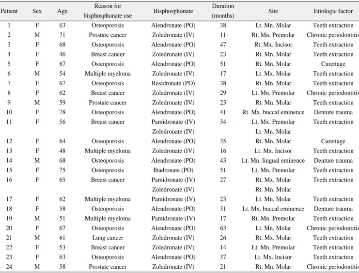
BRONJ 환자 24명 모두는 시진에서 괴사된 골의 노출을 나타내었고(Fig. 1), 통증을 호소하였으며, 17명의 환자에 서 화농성 삼출물이 관찰되었다.(Fig. 2) 진단을 위해 촬영 한 파노라마에서 발치 후 괴사된 골의 노출을 보인 15명의 환자 모두에서 치아발치 후 발치와의 형태가 사라지지 않 고 치조백선이 잔존하는 특징을 나타내었다.(Fig. 3) 또한 만성 치주염과 소파술 후 괴사된 골의 노출을 보인 4명의 환자에서 치조백선이 소실되면서 치주인대 공간이 넓어지 는 골융해소견이 관찰되었다.(Fig. 4) 컴퓨터단층촬영에서 는 24명의 환자 모두에서 골소주의 변형 및 피질수질접합 부의 불분명한 경계를 가지는 부골화 양상을 관찰할 수 있 었으며(Fig. 5), 양악에 발생한 환자 1명을 포함하여 하악골 에서 발생한 15명의 환자 모두에서 명확한 골막반응 (periosteal reaction)을 관찰할 수 있었다.(Fig. 6) 또한, 12명 의 환자에서 골경화, 8명의 환자에서 골용해, 6명의 환자에 서 골막 신생골, 3명의 환자에서 하악관 침범, 2명의 환자 에서 골절 등이 관찰되었다.
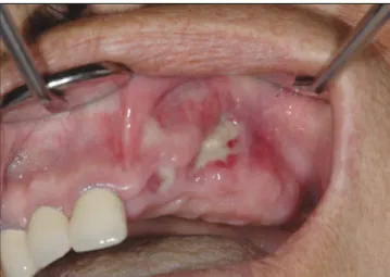
괴사된 골로부터 채취한 연조직과 경조직 생검결과 육아
조직 내에 다수의 형질세포, 호중구, 다핵거대세포 및 모세 혈관의 증식이 관찰되었고(Fig. 7), osteocytic lacuna 내에 골세포가 존재하지 않는 양상을 보였다14.(Fig. 8) 배양결과 18명의 환자에서 Actinomyces israelii, 4명의 환자에서 Bacteroides melanogenicus, 나머지 환자에서 normal oral flo- ra가 나타났다. 또한, 환자들 중 11명이 스테로이드 계열의 약물을 복용하고 있었고 8명의 환자가 당뇨병을 가지고 있 었으며, 7명의 환자가 함암화악요법을 받고 있었다.
환자 24명을 BRONJ stage 기준으로 나누었을 때, 1기는 5 명, 2기는 7명, 3기는 12명이었으며 내과와 협진 후 골주소 의 통증과 전이를 막기 위하여 BP를 유지한 4명을 제외한 20명은 BP를 2-8개월간 중단하면서 BRONJ 치료를 받았다.
모든 환자를 6-30개월 동안 추적조사한 결과 경구용 또는 정주용 BP 투여를 최소 3개월 이상 중단하면서 AAOMS에 서 제시한 BRONJ의 단계별 치료방법을 따랐을 때 노출된 괴사골이 연조직으로 피개되는 만족할 만한 치유양상을 보였다. 또한, BP를 계속 복용하면서 외과적 치료만 받은 경우에 3명의 환자에서 인접 부위에 새로운 괴사골이 나타 났으며, 1명의 환자에서 괴사골의 노출이 계속 유지되었 다. 다른 장기로의 암전이로 인해 사망한 환자 1명을 제외
Fig. 1.Necrotic bone. Fig. 2.Purulent exudate and necrotic bone.
Fig. 3.After extraction, the extraction socket remains. Fig. 4. With disappearing of lamina dura and widening of periodontal space, signs of osteolysis are shown on a panoramic view.
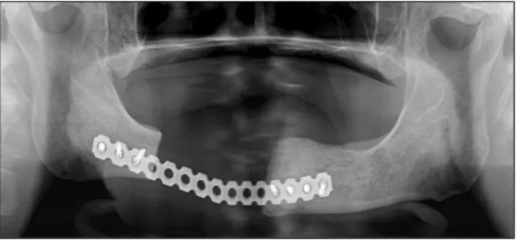
한 나머지 3명의 환자에서는 내과와의 협진하에 3개월 간 BP 대체약물을 사용하면서 전신마취하 분절절제술 및 재 건술을 시행한 결과 완전히 치유되었다.(Figs. 9, 10)
수술 전에 측정한 CTX가 100 pg/mL 미만인 2명의 환자 에게 외과적 치료를 취소하고 3개월간 BP를 중단하면서 항생제와 chlorhexidine 용액을 사용하여 보존적인 치료를 시행하였다. 두 명의 환자 모두 괴사골의 노출은 계속 유지 되었고, 이후 다시 CTX를 측정하여 각각 167 pg/mL, 203 pg/mL의 수치를 얻어 전신마취하 부골적출술을 시행한 결 과 완전히 치유되었다. 또한, 수술 전에 측정한 CTX가 100- 150 pg/mL인 4명의 환자들에게 외과적 수술을 시행하였으 나 2명의 환자에서 인접 부위에 새로운 괴사골의 노출이 나타났고 1명의 환자에서 괴사골의 노출은 계속되었다.
Fig. 5.The bony trabecula deformation of the alveolar bone in the left anterior maxilla and the large sequestrum with poor cor- ticomedullary differntiation are shown on CT scan image.(CT: computed tomography)
Fig. 6.Periosteal reaction is shown with bone sequestration.
Fig. 7. Many plasma cells, neutrophils and capillaries in granulation tissue are seen.(H&E staining, original magnifi- cation ×200).
Fig. 8. Empty osteocytic lacuna.(H&E staining, original magnification ×100)
Ⅳ.
고 찰
BP는 clacium carbonate precipitation의 억제효과를 이용해 파이프의 부식을 막기 위해 사용되던 공업용 재료로, 많은 연구를 통해 피로인산염(pyrophosphate)의 화학구조 변화 에 성공하면서 광화와 골흡수에 관여하는 유사체인 BP를 합성하게 되었다. Pyrophosphate는 쉽게 가수분해가 되는 반면에 BP는 P-O-P 구조 중 산소가 탄소로 치환된 P-C-P 구조로 인해 가수분해가 되지 않아 상대적으로 긴 반감기 를 갖는다.(Fig. 11) 일반적으로 탄소원소에 결합하는 2개 의 사슬 중 R1는 수산화(-OH)기에 의해 골 내의 calcium과 결합하며, R2를 다양하게 변화시킴으로써 골흡수 억제효 과를 증대시킨다.
BP의 R2에 질소가 결합하느냐 하지 않는냐에 따라 크게 nitrogen BP와 non-nitrogen BP로 나눌 수 있으며 구조에 따 라 각각 기능이 다르다15. Nitrogen BP는 non-nitrogen BP에 비해 강한 효과를 지니는데16, 두 형태 모두 골의 광화구조 에 유입된 뒤 파골세포에 의한 흡수 시 유리되어 파골세포 내부로 들어가 작용한다. 차이점은 nitrogen BP는 파골세포 에 의해 흡수된 후 콜레스테롤 합성단계 중 mevalonate 기 전을 교란시키고17,18, 세포 내 소포체 운송이 방해받게 되어 결국 골흡수에 필수적인 ruffled border를 형성하지 못하게 되는19반면에 non-nitrogen BP는 파골세포에 의해 대사되 어 세포 독성을 가진 adenosine triphosphate (ATP) 형태가
되며20, 계속해서 이 물질이 축척되면서 파골세포는 결국 세포사멸을 하게 된다. Nitrogen BP의 종류에는 pamidronate, zoledronate, residronate, alendronate 등이 있으며 non-nitrogen BP의 종류에는 tiludronate, clodronate, etidronate 등이 있다.
현재 BRONJ의 발생기전에 대해 여러 가설이 있으나 아 직 명확하게 규명되어 있지는 않다. 첫 번째 가설은 BP가 파골세포의 기능을 저하해 뼈의 재형성을 억제하고 이로 인해 발치나 감염 등의 국소적 손상이 가해졌을 때 치유되 지 않고 골괴사가 일어난다는 가설이다21, 두 번째 가설은 BP의 혈관형성 억제작용(antiangiogenesis)에 의하여 하악골 에 혈액공급이 저하되어 골괴사가 발생한다는 가설이며22, 세 번째 가설은 BP의 상피세포 독성에 의해 구강 상피세포 아래의 골까지 퍼져 골괴사가 발생한다는 가설이다23. 그 중 BP에 의해 파골세포의 기능억제가 과도하게 진행되어 골교체율을 저하시킴으로써 노화된 골의 흡수와 새로운 골의 형성을 저해하며 국소적 손상이 있는 경우, 치유가 되 지 않고 골괴사가 나타난다는 첫 번째 가설이 가장 받아들 여지고 있다24,25.
최근에 BRONJ가 널리 알려짐에 따라 이에 대한 방사선 학적 소견이 많이 발표되었는데, Phal 등26은 15명의
BRONJ 환자들의 파노라마소견을 분석한 결과 치아발치
후 발치와의 형태가 사라지지 않고 치조백선이 잔존하는 특징을 15예 모두에서 보여 BRONJ의 가장 흔한 방사선학 적 소견이라 하였고 이외에도 BRONJ 초기에 나타나는 치 조백선의 경화 10예, 골용해 7예 등을 보고하였다. 또한, Bisdas 등27은 32명의 BRONJ 환자들의 컴퓨터단층촬영을 분석한 결과 골경화 32예, 골수 용적감소 28예, 골용해 28 예, 골막신생골 20예, 골절 16예, 하악관 침범 14예 등의 소 견을 관찰하였으며, 특히 국소적 골경화, 골소주의 변형, 피질수질접합 부위의 불분명한 경계를 가지는 부골화 양 상, 하악골에서 나타나는 명확한 골막반응 등이 BRONJ의 특징적인 컴퓨터단층촬영소견이라 하였다.
방선균증(actinomycosis)은 구강과 호흡기 및 소화기계에 서 발견되는 혐기성의 그람양성 세균인 Actinomyces israelii에 의한 감염으로, Actinomyces israelii 자체는 점막
Fig. 9. The temporal muscle flap reconstruction was per- formed after segmental resection of the right posterior maxilla. A complete wound healing was achieved.
Fig. 10. The R-plate reconstruction was performed after segmental resection of the right posterior mandible. A complete wound healing was achieved.
Fig. 11.Chemical fomula of basic bisphosphonates structure.
을 투과할 수 없어 발치 등의 외과적 시술, 얇은 점막이나 골융기상의 가철성 보철물이 야기하는 연조직 외상 등의 점막손상에 의해 증상이 나타나게 된다. Actinomyces israelii에 의한 경부감염은 흔치 않은 것으로 알려져 있으 나, BRONJ와 관련된 방선균증의 빈도는 매우 높은 것으로 알려져 있다. Hansen 등28은 악골의 방선균증환자 45명 중 58.7%에서 BRONJ가 나타났고, BRONJ 환자의 93%에서 Actinomyces israelii가 발견되었다고 보고하였으며, Bisdas 등27은 32명의 BRONJ 환자 모두에서 방선균증이 동반되어 있었다고 보고하였다. 본 연구에서도 괴사된 골로부터 채 취된 연조직과 경조직 생검의 배양결과 BRONJ 24명의 환 자 중 18명의 환자에서 Actinomyces israelii가 나타났으며 이를 통해 BRONJ에서 보이는 만성적인 염증이 방선균증 과 밀접한 관계가 있음을 알 수 있었다.
ASBMR에서는 정주용 BP 투여, BP의 장기간 복용, 스테 로이드 복용, 항암요법, 당뇨, 발치 등의 구강 내 수술, 구 강내 위생불량, 잘 맞지 않는 틀니 등이 BRONJ의 위험인 자라고 발표하였으며12, 이번 연구에서도 BRONJ 환자들의 경구용 BP 복용기간은 평균 43.2개월인데 반해 정주용 BP 투여기간은 평균 21.6개월이었고 괴사된 뼈의 노출은 발치 후(15명), 만성 치주염(4명), 지속적인 부적절한 의치에 의 한 외상(3명) 등의 이유로 나타났다. 또한, 11명의 환자가 스테로이드 계열의 약물을 복용하고 있었고 8명의 환자가 당뇨병을 가지고 있었으며, 7명의 환자가 항암화학요법을 받고 있었다.
AAOMS는 2007년 position paper에서 BRONJ의 단계별 치료방법을 제시했으나 현재까지 뚜렷한 효과적인 치료방 법이 정립되지 않은 상태로 무엇보다 예방이 중요하다. BP 를 3년 미만 투여한 환자로서 다른 위험인자가 없는 경우 에는 발치, 치주수술, 임플란트를 실시할 수 있지만, 계속 BP를 복용한다면 BRONJ의 발생위험에 대해 알리고 정기 적으로 추적 관찰하도록 한다. 3년 미만 투여한 환자로서 스테로이드를 동시에 투여하고 있는 경우에는 수술 전 약 3개월 동안 BP 투약을 중단하도록 하고 3년 이상 투여하고 있는 경우에는 가능한 비수술적 치료를 하며 수술이 꼭 필 요한 경우에는 수술 전 약 3개월 동안 BP 투약을 중단하도 록 한다.
일단 BRONJ가 발생하게 되면 Marx 등29은 우선 BP를 중 단하고 중단이 어려울 경우에는 내과의와의 협진을 통해 서 대체약물을 사용하며 항생제치료, 구강세척 및 통증치 료 등의 보존적 치료가 효과적이라고 하였다. 그 이유는 BP가 악골 전체에 작용하여 괴사골과 출혈이 일어나는 신 선한 골의 경계를 찾는 것이 불명확하여 일반적인 골제거 술의 적용이 불가능하기 때문이다. 그러나 보존적 치료에 도 불구하고 괴사골의 노출이 계속되거나 인접 부위에 새 로운 괴사골이 나타나는 경우, 노출된 괴사골이 부골 형태 로 명확하게 분리된 경우에는 외과적 치료를 고려해야 한 다. 이번 연구에서도 경구용 또는 정주용 BP 투여를 최소 3
개월 이상 중단하면서 AAOMS에서 제시한 BRONJ의 단계 별 치료방법을 따랐을 때, 괴사골이 연조직으로 피개되는 만족할만한 치유양상을 보였다. 그러나 BP 복용하면서 외 과적 치료만 받은 경우에 3명의 환자에서 인접 부위에 새 로운 괴사골의 노출, 1명의 환자에서 괴사골의 노출은 계 속되었다. 다른 장기로의 암전이로 인해 사망한 환자 1명 을 제외한 나머지 3명의 환자에서는 내과와의 협진하에 3 개월간 BP 대체약물을 사용하면서 전신마취하 분절절제 술 및 재건술을 시행한 결과 완전히 치유되었다.
CTX는 골흡수의 산물이 혈청과 소변으로 배출되는 양을 측정하는 골흡수 표지자 중 하나이며 type I collagen의 cross-linking peptide를 측정하는 검사로서 골개조를 가장 정확히 반영한다고 알려져 있다30. CTX 수치는 BP를 복용 하지 않는 일반인의 경우 400-500 pg/mL이며, CTX 수치가 낮을수록 정상적인 골개조, 즉 신생골의 형성 정도가 낮음 을 의미한다. Marx 등29은 BRONJ 환자들의 침습적인 외과 적 치료 전에 반드시 CTX를 측정해야 된다고 주장하였는 데, CTX 수치가 150 pg/mL 이상이면 외과적 수술에서 BRONJ의 위험이 최소화되고, 100-150 pg/mL 사이면 중등 도 위험, 100 pg/mL 미만이면 고도 위험군이며 수술을 연 기할 것을 권고하고 있다. 본 연구에서는 수술 전에 측정한 CTX가 100 pg/mL 미만인 2명의 환자에게 외과적 치료를 취소하고 3개월간 보존적 치료를 시행하였으나 괴사골의 노출은 계속 유지되었다. 이후 다시 CTX를 측정하여 각각 167 pg/mL, 203 pg/mL의 수치를 얻었고 전신마취하 부골적 출술을 시행한 결과 완전히 치유되었다. 또한, 수술 전에 측정한 CTX가 100-150 pg/mL인 4명의 환자들에게 외과적 수술을 시행하였으나 2명의 환자에서 인접 부위에 새로운 괴사골의 노출이 나타났고 1명의 환자에서 괴사골의 노출 은 계속되었다. 그러므로 BRONJ 환자들의 외과적 치료 전 에는 반드시 CTX를 측정하고 CTX 수치가 가능한 150
pg/mL 이상일 때 외과적 수술을 시행하는 것이 바람직하
다고 사료된다.
Ⅴ. 결 론
골다공증이나 대사성 질환 치료에 효과적으로 사용되는 BP는 전세계적으로 매년 2억 회 이상 처방되는 보편화된
약제로서31, 최근에 BRONJ가 부작용으로 대두되고 있으나
임상적, 방사선학적 진단기준이 명확하지 않으며 치료에 있어서 아직 뚜렷한 효과적인 방법이 없는 상태이다. 이번 연구에서는 단국대학교 치과대학 부속치과병원에 2005년 1월부터 2009년 12월까지 내원한 환자한 중 BRONJ로 진 단받은 환자 24명을 대상으로 치료한 증례들을 분석하여 다음과 같은 결론을 얻을 수 있었다.
1. BRONJ 환자들은 파노라마에서 치아발치 후 발치와의 형태가 사라지지 않고 치조백선이 잔존하는 특징을 가 졌으며, 컴퓨터단층촬영에서는 국소적 골경화, 골소주
의 변형, 부골화 양상, 하악골에서는 명확한 골막반응 을 가졌다.
2. BRONJ에서 나타나는 만성적인 염증은 방선균증과 밀 접한 관련이 있음을 알 수 있었다.
3. BRONJ는 치료가 쉽지 않고 치료결과에 대한 예측이 어려우므로, 특히 위험인자를 가진 환자에 있어서는 예방이 최선임을 알았다.
4. BRONJ의 치료는 최소한 3개월간 BP를 중단하거나 중 단이 어려울 경우에는 내과의와의 협진을 통해서 대체 약물을 사용하며 가능한 보존적 치료를 시행하였다.
5. BRONJ 환자들의 외과적 치료 전에는 반드시 CTX를
측정하고 CTX 수치가 가능한 150 pg/mL 이상일 때 외 과적 수술을 시행하였다.
References
1.Ruggiero SL, Mehrotra B, Rosenberg TJ, Engroff SL.
Osteonecrosis of the jaws associated with the use of bisphospho- nates: a review of 63 cases. J Oral Maxillofac Surg 2004;62:527- 34.
2.Badros A, Weikel D, Salama A, Goloubeva O, Schneider A, Rapoport A, et al. Osteonecrosis of the jaw in multiple myeloma patients: clinical features and risk factors. J Clin Oncol 2006;24:
945-52.
3.Epstein S. Update of current therapeutic options for the treatment of postmenopausal osteoporosis. Clin Ther 2006;28:151-73.
4.Saad F, Lipton A. Clinical benefits and considerations of bis- phosphonate treatment in metastatic bone disease. Semin Oncol 2007;34(6 Suppl 4):S17-23.
5.Owens G, Jackson R, Lewiecki EM. An integrated approach: bis- phosphonate management for the treatment of osteoporosis. Am J Manag Care 2007;13 Suppl 11:S290-308; quiz S309-12.
6.Migliorati CA, Casiglia J, Epstein J, Jacobsen PL, Siegel MA, Woo SB. Managing the care of patients with bisphosphonate-as- sociated osteonecrosis: an American Academy of Oral Medicine position paper. J Am Dent Assoc 2005;136:1658-68.
7.Woo SB, Hellstein JW, Kalmar JR. Narrative [corrected] review:
bisphosphonates and osteonecrosis of the jaws. Ann Intern Med 2006;144:753-61.
8.Brooks JK, Gilson AJ, Sindler AJ, Ashman SG, Schwartz KG, Nikitakis NG. Osteonecrosis of the jaws associated with use of risedronate: report of 2 new cases. Oral Surg Oral Med Oral Pathol Oral Radiol Endod 2007;103:780-6.
9.Marx RE. Pamidronate (Aredia) and zoledronate (Zometa) in- duced avascular necrosis of the jaws: a growing epidemic. J Oral Maxillofac Surg 2003;61:1115-7.
10.Migliorati CA. Bisphosphanates and oral cavity avascular bone necrosis. J Clin Oncol 2003;21:4253-4.
11.Diel IJ, Bergner R, Gro¨tz KA. Adverse effects of bisphospho- nates: current issues. J Support Oncol 2007;5:475-82.
12.Khosla S, Burr D, Cauley J, Dempster DW, Ebeling PR, Felsenberg D, et al. American Society for Bone and Mineral Research. Bisphosphonate-associated osteonecrosis of the jaw:
report of a task force of the American Society for Bone and Mineral Research. J Bone Miner Res 2007;22:1479-91.
13.Advisory Task Force on Bisphosphonate-Related Ostenonecrosis of the Jaws, American Association of Oral and Maxillofacial Surgeons. American Association of Oral and Maxillofacial Surgeons position paper on bisphosphonate-related osteonecrosis of the jaws. J Oral Maxillofac Surg 2007;65:369-76.
14.Bedogni A, Blandamura S, Lokmic Z, Palumbo C, Ragazzo M, Ferrari F, et al. Bisphosphonate-associated jawbone osteonecro- sis: a correlation between imaging techniques and histopatholo- gy. Oral Surg Oral Med Oral Pathol Oral Radiol Endod 2008;
105:358-64.
15. Green JR. Bisphosphonates: preclinical review. Oncologist 2004;9 Suppl 4:3-13.
16.Mavrokokki T, Cheng A, Stein B, Goss A. Nature and frequency of bisphosphonate-associated osteonecrosis of the jaws in Australia. J Oral Maxillofac Surg 2007;65:415-23.
17. McClung MR. Bisphosphonates. Endocrinol Metab Clin North Am 2003;32:253-71.
18.Lehenkari PP, Kellinsalmi M, Na¨pa¨nkangas JP, Ylitalo KV, Mo¨nkko¨nen J, Rogers MJ, et al. Further insight into mechanism of action of clodronate: inhibition of mitochondrial ADP/ATP translocase by a nonhydrolyzable, adenine-containing metabolite.
Mol Pharmacol 2002;61:1255-62.
19.Alakangas A, Selander K, Mulari M, Halleen J, Lehenkari P, Mo¨nkko¨nen J, et al. Alendronate disturbs vesicular trafficking in osteoclasts. Calcif Tissue Int 2002;70:40-7.
20.Black DM, Delmas PD, Eastell R, Reid IR, Boonen S, Cauley JA, et al.; HORIZON Pivotal Fracture Trial. Once-yearly zole- dronic acid for treatment of postmenopausal osteoporosis. N Engl J Med 2007;356:1809-22.
21.Marx RE, Sawatari Y, Fortin M, Broumand V. Bisphosphonate- induced exposed bone (osteonecrosis/osteopetrosis) of the jaws:
risk factors, recognition, prevention, and treatment. J Oral Maxillofac Surg. 2005;63:1567-75.
22.Reid IR, Bolland MJ, Grey AB. Is bisphosphonate-associated os- teonecrosis of the jaw caused by soft tissue toxicity? Bone 2007;
41:318-20.
23.Sawatari Y, Marx RE. Bisphosphonates and bisphosphonate in- duced osteonecrosis. Oral Maxillofac Surg Clin North Am 2007;
19:487-98, v-vi.
24.Lam DK, Sa′ndor GK, Holmes HI, Evans AW, Clokie CM. A re- view of bisphosphonate-associated osteonecrosis of the jaws and its management. J Can Dent Assoc 2007;73:417-22.
25.Yarom N, Yahalom R, Shoshani Y, Hamed W, Regev E, Elad S.
Osteonecrosis of the jaw induced by orally administered bisphos- phonates: incidence, clinical features, predisposing factors and treatment outcome. Osteoporos Int 2007;18:1363-70.
26.Phal PM, Myall RW, Assael LA, Weissman JL. Imaging findings of bisphosphonate-associated osteonecrosis of the jaws. AJNR Am J Neuroradiol 2007;28:1139-45.
27.Bisdas S, Chambron Pinho N, Smolarz A, Sader R, Vogl TJ, Mack MG. Biphosphonate-induced osteonecrosis of the jaws: CT and MRI spectrum of findings in 32 patients. Clin Radiol 2008;
63:71-7.
28.Hansen T, Kunkel M, Springer E, Walter C, Weber A, Siegel E, et al. Actinomycosis of the jaws-histopathological study of 45 patients shows significant involvement in bisphosphonate-associ- ated osteonecrosis and infected osteoradionecrosis. Virchows Arch 2007;451:1009-17.
29.Marx RE, Cillo JE Jr, Ulloa JJ. Oral bisphosphonate-induced os- teonecrosis: risk factors, prediction of risk using serum CTX test- ing, prevention, and treatment. J Oral Maxillofac Surg 2007;65:
2397-410.
30.Rosen HN, Moses AC, Garber J, Iloputaife ID, Ross DS, Lee SL, et al. Serum CTX: a new marker of bone resorption that shows treatment effect more often than other markers because of low coefficient of variability and large changes with bisphosphonate therapy. Calcif Tissue Int 2000;66:100-3.
31.Bize R, Lamy O, Peytremann-Bridevaux I. Osteoporotic fracture in menopausal women: alendronate reduces the risk. Rev Med Suisse 2008;4:2703.