DOI:10.4078/jkra.2010.17.3.238
<접수일:2010년 5월 7일, 수정일:2010년 5월 31일, 심사통과일:2010년 6월 2일>
※통신저자:김 성 일
부산시 서구 아미동1가
부산대학교 의학전문대학원 내과학교실
Tel:051) 240-7580, Fax:051) 241-7580, E-mail:[email protected]
IL-15에 의한 류마티스관절염 환자 활막 섬유모세포에서의 SDF-1 유도
부산대학교 의학전문대학원 내과학교실1, 가톨릭대학교 류마티스연구센터2
박영은
1ㆍ김성일
1ㆍ박성후
1ㆍ백승훈
1ㆍ오혜좌
2ㆍ허양미
2ㆍ조미라
2= Abstract =
IL-15 Induced an Increased SDF-1 Expression
in the Synovial Fibroblasts of Patients with Rheumatoid Arthritis
Young-Eun Park1, Sung-Il Kim1, Seong-Hu Park1, Seung-Hoon Baek1, Hye-Jwa Oh2, Yang-Mi Heo2, Mi-La Cho2
Department of Internal Medicine, School of Medicine, Pusan National University, Busan
1, The Rheumatism Resarch Center, Catholic Research Institute of Medical Science,
The Catholic University of Korea, Seoul
2, Korea
Objective: Interleukin-15 (IL-15) recruits and activates synovial T cells, and IL-15 plays an important role in amplifying and perpetuating inflammation in the pathogenesis of rheumatoid arthritis (RA). Stromal cell-derived factor-1 (SDF-1) is a potent chemoattractant for memory T cells in the inflamed RA synovium. This study investigated the effect of IL-15 on SDF-1 pro- duction in RA fibroblast-like synoviocytes (FLS).
Methods: The expressions of IL-15 and SDF-1 were determined from the synovium of patients with RA and osteoarthritis (OA) by performing immunohistochemistry. The expressions of SDF-1 was measured from the RA FLS that were cultured with IL-15 and IL-17 by real-time RT-PCR and ELISA. The SDF-1 expression was also measured, via ELISA, from the RA FLS stimulated by IL-15 together with the inhibitors of such intracellular signal molecules as phosphatidylinositol 3-kinase (PI 3-kinase, LY294002), STAT3 (AG490), MAP Kinase (PD98059), NF-κB (parthenolide) and activator protein 1 (AP-1, curcumin).
Results: IL-15 and SDF-1 were mainly expressed in the RA synovium compared to that of the OA synovium. IL-15 increased the SDF-1 expressions and it, and had an additive effect with
IL-17 on the SDF-1 expressions in the cultured RA FLS. The IL-15 induced increase of the SDF-1 expression in the cultured RA FLS was blocked by the inhibitors of PI 3-kinase, NF-κB and AP-1.
Conclusion: The SDF-1 expression was increased in the RA synovium and it was up-regulated by IL-15 in the RA FLS through the PI 3-kinase, NF-κB, and AP-1 pathways. These results imply that the IL-15 induced increase of the SDF-1 expressions may be involved in the immunopathogenesis of RA.
Key Words: IL-15, Stromal cell-derived factor-1, IL-17, Fibroblast-like synoviocytes, Rheumatoid arthritis
서 론
류마티스관절염(rheumatoid arthritis, RA)은 관절 활 막의 지속적인 염증으로 연골 및 골을 포함한 관절 조직의 파괴가 발생하는 전신성 만성 염증성 자가면 역 질환으로, 명확한 병인은 밝혀져 있지 않으나, 선 천면역 세포(항원인지 세포), 후천면역세포(T 및 B 림프구) 및 활막섬유모세포(fibroblast-like synoviocyte, FLS) 등의 활성과 이들 세포에서 유리되는 사이토카 인 및 케모카인들을 포함한 염증 매개물질들이 병인 에 중요한 역할을 한다 (1).
Stromal cell-derived factor 1 (SDF-1; CXCL12)은 T 및 B 림프구, 단핵세포/대식세포들을 염증성 활막 내로 이동하도록 촉진하는 케모카인이다. SDF-1은 RA 환자의 골수 기질세포, FLS, 대식세포 및 혈관 내막세포에서 생성되며, RA 환자의 혈액, 활액 및 활막에서 발현이 증가된다 (2-5). SDF-1은 CD4+ T 세포의 이동 및 축적과 단핵세포의 활막 내 이동을 촉진하고, 혈관생성 유도 및 연골세포와 파골세포를 활성화시켜 관절파괴를 악화시킨다 (2,6). SDF-1 및 SDF-1 수용체인 CXCR4의 억제제들은 collagen-induced arthritis (CIA)에서 관절염 발생 빈도 및 중증도를 감 소시킨다 (7,8).
IL-15는 RA의 병인에 중요한 염증성 사이토카인 으로, RA 환자의 활액 및 활막에서 발현이 증가되 고, 주로 대식세포, FLS 및 혈관 내막세포에서 발현 된다 (9-11). IL-15는 대식세포와 memory T 세포의 상호 접촉에 의해 T 림프구를 염증반응 부위로 이 동시키고 증식시킨다 (9). IL-15에 의하여 T 림프구 에서 생성이 증가된 tumor necrosis factor (TNF), in-
terferon (INF)-γ 및 interleukin (IL)-17은 FLS에서 IL- 15, IL-8 및 IL-6의 생성을 자극하여 염증을 악화시 킨다 (12,13). CIA에서 IL-15의 기능 차단 시 관절염 발생 및 TNF, IL-1β, IL-6 및 IL-17 생성이 감소하 고, RA 환자에게 IL-15에 대한 항체 투여는 질병 활 성을 감소시킨다 (14-16). 이러한 결과들은 IL-15가 RA에서 염증반응을 지속시키고 확장시키는데 중요 한 역할을 함을 암시하며, 치료 전략의 표적 사이토 카인으로서의 가능성을 시사한다.
저자들은 RA 환자의 활막에서 IL-15가 SDF-1의 발현에 미치는 영향에 대하여 조사하고자 하였다.
대상 및 방법 1. 대상
미국 류마티스학회(American College of Rheumatol- ogy)의 RA의 진단 기준에 부합되는 환자 중 무릎관 절 치환술을 받는 4명(평균 연령은 51±11세)에서 활 막 조직을 채취하였고, 대조군은 성별, 연령이 일치 하는 골관절염(osteoarthritis, OA) 환자 4명에서 활막 조직을 채취하였다. 본 연구는 임상연구 윤리위원회 로부터 승인 받았으며, 모든 환자로부터 동의서를 획득하였다.
2. 시약
Recombinant human IL-15, IL-17, IL-1β, anti-IL-15 antibody (Ab) 및 anti-SDF-1 Ab는 R&D System (Minnea- polis, MN)으로부터 구입하였다.
3. 면역조직화학염색
파라포름알데하이드(4%)에 고정된 RA 및 OA 활
막을 파라핀에 포매한 후 절편기를 이용하여 7μm 절편을 만들어 슬라이드에 부착하였다. 면역조직화 학염색은 ABC (Vector laborites, Burlingame, CA, USA) kit를 사용하여 염색하였으며, 슬라이드에 부착된 절 편을 자일렌과 에탄올로 탈파라핀과 함수를 시킨 후 3% H2O2로 내인성과산화효소를 차단시키고, 인산화 완충액(phosphate buffered saline, PBS)으로 수세한다.
비 특이적인 반응을 차단할 목적으로 anti-mouse se- rum을 30분 반응시킨 후 일차 항체인 recombinant human anti-IL-15 Ab와 anti-SDF-1 Ab (R&D systems, 1:100)를 4oC에서 다음날(16시간)까지 반응시켰다.
일차 항체 반응 후 결합이 안 된 항체를 PBS로 수 세하고 바이오틴이 결합된 이차 항체와 과산화효소 가 결합된 streptavidin반응을 시킨 후 DAB으로 발색 시켰다. Mayer’s 해마톡실린 대조염색 한 후 수세하 고 봉입하여 광학현미경으로 관찰하였다. 음의 대조 조직은(negative control tissue)은 isotype control anti- body (Mouse IgG1:R&D Systems, Minneapolis, MN) 를 사용하였다.
4. 활막 섬유모세포(fibroblast-like synoviocyte, FLS)의 분리 및 자극
FLS는 RA 및 OA 환자의 활막에서 효소를 이용하 여 분리하였으며, 조직은 2∼3 mm 조각으로 잘게 자 른 후 Dulbecco’s modified Eagle’s medium (DMEM) 에 type I collagenase (Worthington, Freehold, NJ)와 함께 37oC, 5% CO2에서 4시간 동안 반응시켰다. 분 리된 세포를 500 g에서 원심분리 하여 10% fetal calf serum (FCS), 2 mM L-glutamine, penicillin (100 units/mL), streptomycin (100 μg/mL)를 가한 DMEM 에 재부유시켜 75 cm2 플라크스에 분주하였다. 하루 가 지난 후 배양기에 부착되어 있는 세포만을 10%
FBS를 가한 DMEM에 넣어 5% CO2와 37oC의 조건 에서 배양하고 배양액은 매 3일마다 교환하였다. 플 라스크 바닥의 90∼95%가 FLS로 충만되면 신선한 배양액으로 1:3 희석하여 계대 배양하였다. 실험에 는 4∼8 계대의 FLS를 사용하였다. 배양된 세포가 FLS임을 확인하기 위해 항 CD14 PE 항체(PharMingen, SanDiego, CA), 항 CD3 FITC 항체 또는 항 Thy-1 (CD90) 항체(PharMingen)를 이용하여 유세포 분석을 시행하였다. 세포를 카운트하여 5×10⁴ cells/well을 1
mL의 serum free DMEM/insulin-transferrin-selenium A (Life Technologies, Gaithersburg, MD) 배양액에 섞어 24-well plate에 분주 하여 준다. 다양한 농도의 IL-15 및 IL-17을 각각 혹은 함께 자극하거나, 신호 전달 억제 물질들(LY294002, AG490, PD98059, partheno- lide, 그리고 curcumin)을 처리 후 IL-15 (10 ng/mL)로 자극하였다.
5. SDF-1의 단백질 발현 측정(ELISA)
FLS 자극 후 48시간째에 세포 배양 상층액을 모아 SDF-1 정량을 위한 ELISA를 시행하였다. Sandwich ELISA용 96 well plate (Nunc, Roskilde, Denmark)에 human anti-SDF-1 Ab (4 ug/mL; R&D Systems)에 넣 고 4oC, 밤새 반응시킨 후, 차단 용액(1% bovine se- rum albumin (BSA)/0.05% Tween200)이 함유된 pho- sphate buffered saline (PBS)를 첨가하여 실온에서 2 시간 반응시켰다. Biotinylated SDF-1 polyclonal anti- body (400 ng/mL; R&D Systems), streptavidin-alkaline phosphate (1:2,000, Sigma)와 avidin-peroxidase (50 μL, 1:2,000), 501 tetramethylbenzidine substrate solution (Kirkegaard & Perry, Guildford, UK)을 차례대로 각 well에 첨가하여 반응시켰다.
6. SDF-1 mRNA 측정(real-time RT-PCR)
FLS를 다양한 농도의 IL-15로 자극 후 16시간째에 세포의 mRNA를 RNAzol B (Biotecx, Houston, TX)를 이용하여 추출하였다. 총 2 μg의 추출된 mRNA를 Superscript reverse transcription system (Takara, Shiga, Japan), 42oC에서 역 전사 시켰다. SYBR Green I (Roche Diagnostic, Mannheim, Germany)를 이용하여 real-time PCR을 시행하였다. Real-time PCR 전용 capillary tube 를 사용하여 LightCycler FastStart DNA mastermix (SYBR Green I) 2 μL, 각각의 primer 0.5 μM, MgCl2 4 mM, 그리고 template DNA 2 μl을 포함하여 총 20 μL가 되도록 하였다. SDF-1과 GAPDH 시발체의 염 기 서열은 다음과 같았다; SDF-1 (5’-ATG-AAC-GCC- AAG-GTC-GTG-GTC-3’ (sense), 5’-TGG-CTG-TTG-TGC- TTA-CTT-GTT-T-3’ (antisenss)), GAPDH (5’-CGA-TGC- TGG-GCG-TGAGTA-C-3’ (sense), 5’-CGT-TCA-GCT-CAG- GGA-TGACC-3’ (antisense)). SDF-1과 GAPDH에서, 변 성을 위해 94oC에서 30초, annealing은 56oC에서 1분,
Fig. 1. Immunohistochemical staining for stromal-cell derived factor-1 (SDF-1) and interleukin-15 (IL-15) on the synovium of patients with rheumatoid arthritis (RA) and osteoarthritis (OA). IL-15 and SDF-1 were significantly more expressed in the RA synovium compared to that of the OA synovium (original magnification, ×400).
신장을 위해 72oC에서 1분의 과정을 30회 반복하였 다. 형광커브는 LightCycler software v. 3.0에서 분석 하였다.
7. MTT assay
96 well plate에 1×10³/mL의 농도로 세포를 배양하 여, ITSA가 포함된 serum free DMEM 배양액으로 star- vation한다. LY294002, AG490, PD98059, parthenolide 혹은 curcumin을 처리 후 IL-15는 10 ng/mL의 농도 로 처리하였으며, 48시간 동안 37oC, 5% 배양기에서 배양 하였다. 여기에 MTT 용액(Thiazolyl blue, 2.5 mg/mL, indeiodinized H2O) 10 μL를 혼합하여 4시간 동안 반응시킨 후 5분간 원심 분리하고, 0.04 M HCl 100 μL를 첨가하여 5분 동안 실온에 방치하였다가 ELISA 판독기로 595 nm로 측정하였다.
8. 통계적 유의성의 검증
결과는 평균과 표준오차로 표현하였고 통계적 유 의성은 SPSS 통계 프로그램(version 10.0)을 사용하여 Mann-Whitney U test나 Wilcoxon’s signed rank test를 실시하였고 p값이 0.05 이하일 때 통계적으로 유의 하다고 분석하였다.
결 과
1. RA 활막 조직에서 IL-15 및 SDF-1의 발현 증가
RA 환자의 활막 조직에서 IL-15과 SDF- 1의 발현 이 대조군(골관절염 환자 활막 조직)에 비하여 증가 되어 있음을 면역조직화학염색법을 통해서 관찰하였 다. SDF-1과 IL-15은 모두 류마티스관절염 환자 활 막에서 과발현 되어 있었다(그림 1).
2. RA (FLS)에서 IL-15에 의한 SDF-1 발현 증가
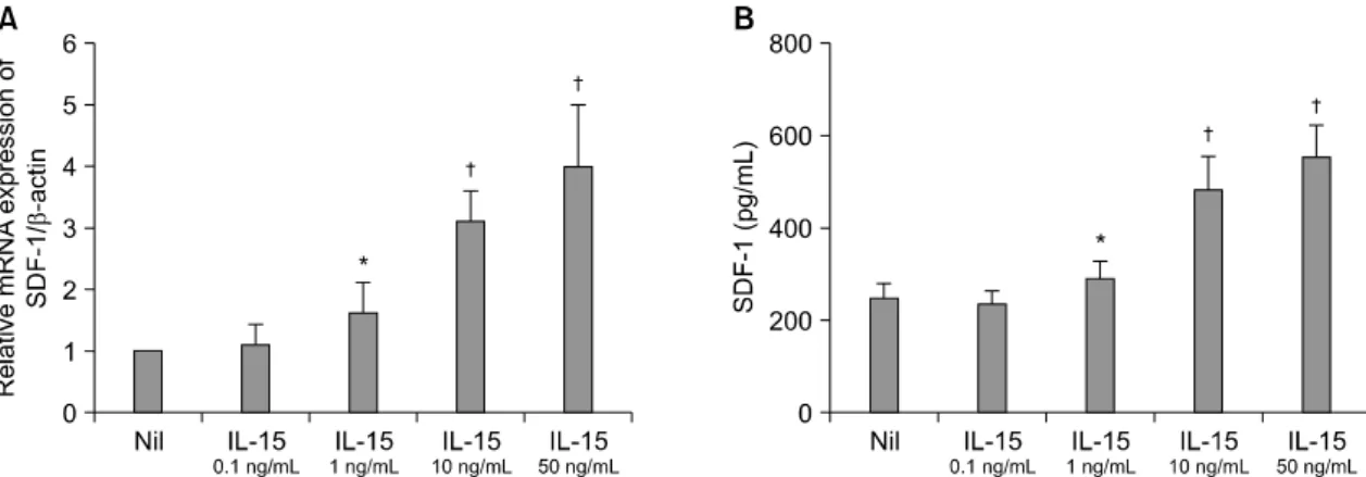
RA 환자 FLS에서 IL-15에 의한 SDF-1 발현이 조 절되는지 알아보고자, FLS 1×10⁵ cell/mL 로 배양된 세포에 IL-15을 0.1, 1, 10, 50 ng/mL의 농도로 처리 하여 12시간의 세포와 48시간 세포배양액에서 각각 SDF-1의 mRNA와 단백질 발현양을 real- time PCR과 ELISA 방법으로 평가하였다. 그 결과 IL-15 농도 의 존적으로 SDF-1의 mRNA (그림 2A)와 단백질 발현 양(그림 2B)이 FLS에서 증가되는 것을 관찰하였다 (*p<0.05 compared to nil, †p<0.01 compared to nil).
IL-15과 IL-17을 함께 자극하였을 때 SDF-1의 단 백질 발현에 부가적인 효과(additive effect)가 관찰되 었다(*p<0.01 compared to nil, †p<0.05 compared to
Fig. 3. The productions of SDF-1 from the RA FLS stimulated with IL-15 and IL-17 were determined by ELISA. IL-15 and IL-17 up-regulated SDF-1 production. IL-17 had an additive effect with IL-15 on the SDF-1 production. *p<0.01 com- pared to nil, †p<0.05 compared to IL-15 1 ng/
mL, ‡p<0.01 compared to IL-15 10 ng/mL.
Fig. 2. The expressions of SDF-1 from the RA synovial fibroblasts stimulated with various concentrations of IL-15 (0, 0.1, 1, 10 and 50 ng/mL) were determined by real-time RT-PCR (A) and ELISA (B). IL-15 increased the SDF-1 expression in a dose-dependent manner. *p<0.05 compared to nil, †p<0.01 compared to nil.
IL-15 1 ng/mL, ‡p<0.01 compared to IL-15 10 ng/
mL) (그림 3).
3. RA FLS에서 IL-15에 의한 SDF-1 발현 증가 관 련 세포 내 신호전달
RA 환자 FLS에서 IL-15에 의한 SDF-1 발현에 관 여하는 신호전달 경로를 조사하기 위하여 세포에 IL-15와 신호전달 물질의 억제제(LY294002; PI3Kinase
inhibitor, AG490; JAK2-STAT3 inhibitor, PD98059;
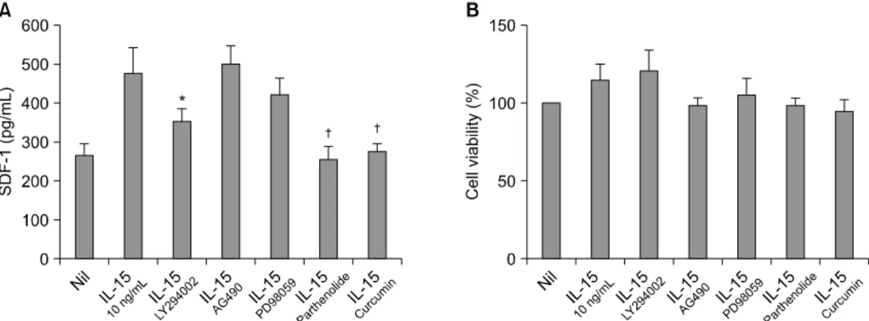
MAPK inhibitor, parthenolide; NF-κB inhibitor, curcu- min; AP-1 inhibitor)를 처리하였다. 이 때 PI3-kinase 억제제(LY294002), NF-κB 억제제(parthenolide) 및 AP-1 억제제(curcumin)는 IL-15에 의한 활막섬유모세 포의 SDF-1 단백질 발현을 유의하게 억제하였으나, STAT3 억제제(AG490) 및 MAPK 억제제(PD98059)는 SDF-1 단백 발현에 영향이 없었다(*p<0.05 com- pared to IL-15 10 ng/mL, †p<0.01 compared to IL-15 10 ng/mL) (그림 4A). 억제제에 의한 세포독성 효과 가 없음은 MTT assay로 확인하였다(그림 4B).
고 찰
저자들은 IL-15와 SDF-1 발현이 OA에 비하여 RA 활막의 lining layer에서 주로 발현되고, IL-15는 RA FLS에서 SDF-1의 mRNA 및 단백 발현을 농도의존 적으로 증가시키며, IL-15와 IL-17은 SDF-1 발현에 부가적인 효과가 있음을 확인하였다. 또한, IL-15에 의한 FLS의 SDF-1 발현 증가는 PI3-kinase, NF-κB 및 AP-1 경로에 의함을 확인하였다.
SDF-1은 CXC 케모카인으로, 단독 수용체인 CXCR4 와 결합하여 세포의 이동 및 정체에 중요한 역할을 한다 (17). SDF-1과 CXCR4의 결합은 인간면역결핍 바이러스(HIV), 암, 제1형 당뇨병 및 RA 등의 다양 한 질병 병인에 관여한다 (18-20). RA에서 SDF-1은 T 림프구의 활막 내 이동 및 혈관 주위의 침윤을
Fig. 4. (A) The IL-15 induced increase of SDF-1 was blocked by the inhibitors of PI3-kinase, NF-κB and AP-1. RA FLS that were, pre-treated with the inhibitors of PI3-kinase (LY294002), STAT3 (AG490), MAPK (PD98059), NF-κB (parthenolide) and AP-1 (curcumin), were cultured with IL-15; the production of SDF-1 was determined by ELISA. Up-regulation of SDF-1 by IL-15 was blocked by the inhibitors of PI3-kinase, NF-κB and AP-1.
(B) MTT assay was done to confirm that the inhibitors did not have cell toxicity. This assay showed that there was no difference in cell viability after all the stimulations and/or inhibitions. *p<0.05 compared to IL-15 10 ng/mL, †p<0.01 compared to IL-15 10 ng/mL.
촉진하고, T 림프구의 활성에 의한 자가사멸(apoptosis) 을 억제하여 활막 내 T 림프구를 축적시킨다 (6). 또 한 활막 내 B 림프구를 축적하여 ectopic germinal center 형성을 촉진하고 FLS 활성, 신생혈관생성 자 극, 연골세포와 파골세포의 matrix metalloprotease-3 (MMP-3)와 MMP-9의 분비를 자극하여 연골 기질 파 괴 및 골 흡수 증가를 야기하여 RA의 병인에서 중 요한 작용을 한다 (2). RA에서 SDF-1의 발현을 조절 하는 기전에 대한 연구는 적은데, 항 CD40 항체, 저 산소증(hypoxia), TGF-1β 및 IL-17은 RA FLS에서 SDF-1 발현을 증가시키고, IL-1β와 TNF-α는 RA FLS에서의 SDF-1 발현에 영향이 없지만 피부와 구강 내 섬유모세포의 SDF-1 발현을 감소시킨다 (6,21,22).
IL-15는 RA에서 염증 지속에 중요한 역할을 한다.
IL-15는 활막 내로 T 림프구 이동 및 활성을 촉진시 키고, 활성화된 T 림프구는 대식세포와 접촉하여 TNF-α의 분비를 유도함으로써 염증 반응을 증폭, 지속시킨다 (9). 또한 IL-15는 TNF와는 독립적으로 auto-reactive CD4+CD28-T cell에서 자연 살상세포 (natural killer cell, NK cell) 수용체인 NKG2Ddml 발 현을 유도하여 RA의 self-perpetuating inflammation에 관여하므로, IL-15가 항 TNF 제제와는 무관하게 memory T 세포의 확장과 유지를 지속시킨다 (23).
RA 활막에서 IL-15 발현이 항 TNF 제제에 의하여 감소하지 않으므로 항 TNF제제에 반응이 불충분한 환자에서 IL-15의 기능 차단은 RA의 새로운 치료법 이 될 수 있음을 시사하였다 (24).
염증성 활막 내의 CD4+T 세포 축적은 RA의 중요 한 병인 중 하나이다. Toshihiro 등은 활막 CD4+
memory T 세포에서 발현된 CXCR4와 활막 세포에 서 발현된 SDF-1의 상호작용이 CD4+T 세포의 이동 에 중심적인 역할을 하며, IL-15가 T 림프구의 CXCR4 의 발현을 증가시킴을 보고하였다 (6). 저자들은 IL- 15가 RA FLS에서 SDF-1의 mRNA 및 단백 발현을 농도 의존적으로 증가시키며, IL-15와 IL-17은 SDF-1 발현에 부가적인 효과가 있음을 확인하였다. 이러한 결과들은 IL-15가 T 림프구의 CXCR4 및 FLS의 SDF-1를 증가시켜 활막 내 T 림프구의 축적에 관여 함을 암시한다. 활막의 활성화 된 T 림프구와 대식 세포 및 단핵세포와의 상호 작용에 의하여 증가된 TNF, IFN-γ 및 IL-17은 FLS를 자극하여 IL- 15를 포함하는 염증성 사이토카인 및 매개 물질의 생성을 증가시킨다. 활막에서 증가된 IL-17과 IL-15는 FLS에 서 SDF-1의 생성을 증가시켜 T 림프구의 활막 내 이동 및 활성화를 더욱 촉진하고, 혈관생성, 연골 파 괴 및 골 흡수를 야기하여 관절 파괴가 지속적으로
진행 됨을 암시한다.
Kim 등은 RA FLS에서 IL-17이 SDF-1의 발현을 증가시키고, 이를 매개하는 세포 내 신호 전달 과정 으로 NF-κB, PI 3-kinase 그리고 AP-1과 연관됨을 보고하였다 (22). IL-15 receptor를 통한 IL-15의 세포 내 신호 전달과정으로는 NF-kB, JAK1, JAK3, STAT3, STAT5 그리고 MAPK 등 여러 신호 전달 물질을 매 개하는 것으로 알려져 있고, 본 연구에서는 RA FLS 에서 IL-15가 NF-κB, PI 3-kinase 및 AP-1과 연관하 여 SDF-1의 발현을 증가시킴을 확인하였다 (25,26).
결 론
본 연구는 SDF-1과 IL-15가 RA 활막에서 발현이 증가하고, IL-15가 RA FLS에서 SDF-1의 발현을 증 가시키며, IL-15에 의한 SDF-1의 증가는 PI-3 kinase, NF-κB 및 AP-1의 신호 전달 과정을 통함을 확인하 였다. 이러한 결과들은 IL-15에 의한 FLS의 SDF-1 증가가 RA의 염증 지속과 관절 파괴에 주요한 역할 을 하며, IL-15 기능 차단을 이용한 치료법 개발에 새로운 근거가 될 것으로 생각된다.
참고문헌
1) Müller-Ladner U, Pap T, Gay RE, Neidhart M, Gay S. Mechanisms of disease: the molecular and cellular basis of joint destruction in rheumatoid arthritis. Nat Clin Pract Rheumatol 2005;1:102-10
2) Mittal GA, Joshi VR, Deshpande A. Stromal cell- derived factor-1 alpha in rheumatoid arthritis. Rheu- matology (Oxford) 2003;42:915-6.
3) Kanbe K, Takagishi K, Chen Q. Stimulation of matrix metalloprotease 3 release from human chondrocytes by the interaction of stromal cell-derived factor 1 and CXC chemokine receptor 4. Arthritis Rheum 2002;
46:130-7.
4) Grassi F, Cristino S, Toneguzzi S, Piacentini A, Facchini A, Lisignoli G. CXCL12 chemokine up- regulates bone resorption and MMP-9 release by human osteoclasts: CXCL12 levels are increased in synovial and bone tissue of rheumatoid arthritis pa- tients. J Cell Physiol 2004;199:244-51.
5) Pablos JL, Santiago B, Galindo M, Torres C, Brehmer MT, Blanco FJ, et al. Synoviocyte-derived CXCL12
is displayed on endothelium and induces angiogenesis in rheumatoid arthritis. J Immunol 2003;170:2147-52.
6) Nanki T, Hayashida K, El-Gabalawy HS, Suson S, Shi K, Girschick HJ, et al. Stromal cell-derived factor-1-CXC chemokine receptor 4 interactions play a central role in CD4+ T cell accumulation in rheu- matoid arthritis synovium. J Immunol 2000;165:
6590-8.
7) De Klerck B, Geboes L, Hatse S, Kelchtermans H, Meyvis Y, Vermeire K, et al. Pro-inflammatory properties of stromal cellderived factor-1 (CXCL12) in collagen-induced arthritis. Arthritis Res Ther 2005;
7:R1208-20.
8) Tamamura H, Fujisawa M, Hiramatsu K, Mizumoto M, Nakashima H, Yamamoto N, et al. Identification of a CXCR4 antagonist, a T140 analog, as an anti-rheumatoid arthritis agent. FEBS Lett 2004;569:
99-104.
9) McInnes IB, al-Mughales J, Field M, Leung BP, Huang FP, Dixon R, et al. The role of interleukin-15 in T-cell migration and activation in rheumatoid arthritis. Nat Med 1996;2:175-82.
10) Thurkow EW, van der Heijden IM, Breedveld FC, Smeets TJ, Daha MR, Kluin PM, et al. Increased expression of IL-15 in the synovium of patients with rheumatoid arthritis compared with patients with Yersinia-induced arthritis and osteoarthritis. J Pathol 1997;181:444-50.
11) Oppenheimer-Marks N, Brezinschek RI, Mohama- dzadeh M, Vita R, Lipsky PE. Interleukin 15 is pro- duced by endothelial cells and increases the transen- dothelial migration of T cells in vitro and in the SCID mouse-human rheumatoid arthritis model in vivo. J Clin Invest 1998;101:1261-72.
12) Miranda-Carús ME, Balsa A, Benito-Miguel M, Pérez de Ayala C, Martín-Mola E. IL-15 and the initiation of cell contact-dependent synovial fibroblast-T lym- phocyte cross-talk in rheumatoid arthritis: effect of methotrexate. J Immunol 2004;173:1463-76.
13) Cho ML, Yoon CH, Hwang SY, Park MK, Min SY, Lee SH, et al. Effector function of type II collagen- stimulated T cells from rheumatoid arthritis patients:
cross-talk between T cells and synovial fibroblasts.
Arthritis Rheum 2004;50:776-84.
14) Ruchatz H, Leung BP, Wei XQ, McInnes IB, Liew FY. Soluble IL-15 receptor alpha-chain administration prevents murine collagen-induced arthritis: a role for IL-15 in development of antigen-induced immuno-
pathology. J Immunol 1998;160:5654-60.
15) Ferrari-Lacraz S, Zanelli E, Neuberg M, Donskoy E, Kim YS, Zheng XX, et al. Targeting IL-15 receptor- bearing cells with an antagonist mutant IL-15/Fc protein prevents disease development and progression in murine collagen-induced arthritis. J Immunol 2004;
173:5818-26.
16) Baslund B, Tvede N, Danneskiold-Samsoe B, Larsson P, Panayi G, Petersen J, et al. Targeting interleukin-15 in patients with rheumatoid arthritis: a proof-of- concept study. Arthritis Rheum 2005;52:2686-92.
17) Murdoch C. CXCR4: chemokine receptor extraordi- naire. Immunol Rev 2000;177:175-84.
18) Mbemba E, Benjouad A, Saffar L, Gattegno L.
Glycans and proteoglycans are involved in the in- teractions of human immunodeficiency virus type 1 envelope glycoprotein and of SDF-1α with mem- brane ligands of CD4+ CXCR4+ cells. Virology 1999;265:354-64.
19) Burger JA, Kipps TJ. CXCR4: a key receptor in the cross talk between tumor cells and their microen- vironment. Blood 2006;107:1761-7.
20) Dubois-Laforgue D, Hendel H, Caillat-Zucman S, Zagury JF, Winkler C, Boitard C, et al. A common stromal cell-derived factor-1 chemokine gene variant is associated with the early onset of type 1 diabetes.
Diabetes 2001;50:1211-3.
21) Hitchon C, Wong K, Ma G, Reed J, Lyttle D, El-
Gabalawy H. Hypoxia-induced production of stromal cell-derived factor 1 (CXCL12) and vascular endo- thelial growth factor by synovial fibroblasts. Arthritis Rheum 2002;46:2587-97.
22) Kim KW, Cho ML, Kim HR, Ju JH, Park MK, Oh HJ, et al. Up-regulation of stromal cell-derived factor 1 (CXCL12) production in rheumatoid synovial fibro- blasts through interactions with T lymphocytes: role of interleukin-17 and CD40L-CD40 interaction. Ar- thritis Rheum 2007;56:1076-86.
23) Groh V, Bruhl A, El-Gabalawy H, Nelson JL, Spies T. Stimulation of T cell autoreactivity by anomalous expression of NKG2D and its MIC ligands in rheu- matoid arthritis. Proc Natl Acad Sci USA 2003;100:
9452-57.
24) Ernestam S, af Klint E, Catrina AI, Sundberg E, Engström M, Klareskog L, et al. Synovial expression of IL-15 in rheumatoid arthritis is not influenced by blockade of tumour necrosis factor. Arthritis Res Ther 2006;8:R18.
25) Giri JG, Kumaki S, Ahdieh M, Friend DJ, Loomis A, Shanebeck K, et al. Identification and cloning of a novel IL-15 binding protein that is structurally related to the alpha chain of the IL-2 receptor. EMBO J 1995;14:3654-63.
26) Waldmann T, Tagaya Y, Bamford R. Interleukin-2, interleukin-15, and their receptors. Int Rev Immunol 1998;16:205-26.