새로운 모형의 Cephalomedullary Nail을 이용한 근위 대퇴골 골절에서의 치료: 전향적 결과 연구
노영호ㆍ노조셉ㆍ남광우
제주대학교병원 정형외과
Treatment of the Proximal Femoral Fracture Using the New Design Cephalomedullary Nail: Prospective Outcomes Study
Young Ho Roh, M.D., Joseph Rho, M.D., Kwang Woo Nam, M.D., Ph.D.
Department of Orthopaedic Surgery, Jeju National University Hospital, Jeju, Korea
Received November 29, 2018 Revised December 20, 2018 Accepted December 31, 2018 Correspondence to:
Kwang Woo Nam, M.D., Ph.D.
Department of Orthopaedic Surgery, Jeju National University Hospital, 15 Aran 13-gil, Jeju 63241, Korea Tel: +82-64-717-1690 Fax: +82-64-717-1131 E-mail: [email protected] Financial support: This study was supported with grants from ZimmerBiomet Korea and by the Academic Research Foundation of Jeju National University Institute of Medical Science.
Conflict of interests: None.
Purpose: The aim of this study is to investigate the clinical performance and safety of Zimmer® natural nail cephalomedullary nail (ZNN CM nail) in the treatment of proximal femur fractures.
Materials and Methods: The following research was conducted as a prospective, non-comparative, single center outcome study. Upon providing written informed consent, enrolled patients’ data were collected and analyzed. Postoperative follow-up visits were scheduled at 6 weeks, 3 months, 6 months, and 1 year. Follow-up evaluation included radiographic assessment, physical examination, and quality of life and adverse events reports.
Results: Thirty-nine patients were available for evaluation at one year postoperative. The patients re- ported the mean EuroQol-5 Dimension score increased after surgery: from 0.4 points at discharge (n=49) to 0.6 points at 1-year post-surgery (n=39). The mean Harris hip score also increased after surgery: from 56.3 points at discharge (n=49) to 72.1 points at 1 year (n=12). Bone union was seen in 64% (n=16) in 6 months and 95% (n=37) in 1 year.
Conclusion: The results of this 1-year follow-up study affirmed the effectiveness and safety of the ZNN CM nail in the treatment of proximal femur fractures.
Key Words: Femur, Femoral fractures, Hip fractures, Fracture fixation, Intramedullary nailing
Copyright © 2019 The Korean Fracture Society. All rights reserved.
This is an Open Access article distributed under the terms of the Creative Commons Attribution Non-Commercial License (http://creativecommons.org/licenses/by-nc/4.0) which permits unrestricted non-commercial use, distribution, and reproduction in any medium, provided the original work is properly cited.
Introduction
A higher incidence of proximal femur fractures has re- sulted from increasing elderly population.1) The 1-year mortality rate of the proximal femur fracture in the elderly population was 2.18 times higher for women and 3.17 times
higher for men when compared to general population.2) Al- though large numbers of different implants are available for fixation, the ideal implant for treatment of proximal femur fractures is still a matter for discussion. The imperative goals of treatment are early mobilization by stable fixation using minimally invasive procedure and lower complication rates.3)
To accomplish this goal, optimal treatment implants with fixed stability and strength should be chosen. Intramedul- lary devices proves to be most appropriate in view of their biomechanical advantage over extramedullary implants.4) However, perioperative and postoperative technical compli- cations are common in somecases, requiring revisional op- eration.5-7) Currently, Zimmer® natural nail system cepha- lomedullary nail (ZNN CM nail; Zimmer® Inc., Warsaw, IN, USA) has become one of the emerging alternatives. This new implant design was intended to improve the treatment of proximal femur fractures, especially in elderly patients.
Short-term results of the ZNN CM nail regarding compli- cations, outcomes, and advantages are herein described.
Materials and Methods
1. Target population and study design
This study enrolled patients with age of 50 or above di- agnosed with either stable or unstable, intertrochanteric or subtrochanteric femur fractures. Patients with poor pre- injury walking ability, sustaining pathologic fractures or multiple fractures, and candidates for revision surgery were ineligible for participation. This study was approved by our Institutional Review Board of Jeju National University Hos- pital (2014-02-003).
Following study was conducted as prospective, non- comparative, post-market, clinical follow-up study of ZNN CM nail. Patients were enrolled consecutively into the study postoperatively. Written consent was obtained after informing each patient about the purpose of the study. Upon signing the informed consent form, relevant preoperative and operative data, radiographic assessment and discharge data were collected and analyzed. Each patient will return to the clinic for follow-up at 6 weeks, 3 months, 6 months, and 12 months postoperatively. Follow-up evaluation includes ra- diographic assessment, physical examination, quality-of-life questionnaire completion and reporting of adverse events.
Demographic characteristics such as gender, age, body mass index, and bone marrow density (BMD) collected from the baseline information of patients were analyzed.
The lowest value of bone mineral density was assigned, excluding the Ward’s portion measured in the hip joint.
Trauma mechanism and fracture types using the AO clas- sification were also analyzed.
2. Surgical techniques
Surgery was performed with the patient in supine position on a radiolucent traction table under fluoroscopic control.
All procedures were performed under aseptic conditions in accordance with the established procedure specified in the appropriate surgical technique manual. A routine method of closed reduction was performed to achieve fracture align- ment with optional assistance from percutaneous bone hooks or long forceps. The entry point of the nail is located at the tip of the greater trochanter, in antero-posterior (AP) view. In the lateral (LAT) view, the entry point should be at the midpoint of the greater trochanter. The specific lag screw was introduced into the center of the femoral head by drilling in both AP and LAT view. A set screw should also be used to prevent lag screw rotation. All procedures were performed by a single experienced surgeon.
3. Clinical outcomes measurement
One of the objectives of this study was to evaluate the clinical outcomes of the ZNN CM nail in the treatment of proximal femur fracture. Patient quality of life was deter- mined by evaluation of the EuroQol-5 Dimension (EQ-5D) self-completed questionnaire. Patient clinical outcome was determined using the Harris hip score (HHS). HHS was categorized as excellent (90-100), good (80-89), fair (70- 79), or poor (<70). Patients completed the two question- naires at the time of discharge and at 6 weeks, 3 months, 6 months, and 1 year postoperatively. The surveys were administered by a trained research nurse blinded to clinical information and not involved in the study. Operation time, length of stay, weight bearing without pain, resumption of pre-injury activity level, and patient work status were also measured.
4. Radiographic outcomes measurement
Implant-specific complications, implant position, and fracture union were evaluated for all participating patients through plain radiographs at immediate postoperative, 6 weeks, 3 months, 6 months, and 1 year postoperatively.
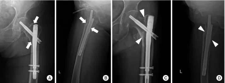
Tip-apex distance (TAD) was defined as the distance be- tween the tip of the screw and the apex of the femoral head on both AP and LAT views with consideration of the radiographic magnification.8) Radiologic fracture union is defined as bridging of the fracture site by callus or bone at a minimum of three cortices. Cortical healing is assessed in four anatomic proximal femur regions (anterior, posterior, medial and LAT) using AP and LAT views of plain hip joint radiography (Fig. 1).9,10)
5. Statistical analysis
Statistical analyses were performed using IBM SPSS statistical software version 20 (IBM Corp., Armonk, NY, USA). Clinical scores, such as EQ-5D, HHS were com- pared at the time of discharge and 1 year postoperatively using a paired samples t-test. A p-value <0.05 was consid- ered statistically significant.
Results
1. Patient baseline characteristics information
The study was originally planned to enroll 50 patients at the Jeju National University Hospital. However, due to the slow recruitment rate, the study was conducted with 49 patients. Following the manufacturer’ surgical technique, all enrolled patients received the CM Asia nail (short) or CM long nail for treatment of proximal femur fracture.
The numbers of patients available for the postoperative follow-ups were 41, 30, 25, and 39 at 6 weeks, 3 months, 6 months, and 1 year, respectively. Five patients withdrew their consent and other 5 patients were not available for the final follow-up (Fig. 2). The mean follow-up period for patients involved in the study was 16.04±7.51 months.
Preoperative BMD was measured in all patients. Only 2.1% of the patients (n=1) had normal value of T-score –1.0 to +1.0, while 85.7% of the patients (n=42) showed osteoporosis with T-score of –2.5 or less and 12.2% (n=6) showed osteopenia with T-score of –1.0 to 2.5. There were 6.1% AO 31-A1 simple petrochanteric fractures (n=3), 77.6% AO 31-A2 multifragmentary petrochanteric frac- tures (n=38), 12.2% AO 31-A3 intertrochanteric fractures
A B C D
Fig. 1. (A) Hip antero-posterior (AP) view of left intertrochanteric fracture immediately postoperation. Medial and lateral (LAT) cortex fracture gap at the fracture site is depicted by white arrows. (B) Hip left LAT view of intertrochanteric fracture immediately postoperation. Anterior and poste- rior cortex fracture gap at the fracture site is depicted by white arrows. (C) Hip AP view of left intertrochanteric fracture 6 months postoperation.
Medial cortex shows no fracture gap (white triangles) and callus formation is observed. (D) Hip left LAT view of femur intertrochanteric fracture 6 months postoperation. Anterior and posterior cortex shows no fracture gap (white triangles) and callus formation is observed.
(n=6), and 4.1% AO 32-A3.1 subtrochanteric fractures (n=2). ZNN CM Asia nail (short) was used in 95.9%
fractures (n=47) while the CM long nail was used in only 4.1% cases (n=2). The 2 cases in which the CM long nail was used were subtrochanteric fractures with fracture lines that were distal to the lesser trochanter. A summary of the demographic, injury, operative and device information are outlined in Table 1 and 2, respectively.
2. Clinical outcomes
The mean EQ-5D score increased after discharge: from 0.4±0.3 points at discharge (n=49) to 0.6±0.3 points at 1 year follow-up (n=39) (Fig. 3). The difference in EQ-5D
Fig. 2. Patient enrollment and follow-up flow chart. This study involved intertrochanteric or subtrochanteric fractures in 39 patients requiring cephalomedullary nailing. FUP: follow-up percentage.
Table 1. Baseline Characteristics Included in This Study
Characteristic Value
Gender
Female 38 (77.6)
Male 11 (22.4)
Age (yr) 79.1±9.1 (49)
Height (cm) 155.3±9.3 (49)
Weight (kg) 56.5±11.5 (49)
Body mass index (kg/m2) 23.4±4.2 (49) Bone quality
Normal 1 (2.1)
Osteopenia 6 (12.2)
Osteoporosis 42 (85.7)
Bone mineral density (T score) −3.41±1.05 Mechanism of injury
Motor vehicle accident 3 (6.1)
Fall 46 (93.9)
AO/OTA classification
31-A1 3 (6.1)
31-A2 38 (77.6)
31-A3 6 (12.2)
32-A3.1 (subtrochanteric fracture) 2 (4.1)
Values are presented as number (%) or mean±standard deviation (number).
Fig. 3. Mean EuroQol-5 Dimension (EQ-5D) score. Mean value of EQ- 5D score measured in outpatient clinic using questionnaires at each follow-up period. Scores tend to increase from the EQ-5D baseline over time.
Table 2. Intra- and Postoperative Variables
Variable Value
Operative side
Left 24 (49.0)
Right 25 (51.0)
Operation time (min) 63.9±23.3 (49)
Hospital stay (d) 27.6±14.3 (49)
Cephalomedullary nail
ZNN cephalomedullary femoral nail, Asia short 47 (95.9) ZNN cephalomedullary femoral nail, long 2 (4.1) No. of distal screws
1 22 (44.9)
2 27 (55.1)
Anti-rotation pin use
None 41 (83.7)
Screw 8 (16.3)
Tip-apex distance (mm) 14.4±3.2 (49)
Values are presented as number (%) or mean±standard deviation (number).
score observed at discharge and at 1 year follow-up was statistical significant (p<0.001).
The mean health state score also increased from 61.2±
15.0 points at discharge (n=49) to 80.7±11.6 points, at 1 year follow-up (n=39). There was a statistically signifi- cant difference between mean health state scores measured at discharge and at 1 year follow-up (p<0.001). At final follow-up, 41% of the cases achieved a good to excellent grade in the HHS. The mean HHS increased from 56.3±
16.0 points, at discharge (n=49) to 72.1±16.9 points, at 1 year follow-up (n=12) (Fig. 4). There was a statistically sig- nificant difference between HHS scores at discharge and at 1 year follow-up (p<0.001). The mean operation time was 63.9±23.3 minutes, length of stay was 27.6±14.3 days. At 1 year follow-up, 77% of patients (n=13) achieved full weight bearing without pain compared to 56% (n=41) at postop- erative 6 weeks (Fig. 5). At 1 year postoperative, 54% of the patients indicated that they were able to resume the activity level they had before the accident. At the same time, 8% of the patients were able to stay employed compared to 3% at 3 months follow-up.
3. Radiographic outcomes
The mean TAD of the implanted CM nail was 11.2- 17.6 mm and anatomic nail fit was assessed to be good in all cases (Table 2). At postoperative 1 year, 95% of patients (n=37) who continued follow-up with X-rays achieved bone union without soft tissue complication (Fig. 6). The 5% (n=2) who did not achieve radiological union at 1 year postoperative showed callus in only 2 cortices. Bony union
was confirmed on subsequent follow-up X-rays. Radiologic union was achieved at an average rate of 44.84±15.6 weeks postoperatively. No adverse events such as implant specific complication were reported.
Discussion
The proximal femoral fractures tend to increase the fre- quency of occurrence in older patients, and the outcomes are usually not good in the conservative treatment. Proximal
Fig. 4. Mean Harris hip score (HHS). Mean value of HHS measured in outpatient clinic questionnaires at each follow-up period. Scores tend to increase from the HHS baseline over time.
Fig. 5. Weight bearing without pain. Proportion of weight bearing without pain observed in outpatient clinic at each follow-up period.
Proportion of weight bearing without pain increases over the follow- up period.
Fig. 6. Radiologic fracture healing. Radiologic bone union rate con- firmed on plain radiographs at each follow-up period. Radiologic bone union rate increases over the follow-up period.
femur fractures developed elderly patients, usually present poor outcome when treated conservatively. Early mobiliza- tion via surgical fixation should be the first line of choice when treating proximal femur fracture in elderly. However, the choice of implant remains controversial. Currently, cephalomedullary devices are widely being used. From the mechanical point of view, a cephalomedullary nail inserted by minimally invasive technique appears to be optimal in the elderly patients.11-13) Closed reduction of the fracture contains the hematoma, an essential element in fracture healing.14) Proximal femoral nailing allows minimal soft- tissue dissection, reduce surgical trauma, blood loss, infec- tion, and wound complications.15)
Some of disadvantages were also reported, including femoral head bone penetration, excessive sliding of lag screw and fatigue fractures of the underside of metal nail.6,13,16) If the lag screw pull out excessive, the Z-effect may cause a postoperative pain.17) Also point out that lag screw migra- tion of more than 10 mm can cause subcutaneous irritation and bursitis.18) In particular, Asian populations tend to show not only smaller average height than that of westerners, but also shorter length and diameter of the proximal femur.19) Usage of conventional implant in femur with ante-curva- ture and excessive bowing can develop irritation and dam- age of anterior cortex of proximal femur. Problems with the technical difficulty due to proximal implant geometry–
patient anatomy mismatch and implant fixation loosening were noted.20,21) Thus, invention of newly designed implant for Asian anatomical structures were in need. New implants with improved designs such as the ZNN CM nail were de- veloped for elderly patients.
The ZNN CM nail is an implant that improvements in- tertrochanteric/subtrochanteric intramedullary nail (ITST;
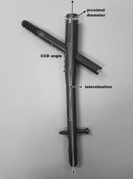
Zimmer®). The proximal diameter of the instrument is re- duced from 16 to 15.5 mm, the front slope is designed to be 15 degrees. Proximal lateralization angle of the instrument is decreased from 5 to 4 degrees and the various caput- column-diaphyseal (CCD) angles are more anatomical. The CCD angle is the projection of the angle between diaphysis and the femoral neck onto an X-ray-image. In addition, the surface of the distal instrument has a floating design for
easy insertion. The length of the lag screw thread is longer than 35 mm and the set screw can be inserted proximal part of the lag screw to resist sliding pull-out and provide a firm fixation force (Fig. 7).
In our clinical cases, the results showed that all per- formance measurements of the postoperative recovery in- cluding EQ-5D, the health state score, and the HHS were improved. In addition, in 1 year follow-up compared to discharge time, all 3 clinical scores show statistically signifi- cant improvements. Improvement in walking, joint exer- cise angles, as well as other sub-introducts including daily living, and reduction of post-traumatic stress levels were noted. The HHS showed similar level of functional recovery when compared to previously published literature.22-24)
There were no adverse events and the overall incidence is zero. The incidence of technical complications in our study, represented a lower rate than that published in dif- ferent studies on the use of the other intramedullary nail device.24-26) However, since this study is a non-comparative study with a short follow-up period; it does not necessar- ily support that ZNN CM nail is superior to other implants because complications did not occur.
Fig. 7. Example of the Zimmer natural nail. The proximal diameter of the instrument is reduced from 16 to 15.5 mm as depicted by the dot- ted outline. Proximal lateralization angle of the instrument is decreased from 5 to 4 degrees as depicted by the square-ended outlines. The caput-columndiaphyseal (CCD) angle is depicted by the dotted-arrow lines.
The advantage of our research is that it is single cen- ter research. All operations were performed by one skilled orthopedic surgeon. This will produce consistent results in surgery compared to studies involving multiple orthope- dic surgeons in multicenter research. It can also reduce the number of variables that can occur during the treatment period, thereby increasing the reliability of research. An- other advantage of our research is that it collected data from 44 patients of 49 patients who were enrolled in the study up to 12 months. This is a 80% follow-up rate, which is rela- tively higher than other papers. Because there are very few excluded data, this reliability of this research results can be increased.27)
Reference to functional status of patients is a character- istic element of this study. For example, patient progress was observed in returning to daily life including; resumption of pre-injury activity level and patient work status. This point is deemed to be fully utilized in other papers in the future.
However, there are some limitations that need to be ac- knowledged and addressed regarding this study. First, fol- lowing study was not conducted in randomized control trial manner, which the evidence is less appealing than random- ized controlled trial studies. Second, the follow-up period is relatively short, leading to inability to represent long term outcomes of ZNN CM nail. Third, there were no control group to compare the functional outcomes to other im- plants. But compared to similar design studies, it can be said that the majority of the scores tend to be well observed.
Conclusion
The orthopedic surgeon must be meticulous when de- ciding to perform surgical fixation of proximal femoral fracture of elderly patients, for their systemic conditions are often poor and brittle. Considering the result obtained from this study, we conclude that the ZNN CM nail emerges as a valid option in the treatment of proximal femoral fractures via improved nail design, simplicity and lack of aggressive- ness of the surgical technique and the low level of complica- tions.
요 약
목적:
본 연구의 목적은 근위 대퇴골 골절 치료에서 Zimmer® natural nail cephalomedullary nail (ZNN CM nail)의 임상 적 효과와 안전한 사용을 알아보고자 함이다.대상 및 방법:
연구는 전향적으로 비교연구가 아닌 단일 기관 결과 연구로서 시행되었다. 사전 동의서에 따라 등록된 환자 들의 정보를 수집하여 분석하였다. 수술 후 6주, 3개월, 6개 월, 12개월에 외래 추시 관찰하였다. 평가항목으로는 신체 검 사, 방사선 검사, 부작용 여부 및 삶의 질과 관련된 설문조사 가 포함되었다.결과:
수술 후 1년 경과 시 39명의 환자에 대해서 평가하였다.EuroQol-5 Dimension 점수는 퇴원 시 0.4점(n=49)에서 1년 경과 시 0.6점(n=39)으로 증가하였다. Harry hip score도 퇴 원 시 56.3점(n=49)에서 1년 경과 시 72.1점(n=12)로 증가하 였다. 골유합은 수술 후 6개월에 64% (n=16)에서 1년 경과 시 95% (n=37)로 나타났다.
결론:
1년간의 추적 관찰 연구의 결과로 근위부 대퇴골 골절치료에 있어서 ZNN CM nail의 효과와 안전한 사용을 확인 하였다.
색인 단어:
대퇴골, 대퇴골 골절, 고관절 골절, 골절 고정, 골 수강내 금속정ORCID
노영호, https://orcid.org/0000-0002-0703-4970 노조셉, https://orcid.org/0000-0002-7945-4085 남광우, https://orcid.org/0000-0003-1096-149X
References
1. Burge R, Dawson-Hughes B, Solomon DH, Wong JB, King A, Tosteson A: Incidence and economic burden of osteoporosis- related fractures in the United States, 2005-2025. J Bone Miner Res, 22: 465-475, 2007.
2. Center JR, Nguyen TV, Schneider D, Sambrook PN, Eisman JA: Mortality after all major types of osteoporotic fracture in men and women: an observational study. Lancet, 353: 878-882, 1999.
3. Lenich A, Vester H, Nerlich M, Mayr E, Stöckle U, Füchtmeier B:
Clinical comparison of the second and third generation of intra- medullary devices for trochanteric fractures of the hip: Blade vs screw. Injury, 41: 1292-1296, 2010.
4. Kuzyk PR, Lobo J, Whelan D, Zdero R, McKee MD, Schemitsch EH: Biomechanical evaluation of extramedullary versus intramedullary fixation for reverse obliquity intertrochan- teric fractures. J Orthop Trauma, 23: 31-38, 2009.
5. Albareda J, Laderiga A, Palanca D, Paniagua L, Seral F: Com- plications and technical problems with the gamma nail. Int Or- thop, 20: 47-50, 1996.
6. Butt MS, Krikler SJ, Nafie S, Ali MS: Comparison of dynamic hip screw and gamma nail: a prospective, randomized, con- trolled trial. Injury, 26: 615-618, 1995.
7. Herrera A, Domingo LJ, Calvo A, Martínez A, Cuenca J: A comparative study of trochanteric fractures treated with the Gamma nail or the proximal femoral nail. Int Orthop, 26: 365- 369, 2002.
8. Johnson LJ, Cope MR, Shahrokhi S, Tamblyn P: Measuring tip-apex distance using a picture archiving and communication system (PACS). Injury, 39: 786-790, 2008.
9. Corrales LA, Morshed S, Bhandari M, Miclau T 3rd: Variabil- ity in the assessment of fracture-healing in orthopaedic trauma studies. J Bone Joint Surg Am, 90: 1862-1868, 2008.
10. Morshed S: Current options for determining fracture union. Adv Med, 2014: 708574, 2014.
11. Hardy DC, Descamps PY, Krallis P, et al: Use of an intramedul- lary hip-screw compared with a compression hip-screw with a plate for intertrochanteric femoral fractures. A prospective, ran- domized study of one hundred patients. J Bone Joint Surg Am, 80: 618-630, 1998.
12. Leung KS, So WS, Shen WY, Hui PW: Gamma nails and dy- namic hip screws for peritrochanteric fractures. A randomised prospective study in elderly patients. J Bone Joint Surg Br, 74:
345-351, 1992.
13. Rosenblum SF, Zuckerman JD, Kummer FJ, Tam BS: A biome- chanical evaluation of the Gamma nail. J Bone Joint Surg Br, 74:
352-357, 1992.
14. McKibbin B: The biology of fracture healing in long bones. J Bone Joint Surg Br, 60: 150-162, 1978.
15. Radford PJ, Needoff M, Webb JK: A prospective randomised comparison of the dynamic hip screw and the gamma locking nail. J Bone Joint Surg Br, 75: 789-793, 1993.
16. Halder SC: The Gamma nail for peritrochanteric fractures. J Bone Joint Surg Br, 74: 340-344, 1992.
17. Park JH, Park JW, Wang JH, Lee JW, Lee JI, Kim JG: Treat- ment of intertrochanteric fracture: comparison of proximal femoral nail and proximal femoral nail A. Korean Soc Fract, 21:
103-109, 2008.
18. Shin DK, Kwun KW, Kim SK, Lee SW, Choi CH, Kim KM:
Proximal femoral nail(PFN) for femur intertrochanteric fracture.
J Korean Fract Soc, 15: 328-335, 2002.
19. Leung KS, Procter P, Robioneck B, Behrens K: Geometric mis- match of the Gamma nail to the Chinese femur. Clin Orthop Relat Res, (323): 42-48, 1996.
20. Hwang JH, Oh JK, Han SH, Shon WY, Oh CW: Mismatch be- tween PFNa and medullary canal causing difficulty in nailing of the pertrochanteric fractures. Arch Orthop Trauma Surg, 128:
1443-1446, 2008.
21. Pu JS, Liu L, Wang GL, Fang Y, Yang TF: Results of the proxi- mal femoral nail anti-rotation (PFNA) in elderly Chinese pa- tients. Int Orthop, 33: 1441-1444, 2009.
22. Akan K, Cift H, Ozkan K, Eceviz E, Tasyikan L, Eren A: Ef- fect of osteoporosis on clinical outcomes in intertrochanteric hip fractures treated with a proximal femoral nail. J Int Med Res, 39: 857-865, 2011.
23. D’Arrigo C, Carcangiu A, Perugia D, et al: Intertrochanteric fractures: comparison between two different locking nails. Int Orthop, 36: 2545-2551, 2012.
24. Vaquero J, Munoz J, Prat S, et al: Proximal Femoral Nail Anti- rotation versus Gamma3 nail for intramedullary nailing of un- stable trochanteric fractures. A randomised comparative study.
Injury, 43 Suppl 2: S47-54, 2012.
25. Simmermacher RK, Ljungqvist J, Bail H, et al: The new proxi- mal femoral nail antirotation (PFNA) in daily practice: results of a multicentre clinical study. Injury, 39: 932-939, 2008.
26. Wu D, Ren G, Peng C, Zheng X, Mao F, Zhang Y: InterTan nail versus Gamma3 nail for intramedullary nailing of unstable tro- chanteric fractures. Diagn Pathol, 9: 191, 2014.
27. Buecking B, Boese CK, Seifert V, Ruchholtz S, Frink M, Lechler P:
Femoral offset following trochanteric femoral fractures: a pro- spective observational study. Injury, 46 Suppl 4: S88-92, 2015.