Korean J Gastroenterol Vol. 65 No. 3, 173-176 http://dx.doi.org/10.4166/kjg.2015.65.3.173 pISSN 1598-9992 eISSN 2233-6869
CASE REPORT
Korean J Gastroenterol, Vol. 65 No. 3, March 2015 www.kjg.or.kr
골수형성 이상증후군 환자에서 조기위암에 대한 내시경 점막하 박리술
임은주, 심은희, 김병욱, 김종인, 김준성, 지정선, 최 황
가톨릭대학교 인천성모병원 소화기내과
Endoscopic Submucosal Dissection for Early Gastric Cancer in a Patient with Myelodysplastic Syndrome
Eun Joo Lim, Eun Hui Sim, Byung-Wook Kim, Jong In Kim, Joon Sung Kim, Jeong-Seon Ji and Hwang Choi
Division of Gastroenterology, Department of Internal Medicine, The Catholic University of Korea Incheon St. Mary’s Hospital, Incheon, Korea
Endoscopic submucosal dissection (ESD) has been successfully performed in thrombocytopenic conditions such as in patients with liver cirrhosis but successful ESD for early gastric cancer (EGC) in hematologic diseases has rarely been reported. A 52-year-old male patient, who had previously been diagnosed with myelodysplastic syndrome 2 years ago, was admitted to our hospital for ESD of EGC. ESD was performed successfully in this patient after platelet concentrates transfusion on the day of ESD. ESD might be an option for the treatment of EGC in thrombocytopenia due to hematologic diseases when optimal supportive managements are applied. (Korean J Gastroenterol 2015;65:173-176)
Key Words: Stomach neoplasms; Myelodysplastic syndromes; Endoscopy
Received August 13, 2014. Revised September 3, 2014. Accepted September 6, 2014.
CC This is an open access article distributed under the terms of the Creative Commons Attribution Non-Commercial License (http://creativecommons.org/licenses/
by-nc/3.0) which permits unrestricted non-commercial use, distribution, and reproduction in any medium, provided the original work is properly cited.
Copyright © 2015. Korean Society of Gastroenterology.
교신저자: 김병욱, 403-720, 인천시 부평구 동수로 56, 가톨릭대학교 인천성모병원 소화기내과
Correspondence to: Byung-Wook Kim, Division of Gastroenterology, Department of Internal Medicine, The Catholic University of Korea Incheon St. Mary’s Hospital, 56 Dongsu-ro, Bupyeong-gu, Incheon 403-720, Korea. Tel: +82-32-280-5052, Fax: +82-32-280-5987, E-mail: [email protected]
Financial support: None. Conflict of interest: None.
INTRODUCTION
Endoscopic submucosal dissection (ESD) is now accepted as an alternative to surgery for the treatment of early gastric cancer (EGC). Because the incidence of complications, such as bleeding or perforation, are high, the utility of this treat- ment modality can be limited in patients with a high proba- bility of bleeding. Although ESD has been successfully per- formed in thrombocytopenic conditions such as in patients with liver cirrhosis1 successful ESD for EGC in hematologic diseases has rarely been reported. In this case, we report a patient who, after being diagnosed with myelodysplastic syn-
drome (MDS) and accompanying EGC, was successfully treated with ESD.
CASE REPORT
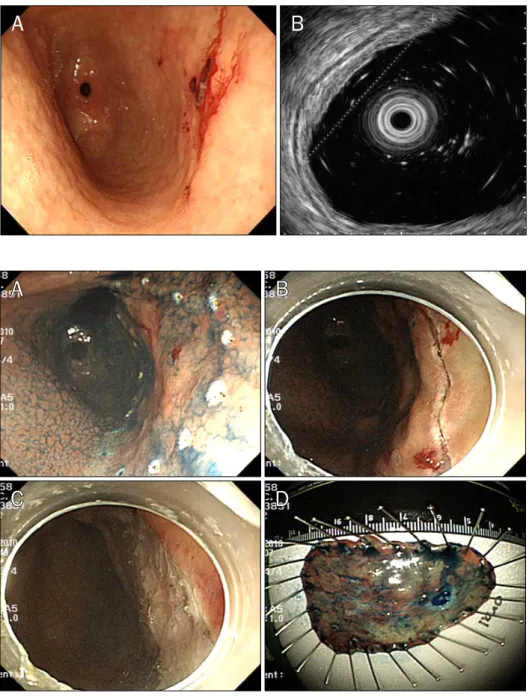
A 52-year-old male patient, who had previously been diag- nosed with MDS 2 years ago, was admitted to our hospital for ESD of EGC. As for MDS, he was in the low risk group accord- ing to the International Prognostic Scoring System2 and was under conservative management with regular follow up. On initial gastroscopy, an EGC lesion was noted at the posterior wall of the gastric antrum (Fig. 1A). A well-differentiated ad-
174 임은주 등. 골수형성 이상증후군에서 내시경 점막하 박리술
The Korean Journal of Gastroenterology
Fig. 1. (A) Initial gastroscopy. An irregularly elevated lesion, which bled easily when touched, is noted at the posterior wall of the antrum. (B) Endoscopic ultrasound. The lesion measured 3.0 cm at the long axis and is limited to the mucosal layer.
Fig. 2. Endoscopic submucosal dissec- tion procedures. (A) The lesion is marked with an argon plasma laser after the spraying of indigo carmine.
(B) The precut is completed with a hook knife. (C) The lesion is com- pletely dissected en bloc. (D) The dissected lesion measured 6.0×4.5 cm.
enocarcinoma was confirmed histopathologically. The pa- tient underwent endoscopic ultrasound, which revealed a 1.5×3.0 cm-sized isoechoic mass in the mucosal layer (Fig. 1B). No extragastric metastatic lesions were found by abdominal computed tomography. The patient’s laboratory data showed a hemoglobin level of 8.6 g/dL and a platelet count of 21,000/mm3. Other laboratory values were within normal limits.
Three hours before ESD, six units of platelet concentrates were transfused, and the procedure was performed success- fully without any complications (Fig. 2). The entire procedure took 44 minutes. On the next day, laboratory data showed a
hemoglobin level of 7.5 g/dL and a platelet count of 77,000/mm3. An additional six units of platelet concentrates were administered to the patient. The resected lesion meas- ured 6.0×4.5 cm. A well-differentiated adenocarcinoma lim- ited to the mucosa was confirmed histopathologically.
Intravenous pantoprazole 80 mg was administered before the procedure and then 8 mg/hour for 3 days. Oral feeding was started on the third day of the procedure along with oral pan- toprazole 40 mg per day for six weeks. The post-ESD course was uneventful and the patient was discharged on the fifth day.
A follow-up gastroscopy, which was performed eight weeks after ESD, showed complete healing of the lesion (Fig. 3).
Lim EJ, et al. ESD in Myelodysplastic Syndrome 175
Vol. 65 No. 3, March 2015 Fig. 3. Follow-up gastroscopy eight weeks after endoscopic submu-
cosal dissection. A red ulcer scar is noted at the site of the dissection.
After seven months, laboratory data showed a hemoglobin level of 10.3 g/dL and a platelet count of 22,000/mm3. The patient is in good condition without evidence of disease re- currence over 4 years.
DISCUSSION
Bleeding and perforation are common complications of ESD: bleeding is observed in 15.6% of patients, and perfo- ration is observed in 1.2%.3 Despite the high rate of bleeding, this procedure has been performed successfully in patients with bleeding diatheses, such as liver cirrhosis or chronic re- nal failure.4,5 However, there have been no reports on suc- cessful ESD for EGC in patients with thrombocytopenia due to hematologic diseases.
MDS comprise a group of biologically and clinically hetero- geneous clonal hematopoietic neoplasms characterized by aberrant myeloid differentiation, dysplastic changes, in- effective hematopoiesis and increasing genomic instability that manifest clinically into peripheral blood cytopenias and variably increased rates of leukemic progression. In the low risk groups, life expectancy is considerable and malig- nancies of other organs should be treated properly.6
In patients with hematologic diseases, severe thrombocy- topenia frequently develops as a consequence of the disease or its treatment. In most cases, platelet concentrates are ad- ministered as prophylaxis, to increase low platelet counts and reduce the risk of bleeding. However, the degree to which
prophylactic platelet transfusions benefit patients with se- vere thrombocytopenia has been unclear. A recent trial sug- gested that a policy of giving platelet concentrates only as treatment for bleeding might become a new standard of care in selected patients, although the primary end point was a re- duction in the number of platelet transfusions, not clinical outcome such as bleeding.7
In general platelet counts of 50,000/mm3 are considered sufficient for surgery in most cases.8 However, guidelines of optimal platelet counts for endoscopic resection have not been suggested. One systematic review on ESD of gastric ne- oplasms in liver cirrhosis patients reported that complication rates were not different when platelet counts were over 50,000/mm3.9 Considering these reports, we think that pla- telet counts of 50,000/mm3 might be considered sufficient for endoscopic resection for gastric neoplasms.
To prevent ESD-related bleeding, pharmacological treat- ment with proton pump inhibitors (PPIs) as well as endo- scopic hemostasis should be considered. Re-bleeding up to 72 hours after endoscopic treatment is often caused by the dissolution of formed fibrin clots by gastric acid. Because pla- telet aggregation, coagulation, and fibrinolysis on gastric hemorrhagic ulcers strongly depend on intragastric pH lev- els, ways to neutralize pH levels should be considered.
Studies concerning post-ESD ulcer healing and post-oper- ation hemorrhage have reported that PPI therapy gives good healing rates for post-ESD ulcers, and it is also effective for the prevention of post-operative hemorrhage.10,11
In this case, the patient was diagnosed with MDS, and the patient’s platelet count was 21,000/mm3 on the day of the ESD, but the transfusion of platelet concentrates enabled us to perform the procedure without bleeding. PPI admin- istration also might have contributed to the prevention of post-ESD bleeding.
In conclusion, ESD can be performed successfully in pa- tients with thromobcytopenias due to hematologic diseases such as MDS, when platelet concentrates and PPIs are ad- ministered properly.
REFERENCES
1. Ogura K, Okamoto M, Sugimoto T, et al. Efficacy and safety of en- doscopic submucosal dissection for gastric cancer in patients with liver cirrhosis. Endoscopy 2008;40:443-445.
2. Greenberg P, Cox C, LeBeau MM, et al. International scoring sys-
176 임은주 등. 골수형성 이상증후군에서 내시경 점막하 박리술
The Korean Journal of Gastroenterology tem for evaluating prognosis in myelodysplastic syndromes.
Blood 1997;89:2079-2088.
3. Chung IK, Lee JH, Lee SH, et al. Therapeutic outcomes in 1000 cases of endoscopic submucosal dissection for early gastric ne- oplasms: Korean ESD Study Group multicenter study. Gastroin- test Endosc 2009;69:1228-1235.
4. Kwon YL, Kim ES, Lee KI, et al. Endoscopic treatments of gastric mucosal lesions are not riskier in patients with chronic renal fail- ure or liver cirrhosis. Surg Endosc 2011;25:1994-1999.
5. Villias C, Gourgiotis S, Veloudis G, Sampaziotis D, Moreas H.
Synchronous early gastric cancer and gastrointestinal stromal tumor in the stomach of a patient with idiopathic thrombocyto- penic purpura. J Dig Dis 2008;9:104-107.
6. Fenaux P, Adès L. How we treat lower-risk myelodysplastic syndromes. Blood 2013;121:4280-4286.
7. Stanworth SJ, Estcourt LJ, Powter G, et al; TOPPS Investigators.
A no-prophylaxis platelet-transfusion strategy for hematologic
cancers. N Engl J Med 2013;368:1771-1780.
8. Rebulla P. Platelet transfusion trigger in difficult patients.
Transfus Clin Biol 2001;8:249-254.
9. Repici A, Pagano N, Hassan C, et al. Endoscopic submucosal dis- section of gastric neoplastic lesions in patients with liver cir- rhosis: a systematic review. J Gastrointestin Liver Dis 2012;21:
303-307.
10. Uedo N, Takeuchi Y, Yamada T, et al. Effect of a proton pump in- hibitor or an H2-receptor antagonist on prevention of bleeding from ulcer after endoscopic submucosal dissection of early gas- tric cancer: a prospective randomized controlled trial. Am J Gastroenterol 2007;102:1610-1616.
11. Yang Z, Wu Q, Liu Z, Wu K, Fan D. Proton pump inhibitors versus histamine-2-receptor antagonists for the management of iatro- genic gastric ulcer after endoscopic mucosal resection or endo- scopic submucosal dissection: a meta-analysis of randomized trials. Digestion 2011;84:315-320.