144
Received:August 11, 2017, Revised:August 29, 2017, Accepted:October 25, 2017 Corresponding to:Hyoun-Ah Kim http://orcid.org/0000-0003-2609-3367
Department of Rheumatology, Ajou University School of Medicine, 164 WorldCup-ro, Yeongtong-gu, Suwon 16499, Korea.
E-mail:[email protected]
Copyright ⓒ 2018 by The Korean College of Rheumatology. All rights reserved.
This is a Open Access article, which permits unrestricted non-commerical use, distribution, and reproduction in any medium, provided the original work is properly cited.
Case Report
pISSN: 2093-940X, eISSN: 2233-4718
Journal of Rheumatic Diseases Vol. 25, No. 2, April, 2018 https://doi.org/10.4078/jrd.2018.25.2.144
Patient with Systemic Lupus Erythematosus Combined with Erosive Arthritis was Treated Successfully with Tocilizumab:
A Case Report
Moon Young Kim1, Sunghoon Park2, Chang-Hee Suh1, Ju-Yang Jung1, Hyoun-Ah Kim1
Departments of 1Rheumatology and 2Radiology, Ajou University School of Medicine, Suwon, Korea
Systemic lupus erythematosus (SLE) is a systemic autoimmune inflammatory disease that frequently involves the joints at some stage during the disease course. Although lupus arthritis is usually non-erosive, approximately 5% of patients develop erosions.
This paper reports a patient with SLE combined with erosive arthritis, who was treated successfully with tocilizumab. A 20-year-old female, who was first diagnosed with SLE at the age of 13 years, had been admitted frequently to hospital with dis- ease flare-ups during the previous 3 years. Despite aggressive treatment, her arthritis became aggravated, particularly in both wrists and the right knee. Radiologically, erosive arthritis was noted in the wrists.The serum interleukin (IL)-6 level was elevated significantly along with the other inflammatory markers. After the tocilizumab treatment, the arthritis subsided with a normal- ization of the IL-6 level, suggesting that tocilizumab may be a suitable treatment for lupus erosive arthritis if the IL-6 level is high.
(J Rheum Dis 2018;25:144-147)
Key Words. Systemic lupus erythematosus, Arthritis, Interleukin-6 inhibitor, Interleukin-6
INTRODUCTION
Systemic lupus erythematosus (SLE) is a systemic auto- immune inflammatory disease that frequently involves the joints, which are affected in about 90% of patients at some stage of disease [1,2]. Lupus arthritis does not in- duce severe bone damage or erosion, unlike rheumatoid arthritis (RA). However, about 5% of patients with lupus arthritis exhibit erosions, rendering the boundary be- tween SLE and RA obscure [2]. This apparent clinical co-existence of RA and SLE has been termed “rhupus” [3].
Recently, serological markers including interleukin-6 (IL-6), C-reactive protein (CRP), and anti-citrullinated (anti-CCP) antibody have been evaluated as predictors of joint erosion in SLE [4]. IL-6 is a pleiotropic cytokine in- volved in various cellular activities including inflam- mation, the immune response, and B-cell differentiation.
Several studies have suggested that IL-6 may play an im-
portant role in SLE, inducing tissue damage [5-7]. Serum IL-6 levels were elevated in lupus patients and correlated with the extent of disease activity. B-cells from lupus pa- tients spontaneously produce large amounts of im- munoglobulins; IL-6 neutralization reduces such pro- duction [7]. Also, blocking of IL-6 inhibited the pro- duction of anti-double-stranded DNA (dsDNA) antibody in vitro [6].
Tocilizumab, a humanized IL-6 receptor-inhibiting mo- noclonal antibody, was first approved by the Food and Drug Administration in 2010 as a treatment for RA. As no biological agent other than belimumab has been licensed as an SLE treatment, tocilizumab has been tested in cer- tain SLE patients, especially those who failed standard therapy. Herein, we report a case of successful use of toci- lizumab to treat a patient with lupus erosive arthritis that progressed despite aggressive treatment. Consent, for the publication of the case report and any additional related
SLE with erosive arthritis
www.jrd.or.kr 145
Figure 1. (A) Axial T2-weighted fat-suppressed and T1-weighted images reveal osteonecrosis with the double-line sign, represent- ing the outer rim of the sclerosis (arrow) and the inner rim of the high-signal intensity region in the distal femoral condyles. (B) Both anteroposterior and obli- que images of the left hand re- veal erosion with narrowing of the joint space, and ankylosis between the trapezoid, cap- itate, and hamate; and the sec- ond, third, and fourth meta- carpal bases.
information was taken from the patient involved in the study.
CASE REPORT
A 20-year-old female was first diagnosed with SLE at the age of 13 years when she presented with a malar rash, a vasculitic skin rash of the hand, leukopenia, a positive an- tinuclear antibody (>1:2,560, speckled type), a positive anti-dsDNA antibody (30.9 IU/mL), and hypocomple- mentemia (C3: 54 mg/dL, C4: 7 mg/dL). The tests for rheumatoid factor and anti-CCP antibody were negative.
She has been admitted to hospital with flare-ups six times during recent 3 years; the problems included fever, arthri- tis, and lymphadenopathy, despite aggressive therapy in- cluding hydroxychloroquine (300 mg/day), moderate levels of prednisolone (20∼30 mg/day), and methotrex-
ate (10 mg/week). Lately, her arthritis had become ag- gravated, especially in both wrists and the right knee. We prescribed oral methotrexate (10 mg/week) and tacroli- mus (2 mg/day) for 3 months, but the arthritis continued to worsen, now accompanied with fever. Magnetic reso- nance imaging of the right knee revealed an osteonecrotic area surrounded by a rim of low signal intensity, presum- ably reflecting the long-term corticosteroid treatment (Figure 1A). On hand radiography, joint space narrowing with erosion was apparent, and juxta-articular osteopo- rosis was also evident, similar to that associated with RA (Figure 1B). These pathologies were particularly obvious between the trapezoid, capitate, and hamate; and the sec- ond, third, and fourth metacarpal bases of the left hand.
The erythrocyte sedimentation rate (ESR) was elevated to 57 mm/hour, the CRP level to 7.7 mg/dL, the C3 level to 125 mg/dL, and the C4 level to 34 mg/dL, indicating
Moon Young Kim et al.
146 J Rheum Dis Vol. 25, No. 2, April, 2018
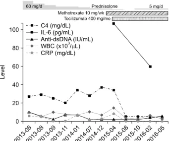
Figure 2. The time courses of the levels of complement 4 (C4), interleukin-6 (IL-6), anti-double-stranded DNA (anti-dsDNA) antibodies, white blood cells (WBCs), and C-reactive protein (CRP).
that active inflammation was in play. The IL-6 level was significantly elevated to 106.7 pg/mL (reference range, 0∼7 pg/mL). SLE disease activity index (SLEDAI) was 7, and disease activity score for 28 joints (DAS28) was 5.7.
With intravenous tocilizumab treatment (400 mg/month), the fever and arthritis subsided. The ESR and CRP levels decreased to within the normal ranges, and the C3 and C4 levels fell to 56 mg/dL and 5 mg/dL (30 and 10% of their peak levels, respectively). Six months later, the IL-6 level had decreased to 59.5 pg/mL (Figure 2) with monthly in- travenous tocilizumab treatment. The corticosteroid dose was reduced to 8 mg methylprednisolone daily and tacrolimus was discontinued. After discharge, the oral corticosteroid was gradually tapered, and ultimately dis- continued, over a period of 2 months, but then restarted 2 months later because of relapse of the malar rash. The average disease activity score of 28 joints decreased from 5.78 to 0.6 after 10 months of tocilizumab treatment. At recent (18-month) follow-up, the arthritis was well con- trolled; the counts of swollen and tender joints were both zero. No manifestation of active lupus except for the ma- lar rash developed during the period of monthly tocilizumab treatment. SLEDAI was 4, and DAS28 was 0.8. Currently, her medication includes tocilizumab (400 mg/month), methotrexate (10 mg/week), hydroxychloroquine (200 mg/day), and methylprednisolone (3 mg/day).
DISCUSSION
SLE is an autoimmune disease associated with diverse clinical and serological findings. The joints are commonly involved; both the small- and medium-sized joints are symmetrically affected at the time of disease onset.
Although SLE may be initially confused with RA, lupus arthritis is usually well controlled with low-dose cortico- steroids and antimalarial drugs. In late stages of the dis- ease, however, the joints may develop reducible non-ero- sive changes termed “Jaccoud’s arthritis”. Unlike RA, de- forming arthropathy associated with SLE is usually attrib- utable to ligamentous rather than erosive change [8].
Efforts have been made to explain the lack of erosion in SLE arthritis compared to RA. Upregulation of interfer- on-inducible genes and downregulation of genes involved in extracellular matrix homeostasis have been reported in patients with SLE arthritis [9]. However, up to 3%∼5%
of examples of SLE arthritis are erosive, and such patients may meet the American College of Rheumatology (ACR) criteria for RA also [3]. Whether this reflects true disease overlap or, rather, the existence of a rare subtype of SLE arthritis, remains unclear. Recently, accumulating evi- dence has suggested that ‘rhupus’ reflects an overlap be- tween RA and SLE. Budhram et al. [10] suggested that SLE-associated erosions alone do not reflect a true over- lap between SLE and RA, but any additional common- ality, such as elevated levels of anti-CCP antibody (a well-known marker of RA), may indicate co-existence of the two diseases.
IL-6 may play an important role in SLE, including the arthritis. One study found that plasma IL-6 levels were higher in lupus patients with than without arthritis, and correlated with both clinical and ultrasound measures of arthritis activity [11]. Joint erosion in SLE patients corre- lated with higher levels of IL-6 and CRP. Tocilizumab re- duced SLE disease activity attributable to arthritis, sug- gesting that IL-6 blockade may be useful when treating drug-resistant lupus arthritis [12]. Some case reports have described SLE patients with cutaneous symptoms, hemolytic anemia or pericardial effusions intractable to standard therapy but that responded to tocilizumab [13-15]. However, any effect of tocilizumab on erosive ar- thritis remains unclear. Here, we report successful tocili- zumab treatment of a patient with SLE combined with erosive arthritis. She did not respond to standard thera- pies including hydroxychloroquine, prednisolone, me- thotrexate, and tacrolimus. Moreover, continuous steroid
SLE with erosive arthritis
www.jrd.or.kr 147
usage had triggered avascular necrosis of the right knee.
After tocilizumab treatment, disease activity (including the arthritic component) became well controlled and the prednisolone dose could be reduced. Also, the levels of complement acute phase reactants were elevated before tocilizumab treatment, perhaps associated with the rise in IL-6 and CRP [16]. After tocilizumab infusion, the complement levels decreased to C3 56 mg/dL and C4 5 mg/dL, similar to those of other lupus patients and initial her manifestations of SLE including malar rash, suggesting that elevated IL-6 levels may mask hypocomplementemia in SLE patients. The reported side-effects of tocilizumab (neutropenia, liver enzyme abnormalities, alterations in lipid metabolism, and infection) were not noted in our pa- tient [12].
SUMMARY
To the best of our knowledge, this is the first report of the successful use of tocilizumab to treat erosive arthritis in a patient with SLE. The case suggests that tocilizumab could be a treatment of choice for lupus erosive arthritis if the IL-6 level is high.
CONFLICT OF INTEREST
No potential conflict of interest relevant to this article was reported.
REFERENCES
1. Aptekar RG, Lawless OJ, Decker JL. Deforming non-erosive arthritis of the hand in systemic lupus erythematosus. Clin Orthop Relat Res 1974;(100):120-4.
2. Richter Cohen M, Steiner G, Smolen JS, Isenberg DA.
Erosive arthritis in systemic lupus erythematosus: analysis of a distinct clinical and serological subset. Br J Rheumatol 1998;37:421-4.
3. Fernández A, Quintana G, Rondón F, Restrepo JF, Sánchez A, Matteson EL, et al. Lupus arthropathy: a case series of pa- tients with rhupus. Clin Rheumatol 2006;25:164-7.
4. Amezcua-Guerra LM, Márquez-Velasco R, Bojalil R. Erosive arthritis in systemic lupus erythematosus is associated with high serum C-reactive protein and anti-cyclic citrullinated
peptide antibodies. Inflamm Res 2008;57:555-7.
5. Chun HY, Chung JW, Kim HA, Yun JM, Jeon JY, Ye YM, et al. Cytokine IL-6 and IL-10 as biomarkers in systemic lupus erythematosus. J Clin Immunol 2007;27:461-6.
6. Linker-Israeli M, Deans RJ, Wallace DJ, Prehn J, Ozeri-Chen T, Klinenberg JR. Elevated levels of endogenous IL-6 in sys- temic lupus erythematosus. A putative role in pathogenesis.
J Immunol 1991;147:117-23.
7. Klashman DJ, Martin RA, Martínez-Maza O, Stevens RH. In vitro regulation of B cell differentiation by interleukin-6 and soluble CD23 in systemic lupus erythematosus B cell sub- populations and antigen-induced normal B cells. Arthritis Rheum 1991;34:276-86.
8. Alarcón-Segovia D, Abud-Mendoza C, Diaz-Jouanen E, Iglesias A, De los Reyes V, Hernández-Ortiz J. Deforming arthropathy of the hands in systemic lupus erythematosus.
J Rheumatol 1988;15:65-9.
9. Nzeusseu Toukap A, Galant C, Theate I, Maudoux AL, Lories RJ, Houssiau FA, et al. Identification of distinct gene expression profiles in the synovium of patients with sys- temic lupus erythematosus. Arthritis Rheum 2007;56:
1579-88.
10. Budhram A, Chu R, Rusta-Sallehy S, Ioannidis G, Denburg JA, Adachi JD, et al. Anti-cyclic citrullinated peptide anti- body as a marker of erosive arthritis in patients with sys- temic lupus erythematosus: a systematic review and meta-analysis. Lupus 2014;23:1156-63.
11. Ball EM, Gibson DS, Bell AL, Rooney MR. Plasma IL-6 levels correlate with clinical and ultrasound measures of arthritis in patients with systemic lupus erythematosus. Lupus 2014;
23:46-56.
12. Illei GG, Shirota Y, Yarboro CH, Daruwalla J, Tackey E, Takada K, et al. Tocilizumab in systemic lupus eryth- ematosus: data on safety, preliminary efficacy, and impact on circulating plasma cells from an open-label phase I dos- age-escalation study. Arthritis Rheum 2010;62:542-52.
13. Kamata Y, Minota S. Successful treatment of massive in- tractable pericardial effusion in a patient with systemic lu- pus erythematosus with tocilizumab. BMJ Case Rep 2012.
DOI: 10.1136/bcr-2012-007834.
14. Makol A, Gibson LE, Michet CJ. Successful use of inter- leukin 6 antagonist tocilizumab in a patient with refractory cutaneous lupus and urticarial vasculitis. J Clin Rheumatol 2012;18:92-5.
15. García-Hernández FJ, González-León R, Castillo-Palma MJ, Ocaña-Medina C, Sánchez-Román J. Tocilizumab for treat- ing refractory haemolytic anaemia in a patient with systemic lupus erythematosus. Rheumatology (Oxford) 2012;51:
1918-9.
16. Korkmaz HI, Krijnen PAJ, Ulrich MMW, de Jong E, van Zuijlen PPM, Niessen HWM. The role of complement in the acute phase response after burns. Burns 2017;43:1390-9.