aureus isolated from the oral cavity of patients with periodontitis
Ga-Yeon Kim1, Chong Heon Lee2,*
1Department of Dental Hygiene, Dankook University College of Health Sciences, Cheonan, Korea
2Department of Oral Pathology, Dankook University College of Dentistry, Cheonan, Korea
Research Article
J Periodontal Implant Sci 2015;45:223-228 http://dx.doi.org/10.5051/jpis.2015.45.6.223
Purpose: The goal of this study was to characterize the patterns of antimicrobial resistance and virulence genes in samples of Staphylococcus aureus (S. aureus) isolated from peri- odontitis patients.
Methods: From July 2015 to August 2015, oral saliva was collected from a total of 112 pa- tients diagnosed with periodontitis, including 80 outpatients in dental hospitals and 32 pa- tients in dental clinics located in Seoul and Cheonan. The samples were subjected to a sus- ceptibility test to evaluate the prevalence of antimicrobial resistance, and the pathogenic factors and antimicrobial resistance factors in the DNA of S. aureus were analyzed using polymerase chain reaction.
Results: A susceptibility test against 15 antimicrobial agents showed that 88% of cultures were resistant to ampicillin, 88% to penicillin, and 2% to oxacillin. Resistance to at least two drugs was observed in 90% of cultures, and the most common pattern of multidrug resistance was to ampicillin and penicillin. Enterotoxins were detected in 65.9% of samples.
The cell hemolysin gene hld was detected in 100% of cultures and hla was detected in 97.6% of samples. All strains resistant to penicillin and ampicillin had the blaZ gene. The aph(3′)IIIa gene, which encodes an aminoglycoside modifying enzyme, was detected in 46.3% of samples.
Conclusions: In the treatment of oral S. aureus infections, it is important to identify the pathogenic genes and the extent of antimicrobial resistance. Furthermore, it is necessary to study patterns of antimicrobial resistance and cross-infection in the context of periodon- tological specialties in which antimicrobials are frequently used, such as maxillofacial sur- gery, where the frequency of antimicrobial use for minor procedures such as implant place- ment is increasing.
Keywords: Antimicrobial resistant gene, Pathogenic gene, Periodontitis, Staphylococcus aureus.
Received: Oct. 27, 2015 Accepted: Nov. 30, 2015
*Correspondence:
Chong Heon Lee
Department of Oral Pathology, Dankook University College of Dentistry, 119 Dandae-ro,
Dongnam-gu, Cheonan 31116, Korea E-mail: [email protected] Tel: +82-41-550-1946 Fax: +82-41-559-7902
INTRODUCTION
Staphylococcus aureus is a major causative agent of major human disease; although it is associated with mild symptoms in healthy skin or soft tissue, in some environments, such as hospitals, it is the main source of infections accounting for more than 80% of pyogenic diseases. In the oral tract, S. aureus has been associated with dentoalveolar infections, and oral mucosal lesions. Moreover, staphylococcal colonization has been demonstrated from the tongue, saliva, mucosal surfaces, supragingival tooth surfaces and the periodontal pocket [1,2]. The wide range of virulence factors of S. aureus associated with infection and diseases can be classified into surface-associated factors, degradative enzymes and supe-
This is an Open Access article distributed under the terms of the Creative Commons Attribution Non-Commercial License (http://creativecommons.org/licenses/by-nc/3.0/).
rantigenic toxins; the diversity and great variability in these genes may affect the course of an infection [3]. The enterotoxins of S.
aureus are heat-resistant toxins causing diarrhea and vomiting in humans, and serological analysis has identified five different types based on their antigenicity. Although new types of enterotoxin have recently been reported, their association with food poisoning is not clear [4]. S. aureus has four hemolysins, hla dissolves cells and causes hemolytic necrosis, hlb affects the lung and the cornea, and hld which has been reported to be produced in 97% of S. au- reus samples, dissolves the inner cell structures of red blood cells and various mammalian cells [5]. Due to the increase of the indis- criminate use of penicillin, which was discovered in 1928 by Alex- ander Fleming, more penicillin-resistant strains had emerged by approximately 1950, when the resistance rate reached 80%, lead- ing to a gradual loss of the effectiveness of penicillin. The vicious cycle of misuse and abuse of antimicrobials and the emergence of antimicrobial-resistant bacteria has become a problem that has not shown meaningful improvement from the early days of anti- microbial use through the present days [6].
Periodontitis involves the progressive loss of alveolar bone around the teeth, and if left untreated, can lead to the loosening and sub- sequent loss of teeth. Periodontitis is caused by microorganisms that adhere to and grow on the surfaces of teeth, along with an over-aggressive immune response to those microorganisms. Studies of the causative pathogens of periodontal diseases isolated from Korean periodontitis patients have demonstrated that S. aureus plays a role in exacerbating dental diseases by forming a biofilm with the causative pathogens of periodontal diseases [7-10]. The frequency of antimicrobial use in oral surgical procedures and mi- nor procedures such as implant placement has recently increased, which is a trend that can be directly associated with the problem of increased antimicrobial resistance. This study characterized the dis- tribution of antimicrobial resistance, antibiotic resistance genes, and virulence genes in S. aureus cultures isolated from Korean peri- odontitis patients. The findings of this study are relevant for the ongoing treatment and prevention of periodontitis.
MATERIALS AND METHODS
Sample collection
From July 2015 to August 2015, oral saliva was collected from a total of 112 patients diagnosed with periodontitis, including 80 outpatients in dental hospitals and 32 patients of dental clinics lo- cated in Seoul and Cheonan. The subjects of this study were pa- tients who visited a dental hospital or dental clinic and were diag- nosed with periodontitis, and were mostly chronic periodontitis pa- tients (K05.3 in the International Classification of Diseases-10th Re- vision). They had periodontal pockets deeper than 4 mm and alveo- lar bone loss visible on X-ray films. Scaling only is unlikely to be an adequate treatment, so all patients who required periodontal treat- ment were screened. Samples were collected in the form of oral sa- liva prior to scaling and medication and in the form of plaque from
the tooth surface obtained with sterile swabs when patients were in the hospital, and the samples were stored in sterile containers. This study was approved by the Institutional Review Board of Dankook University (IRB No: DKU 2015-06-003-001).
Isolation and identification of S. aureus
The isolation of S. aureus was performed in accordance with the methods described by the Korea National Research Institute of Health [11] and Murray et al. [12]. Polymerase chain reaction (PCR) was used to identify S. aureus [13,14]. A Bio-Rad (Hercules, CA, USA) C1000 thermal cycler was utilized and the final product was subjected to electrophoresis followed by confirmation of the elec- trophoresis pattern using a transilluminator (Model: Gel Doc XR+
Bio-Rad, Hercules, CA, USA). A 100-base pair ladder (Bioneer, Dae- jeon, Korea) was used as a standard size maker.
Antimicrobial susceptibility test
Antimicrobial susceptibility tests were performed in accordance with the guidelines of the World Health Organization and the Clinical and Laboratory Standards Institute [15,16].
PCR analysis of pathogenic factors and antimicrobial resistance factors
PCR was performed to identify pathogenic factors and antimi- crobial resistance factors present in the DNA of S. aureus. DNA was extracted from the samples of S. aureus and the presence or ab- sence of the pathogenic gene factors of interest was determined using the appropriate primers [3,6,17-19].
RESULTS
Isolation rates of S. aureus from the oral cavity of patients with periodontitis
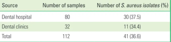
The results regarding the isolation of S. aureus from outpatients in dental hospitals and clinics located in Seoul and Cheonan are presented in Table 1. 41 strains of S. aureus (36.6%) were isolated from the test samples of oral saliva collected from a total of 112 patients diagnosed with periodontitis which include 80 outpa- tients in dental hospitals and 32 outpatients in dental clinics lo- cated in Seoul and Cheonan from July to August of 2015, and 30 strains (37.5%) were isolated from the outpatients in dental hospi- tals and 11 strains (34.4%) from the patients of dental clinics.
Table 1. Isolation rates of Staphylococcus aureus in the oral cavity of patients with periodontitis.
Source Number of samples Number of S. aureus isolates (%)
Dental hospital 80 30 (37.5)
Dental clinics 32 11 (34.4)
Total 112 41 (36.6)
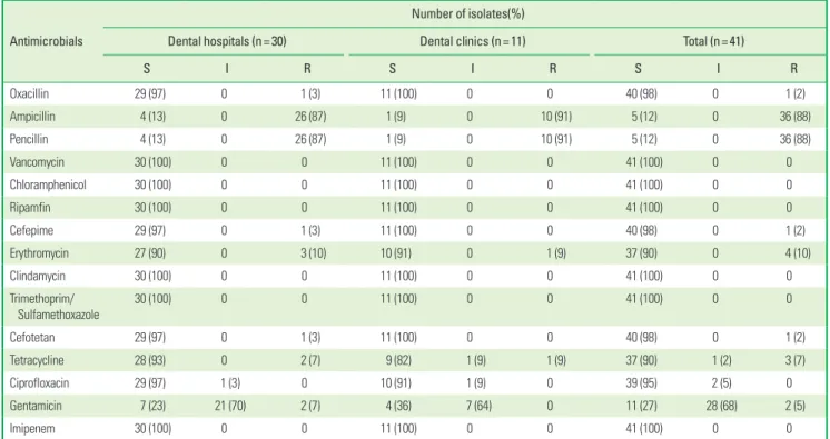
Antimicrobial susceptibility of S. aureus isolated from patients with periodontal diseases
Data regarding the antimicrobial susceptibility of S. aureus iso- lated from the oral cavity of outpatients in dental hospitals and clinics are presented in Table 2. Among the total number of 41 S.
aureus isolates, the susceptibilities to vancomycin, chlorampheni- col, rifampin, clindamycin, sulfamethoxazole, and imipenem were all (100%). High susceptibility was found to oxacillin, cefepime, and cefotetan 40 isolates (98%), ciprofloxacin 39 isolates (95%), and erythromycin and tetracycline 37 isolates (90%). In contrast, 11 iso- lates (27%) were susceptible to gentamicin and 5 isolates (12%) were susceptible to ampicillin and penicillin. The antimicrobial re- sistance rate was 88% (36/41) each for ampicillin and penicillin, 10% (4/41) for erythromycin, 7% (3/41) for tetracycline, 5% (2/41) for gentamycin, and 2% (1/41) each for cefepime and cefotetan.
Patterns of multiple antimicrobial resistance
The results regarding multiple antimicrobial resistance for the 41 strains isolated from the oral cavity of patients with periodontal disease are presented in Table 3. Of the 41 strains, four (10%) showed susceptibility to all antimicrobials tested, while 37 (90%) were resistant to at least two antimicrobials. Resistance to two an- timicrobials was found in 29 strains (71%), seven strains (14%) showed resistance to three antimicrobials, one strain (2%) showed resistance to four antimicrobials, and one strain (2%) was resistant to six antimicrobials.
Analysis of antimicrobial resistance genes
In order to determine the presence or absence of antimicrobial resistance genes in the 41 S. aureus strains isolated from patients with periodontal disease, PCR was performed, and the results are presented in Table 4. The aac(6′)/aph(2″) gene was identified in two strains (5%), the aph(3′)-IIIa gene in 19 strains (46%), and the ant(4′)-Ia gene was isolated in one strain (2%). The methicillin re- sistance gene mecA was isolated from one strain (2%) and tem, an Table 2. Antimicrobial resistance rates in samples of Staphylococcus aureus isolated from the oral cavity of patients with periodontitis.
Antimicrobials
Number of isolates(%)
Dental hospitals (n=30) Dental clinics (n=11) Total (n=41)
S I R S I R S I R
Oxacillin 29 (97) 0 1 (3) 11 (100) 0 0 40 (98) 0 1 (2)
Ampicillin 4 (13) 0 26 (87) 1 (9) 0 10 (91) 5 (12) 0 36 (88)
Pencillin 4 (13) 0 26 (87) 1 (9) 0 10 (91) 5 (12) 0 36 (88)
Vancomycin 30 (100) 0 0 11 (100) 0 0 41 (100) 0 0
Chloramphenicol 30 (100) 0 0 11 (100) 0 0 41 (100) 0 0
Ripamfin 30 (100) 0 0 11 (100) 0 0 41 (100) 0 0
Cefepime 29 (97) 0 1 (3) 11 (100) 0 0 40 (98) 0 1 (2)
Erythromycin 27 (90) 0 3 (10) 10 (91) 0 1 (9) 37 (90) 0 4 (10)
Clindamycin 30 (100) 0 0 11 (100) 0 0 41 (100) 0 0
Trimethoprim/
Sulfamethoxazole
30 (100) 0 0 11 (100) 0 0 41 (100) 0 0
Cefotetan 29 (97) 0 1 (3) 11 (100) 0 0 40 (98) 0 1 (2)
Tetracycline 28 (93) 0 2 (7) 9 (82) 1 (9) 1 (9) 37 (90) 1 (2) 3 (7)
Ciprofloxacin 29 (97) 1 (3) 0 10 (91) 1 (9) 0 39 (95) 2 (5) 0
Gentamicin 7 (23) 21 (70) 2 (7) 4 (36) 7 (64) 0 11 (27) 28 (68) 2 (5)
Imipenem 30 (100) 0 0 11 (100) 0 0 41 (100) 0 0
S, sensitivity; I, intermediate resistance; R, resistance.
Table 3. Patterns of multiple antimicrobial resistance in Staphylococcus au- reus isolated from the oral cavity of patients with periodontitis.
Multiple resistance patterns
Number of multiple resistance patterns (%) Dental hospitals
(n=30) Dental clinics
(n=11) Total
(n=41)
2 AM-P 21 (70) 8 (73) 29 (71) 29 (71)
3 AM-P-E 2 (7) 1 (9) 3 (7) 6 (14)
AM-P-TE 1 (3) 1 (9) 2 (5)
AM-P-GM 1 (3) 0 1 (2)
4 AM-P-E-GM 1 (3) 0 1 (2) 1 (2)
6 AM-P-OX-CTT-CC-FEP 1 (3) 0 1 (2) 1 (2)
None 3 (10) 1 (9) 4 (10) 4 (10)
Total 30 (100) 11 (100) 41 (100)
AM, ampicillin; OX, oxacillin; CTT, cefotetan; P, penicillin; GM, gentamicin; E, erythromycin;
CC, clindamycin; FEP, cefepime.
ampicillin resistance gene, was isolated from 21 strains (51%), but ampicillin resistance was found in 36 strains (58%). Resistance to erythromycin is derived from the ermB, ermTR, and mefA/E genes, none of which was detected. The β-lactamase resistance gene blaZ, was detected in 37 strains, which was consistent with the results of the disk expansion method. Moreover, the gene encoding peni- cillin-binding protein was not detected.
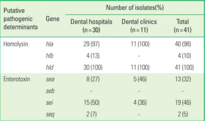
Analysis of pathogenic genes
In order to determine whether the virulence genes encoding for hemolysins and enterotoxins were present in the 41 S. aureus strains isolated from patients with periodontal disease, PCR was performed and the results are presented in Table 5. The hemolysin gene hla was present in 40 strains (97.6%), hlb was present in 4 strains (10%), and hld was present in 41 strains (100%). The entero- toxin gene sea was present in 13 strains (32%), seb was present in no strains (0%), sei was present in 19 strains (46%), and seq was present in two strains (5%).
DISCUSSION
Periodontal disease represents a possible reservoirs for opportu- nistic bacteria in the oral cavity. The use of antibiotics to treat periodontal disease or other infections may lead to the increase of Staphylococcus spp. in the oral cavity. Strains of S. aureus may easily become resistant to antibiotics, potentially resulting in peri- odontitis that persists despite antibiotic treatment. The presence of a higher proportions of S. aureus in the oral cavity may lead to a significant increase in the incidence of periodontal infections and the difficulty of treating those that do occur.
The isolation rate of S. aureus from patients with periodontitis
in this study was lower than the figure of 41.9% reported by Kim [20], but higher than the rate of 4.8% reported by Min et al. [21], who investigated the nasal cavities of hospital clinical staff; the figure of 20% reported by Han et al. [22]; and the rate of 23% re- ported by Kang [23], who studied dental students. The rate that we found was higher than the rate of 30% reported by Kim [24]
among patients with oral infections, but similar to the rate of 36%
that they observed in the general population. Notably, the rate of 60.4% reported by Lee et al. [25] was much higher than the results of the present study. The previously published literature indicates that S. aureus is thought to play an important role in causing peri- odontal diseases by forming a biofilm on dental plaque. Previous studies have suggested that S. aureus exhibits a diverse range of pathogenicity. In dental patients, S. aureus is thought to be trans- mitted through the nasal cavity and the oral cavity, where it is most highly distributed in the human body. Kim et al. [24] raised the possibility of cross-infection with multiple strains of S. aureus in the dental field, mediated either by contact of effusions, skins, and saliva between dental care staff and patients or by contact with S. aureus floating in the form of aerosols and contaminating the medical instruments and unit chairs. The general use of anti- microbials is therefore necessary to treat nosocomial S. aureus in- fections. Dental patients take antibiotics primarily to prevent post- operative infections and to prevent secondary infections, for which purpose antimicrobials are administered prior to surgery. Antimi- crobials are primarily used therapeutically to treat abscess forma- tion, osteomyelitis of the jaw, inflammation of the maxillary sinus, and acute orofacial infections that are caused by dental infections;
in such cases, antimicrobials have been reported to be prescribed for an average of approximately 6.9 days [26]. Kim [20] reported that S. aureus strains isolated from dental patients with periodon- tal disease showed antimicrobial resistance rates of 92.6% to am- picillin, 90.7% to penicillin, 11.1% each to oxacillin and cefotetan, and 5.6% to erythromycin, which is consistent with the results of our study. This profile of antimicrobial resistance may have resulted from the fact that the samples were isolated from the oral cavity Table 4. Antimicrobial resistance genes in Staphylococcus aureus samples
isolated from periodontitis patients in dental hospitals and dental clinics.
Putative antimicrobial resistance
determinants Gene
Number of isolates (%) Dental hospital
(n=30) Dental clinic (n=11) Total
(n=41)
Aminoglycosides aac (6')/aph (2") 2 (7) - 2 (5)
aph (3")-IIIa 16 (53) 3 (27) 19 (46)
ant (4')-Ia 1 (3) 1 (9) 2 (5)
Methicillin mecA 1 (3) - 1 (2)
Ampicillin tem 17 (57) 4 (36) 21 (51)
Erythromycin and ermB - - -
clindamycin ermTR - - -
mefA/E - - -
Beta-lactamse blaZ 27 (90) 10 (91) 37 (90)
Penicillin binding pbp1a - - -
protein precursor pbp2b - - -
pbp2X - - -
Table 5. Distribution of hemolysin and enterotoxin gene in Staphylococcus aureus samples isolated from periodontitis patients in dental hospitals and dental clinics.
Putative pathogenic determinants Gene
Number of isolates(%) Dental hospitals
(n=30) Dental clinics
(n=11) Total
(n=41)
Hemolysin hla 29 (97) 11 (100) 40 (98)
hlb 4 (13) - 4 (10)
hld 30 (100) 11 (100) 41 (100)
Enterotoxin sea 8 (27) 5 (46) 13 (32)
seb - - -
sei 15 (50) 4 (36) 19 (46)
seq 2 (7) - 2 (5)
of patients with periodontal disease. In contrast, Jung and Lee [27]
reported that strains of S. aureus isolated from the nasal cavity of students and hospital clinical staff showed resistance rates of 90%
to penicillin, 43% to tetracycline, 37% to erythromycin, 10% to cephalothin, and 13% to clindamycin and vancomycin. These find- ings were different from those obtained in the present study, and may have been related to the fact that nasal cavity is more favor- able for the survival of S. aureus than the oral cavity. In contrast, Kim et al. [28] reported that the antimicrobial resistance rates of S.
aureus collected from pyogenic lesions of outpatients were 97.6%
to penicillin, 9.4% to oxacillin, 8.1% to erythromycin, and 2.4% to clindamycin, and Kim [24] reported that resistance rates of S. au- reus in patients who were hospitalized due to acute oral infections were 100% to penicillin, 68.1% to oxacillin, and 88.1% to erythro- mycin, which are drastically different from the resistance rates ob- served in this study. It is possible that those higher resistance rates were due to the higher likelihood that lesions in acute oral infec- tions and pyogenic lesions may come into contact with antimicro- bials, thereby acquiring resistance.
In the study performed by Kim [20] on S. aureus isolated from patients in dental hospitals and clinics, 3.7% of strains were sus- ceptible to all antimicrobials, 5.6% showed resistance to a single antimicrobial, 74.1% showed resistance to two antimicrobials, 3.7% were resistant to three antimicrobials, 3.7% were resistant to four antimicrobials, 3.7% were resistant to five antimicrobials, 3.7% were resistant to six antimicrobials, and 1.9% were resistant to seven antimicrobials. In these results, strains with resistance to two antimicrobials comprised the majority, accounting for 70% of the samples, which was consistent with the findings of the present study. However, the rate of strains with resistance to three antimi- crobials in the present study was much higher than that reported by Kim [29], indicating that S. aureus isolated from patients with the same periodontal disease may show strikingly different pat- terns of multidrug resistance, which is a topic that requires further research. Kim [20] showed that the detection rates of enterotoxin genes in S. aureus isolated from patients with periodontal disease were 11.6% for sea, 88.9% for seb, and 44.4% for sei, in sharp contrast to the 2006 findings of Jung et al. [30], indicating that methicillin-resistant S. aureus strains isolated from patients in uni- versity hospitals had both sea and seb. This discrepancy may have resulted from the different characteristics of strains originating from dental patients in comparison to patients from general hos- pitals. Peck et al. [19] reported seg and sei to be the main entero- toxins in their study; Hwang et al. [31] reported that the main en- terotoxins were seg, seh, and sei; Cho et al. [32] found that seg, sei, and she were the main enterotoxins; Kim et al. [33] identified sea as the main enterotoxin; Baik et al. [34] reported seg, sei, and sec to be the main enterotoxins; Nashev et al. [18] identified seg, sei and seb as the main enterotoxins; and Bania et al. [35] reported that sec and seg were dominant, suggesting that enterotoxins are very diverse, and vary according to the location where the strains and specimen were isolated. S. aureus produces five toxins that
cause damage to various membranes, including four hemolysins and leukocidin. Alpha hemolysin is secreted by S. aureus and dis- solves bacterial cells and red blood cells by forming complexes on the membranes of the target cells. In addition, it reacts with plate- lets and white blood cells to induce inflammation responses and the secretion of cytokines. The role of beta hemolysin in disease is not yet clear, but its high prevalence in strains derived from ani- mals suggests that producers of beta hemolysin accumulate selec- tive advantages in toxin secretion. 97% of S. aureus strains have been found to produce delta hemolysin. In this study, the hla and hld genes were detected in 97.6% and 100% of samples, respec- tively, confirming that they are important pathogenic factors.
In summary, S. aureus isolated from patients with periodontal diseases showed a significant prevalence of antimicrobial resis- tance and virulence factors. The expression of various pathogenic factors by the virulence genes of S. aureus aggravates periodontal disease and increases a patient’s tolerance to antibiotic treatment by generating a biofilm with the bacteria that cause periodontitis.
This challenge to periodontal treatment can meaningfully exacer- bate the condition of periodontitis patients. S. aureus can current- ly exacerbate periodontal diseases by secreting a variety of patho- genic factors. In particular, antimicrobial-resistant bacteria make treatment difficult, can result in a poorer prognosis, can cause un- expected bacterial infections, and can cause the growth of oppor- tunistic pathogens. Therefore, it is very important to characterize patterns of antimicrobial susceptibility, multidrug resistance, and pathogenic factors in S. aureus.
CONFLICT OF INTEREST
No potential conflict of interest relevant to this article was re- ported.
ACKNOWLEDGEMENTS
This research was supported by Intramural Research Funds from the Dankook University Research in Korea (R201500232).
ORCID
Ga-Yeon Kim http://orcid.org/0000-0001-8751-5055 Chong Heon Lee http://orcid.org/0000-0002-7664-9186
REFERENCES
1. Francis AW. Staphylococcus aureus (including toxic shock syn- drome). In: Mandell GL, Bennett JE, Dolin R, editors. Principles and practices of infectious diseases. 4th ed. Philadelphia: Churchill Livingstone Elsevier; 1995. p.1754-5.
2. Rams TE, Feik D, Slots J. Staphylococci in human periodontal dis- eases. Oral Microbiol Immunol 1990;5:29-32.
3. Dinges MM, Orwin PM, Schlievert PM. Exotoxins of Staphylococ-
cus aureus. Clin Microbiol Rev 2000;13:16-34.
4. Bergdoll MS. Enterotoxins. In: Easton C, Adlam C, editors. Staph- ylococci and staphylococcal infections. London: Academic Press;
1983. p.559-98.
5. Wiseman GM. The hemolysins of Staphylococcus aureus. Bacte- riol Rev 1975;39:317-44.
6. Alanis AJ. Resistance to antibiotics: are we in the post-antibiotic era? Arch Med Res 2005;36:697-705.
7. Cuesta AI, Jewtuchowicz V, Brusca MI, Nastri ML, Rosa AC. Prev- alence of Staphylococcus spp and Candida spp in the oral cavity and periodontal pockets of periodontal disease patients. Acta Odontol Latinoam 2010;23:20-6.
8. Lam OL, McGrath C, Bandara HM, Li LS, Samaranayake LP. Oral health promotion interventions on oral reservoirs of Staphyloco- cus aureus: a systematic review. Oral Dis 2012;18:244-54.
9. Passariello C, Puttini M, Iebba V, Pera P, Gigola P. Influence of oral conditions on colonization by highly toxigenic Staphylococ- cus aureus strains. Oral Dis 2012;18:402-9.
10. Smith AJ, Jackson MS, Bagg J. The ecology of Staphylococcus species in the oral cavity. J Med Microbiol 2001;50:940-6.
11. National Research Institute of Health (KR). Diagnostic laboratory tests of infectious disease I Test methods by disease. Cheongju:
National Research Institute of Health; 2005.
12. Murray PR, Baron EJ, Landry ML, Jorgensen JH, Pfaller MA. Man- ual of clinical microbiology. Washington, DC: ASM Press; 2007.
13. Kumar R, Yadav BR, Anand SK, Singh RS. Molecular surveillance of putative virulence factors and antibiotic resistance in Staphy- lococcus aureus isolates recovered from intra-mammary infec- tions of river buffaloes. Microb Pathog 2011;51:31-8.
14. Martineau F, Picard FJ, Roy PH, Ouellette M, Bergeron MG. Spe- cies-specific and ubiquitous-DNA-based assays for rapid identifi- cation of Staphylococcus aureus. J Clin Microbiol 1998;36:618-23.
15. Clinical and Laboratory Standard Institute (CLSI). Performance standards for antimicrobial disk susceptibility tests; approved standard. MO2-A11 2012. p.17-23.
16. World Health Organization. Manual for the laboratory identifica- tion and antimicrobial susceptibility testing of bacterial patho- gens of public health importance in the developing world. 2003.
17. Fleming A. The antibacterial action of cultures of a Penicillium, with special reference to their use in the isolation of B. influen- zae. Br J Exp Pathol 1929;10:226-36.
18. Nashev D, Toshkova K, Salasia SI, Hassan AA, Lämmler C, Zschöck M. Distribution of virulence genes of Staphylococcus aureus iso- lated from stable nasal carriers. FEMS Microbiol Lett 2004;233:
45-52.
19. Peck KR, Baek JY, Song JH, Ko KS. Comparison of genotypes and enterotoxin genes between Staphylococcus aureus isolates from blood and nasal colonizers in a Korean hospital. J Korean Med Sci 2009;24:585-91.
20. Kim Y. Multiple antimicrobial resistance patterns of Staphylococ-
cus aureus in solated from periodontitis patients in Seoul, Korea.
Korean J Oral Maxillofac Pathol 2012;36:317-22.
21. Min JH, Park SN, Hwang HK, Min JB, Kim HS, Kook JK. Detection of methicillin or vancomycin-resistant Staphylococcus aureus from dental hospital. J Korean Acad Conserv Dent 2007;32:102-10.
22. Han SH, Song IS, Kim JK, Park JG, Park JH, Lee MJ, et al. Monitor- ing of methicillin-resistant Staphylococcus aureus in nasal swabs obtained from dental clinic healthcare providers and medical environment nurses. Int J Oral Biol 2010;35:7-12.
23. Kang HK. Cross infection by MRSA isolates in dental hygiene students. J Korean Acad Oral Health 2010;34:36-40.
24. Kim JK. Distribution and antibiotic resistance of Staphylococcus aureus in dental field. J Korean Dent Assoc 1996;2:110-8.
25. Lee JR, Kim YS, Chang WS, Park OS, Lee YK. Antimicrobial resis- tance of Staphylococcus aureus isolated from Korean oral cavity.
Korean J Oral Maxillofac Pathol 2011;35:31-6.
26. Dental Pharmacology Professors Association in Collaboration.
Drug Manuals for Clinical Dentistry. Seoul: Korea Narae Publish- ing; 2007. p.39-52.
27. Jung YS, Lee KW. Antimicrobial susceptibility of bacteria isolated from Korean patients. Korean J Microbiol 1998;24:10-2.
28. Kim YH, Moon JY, Sun YS, Kim YB, Oh YH. Detection of multidrug resistant patterns and associated-genes of methicillin resistant Staphylococcus aureus (MRSA) isolated from clinical specimens.
J Life Sci 2001;11:24-34.
29 Kim YJ. A study of prevalence and antibiotic susceptibilities of Staphylococcus aureus in the bacterial skin infection of derma- tology outpatients. Korean J Dermatol 2001;39:866-71.
30. Jung HJ, Cho JI, Song ES, Kim JJ, Kim GS. PCR detection of viru- lence genes encoding coagulase and other toxins among clinical methicillin-resistant S. aureus Isolates. Korean J Microbiol Bio- technol 2005;33:207-14.
31. Hwang SY, Park YK, Koo HC, Park YH. spa typing and enterotoxin gene profile of Staphylococcus aureus isolated from bovine raw milk in Korea. J Vet Sci 2010;11:125-31.
32. Cho YS, Lee JY, Lee MK, Shin DB, Kim DH, Park KM. Prevalence and characterization of Staphylococcus aureus pathogenic fac- tors isolated from various foods in Korea. Korean J Food Sci Tech- nol 2001;43:648-54.
33. Kim NH, Chae HS, Son HR, Kim CK, Kim SH, Lee JH, et al. Antimi- crobial susceptibility and detection of enterotoxin by multiplex PCR of Staphlylococcus aureus isolated from dogs and cats in Seoul. Korean J Vet Serv 2010;33:263-9.
34. Baik KS, Ki GS, Choi NH, Park SC, Koh EC, Kim HR, et al. Toxins and antibiotic resistance of methicillin-resistant Staphylococcus aureus isolated from clinical specimens. J Life Sci 2011;21:257-64.
35. Bania J, Dabrowska A, Korzekwa K, Zarczynska A, Bystron J, Chr- zanowska J, et al. The profiles of enterotoxin genes in Staphylo- coccus aureus from nasal carriers. Lett Appl Microbiol 2006;42 315-20.