J Korean Soc Radiol 2018;78(4):249-258 https://doi.org/10.3348/jksr.2018.78.4.249
INTRODUCTION
Time-of-flight magnetic resonance angiography (TOF-MRA) is one of the most commonly used magnetic resonance imag- ing (MRI) for assessing intracranial arterial disease. TOF-MRA is useful to detect and diagnose localized arterial disease, such as steno-occlusive lesions or aneurysms. In addition, given its relatively large field of view and high spatial resolution, TOF- MRA provides a general overview of the morphological char- acteristics of imaged vasculature (1, 2). Information regarding morphological characteristic, such as tortuosity and dilatation,
are well-known morphological characteristics of intracranial arteries (2-6). Previous studies have suggested that these mor- phological characteristics are important imaging phenotypes that reflect the atherosclerotic changes and aging of intracranial arteries (1-3, 5, 7).
Atherosclerotic changes in intracranial arteries are important causes of acute ischemic stroke. Therefore, previous studies have suggested that there is an association between the mor- phological characteristics of intracranial arteries and acute ischemic stroke (3, 4, 8, 9). However, previous reports have only suggested that there is an association between them using cross
The Added Prognostic Value of Intracranial Artery Morphology to Predict Non-Cardioembolic Ischemic Stroke
두개내 동맥의 형태학적 특성이 비심인성 색전 뇌경색 발생 위험에 미치는 추가적 예후 가치
Na Hye Han, MD
1, Jinhee Jang, MD
1*, Hokyun Byun, MD
1, Kijeong Lee, MD
2, Jaseong Koo, MD
2, Hyun Seok Choi, MD
1, So-Lyung Jung, MD
1, Kook-Jin Ahn, MD
1, Bum-soo Kim, MD
1Departments of 1Radiology, 2Neurology, Seoul St. Mary’s Hospital, College of Medicine, The Catholic University of Korea, Seoul, Korea
Purpose: To assess the added prognostic value of the morphologic characteristics of intracranial arteries in the risk modeling of a future non-cardioembolic stroke.
Materials and Methods: This retrospective study included 86 patients without acute ischemic stroke who first underwent magnetic resonance imaging (MRI) in- cluding the time-of-flight magnetic resonance angiography (TOF-MRA) at 3T. Diffu- sion-weighted imaging (DWI) was performed for the follow-up imaging of these patients > 120 days after the initial MRI. The TOF-MRA result was used to analyze three morphological characteristics: dilatation, stenosis, and tortuosity. The presence of acute ischemic stroke was assessed using the follow-up DWI data. We built two prognostic models: model 1 includes the conventional stroke-risk factors, while model 2 includes the conventional risk factors and the morphologic characteristics of the intracranial arteries. We used the likelihood-ratio test to compare these two models. The models’ performances were evaluated using Harrell’s concordance index.
Results: Fourteen patients suffered non-cardioembolic strokes. The performances of the two models differed significantly regarding the future-risk modeling of the non- cardioembolic stroke (p = 0.031). The Harrell’s concordance index of model 2 (0.78 ± 0.05) exceeded that of model 1 (0.72 ± 0.07).
Conclusion: In addition to the conventional stroke-risk factors, the morphologic characteristics of the intracranial arteries were useful in the modeling of the future risk of the non-cardioembolic ischemic stroke.
Index terms Cerebral Arteries Morphology MRI Angiography Brain Infarction
Intracranial Arteriosclerosis
Received May 24, 2017 Revised August 8, 2017 Accepted October 21, 2017
*Corresponding author: Jinhee Jang, MD Department of Radiology, Seoul St. Mary’s Hospital, College of Medicine, The Catholic University of Korea, 222 Banpo-daero, Seocho-gu, Seoul 06591, Korea.
Tel. 82-2-2258-5799 Fax. 82-2-599-6771 E-mail: [email protected]
This is an Open Access article distributed under the terms of the Creative Commons Attribution Non-Commercial License (http://creativecommons.org/licenses/by-nc/4.0) which permits unrestricted non-commercial use, distri- bution, and reproduction in any medium, provided the original work is properly cited.
sectional studies (3, 4, 8-11). There has been limited assessment of causality. One study suggested the prognostic value of the morphological characteristics of the vasculature (12). However, this study only focused on young patients with connective tis- sue disease; therefore, it is difficult to generalize their results to the general population. In this study, we evaluated the morpho- logical characteristics of intracranial arteries using TOF-MRA at 3T. In a retrospective cohort, we then assessed the added prognostic value of the morphological characteristics of intra- cranial arteries to predict future ischemic stroke in addition to conventional stroke risk factors.
MATeRIAlS AND MeThODS
Study Population
We systemically reviewed our radiology database to create a retrospective cohort (Fig. 1). We identified patients according
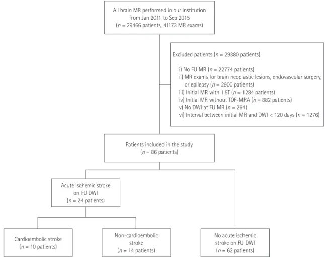
to the following inclusion criteria: 1) patients who first under- went MRI, including TOF-MRA on 3T; 2) no acute ischemic stroke on initial MRI; 3) follow-up MRI with diffusion-weighted imaging (DWI); and 4) follow-up interval ≥ 120 days. We iden- tified 29466 patients (41173 MRIs) who underwent brain MRI between January 2011 and September 2015. A total of 22774 patients were excluded because they underwent MRI without imaging follow-up and 2900 because they had MRI for brain neoplastic lesions, endovascular surgery, or epilepsy. Finally, we identified 3878 patients who underwent two or more MRI.
Among these patients, we excluded the following; 1) patients who underwent first MRI at 1.5T (n = 1284) because better im- age quality and spatial resolution of MRA at 3T than 1.5T, those were critical for diameter measurement and morphological evaluation; 2) patients who did not first undergo TOF-MRA (n = 882); 3) those without DWI on follow-up MRI (n = 264); and 4) those in whom the interval between the initial TOF-MRA and
Fig. 1. Patient selection flow chart. After systematic evaluation of the radiology database, 86 patients were included in this study. Among them, 14 patients had acute non-cardioembolic ischemic strokes.
DWI = diffusion-weighted imaging, FU = follow-up, MR = magnetic resonance, TOF-MRA = time-of-flight magnetic resonance angiography All brain MR performed in our institution
from Jan 2011 to Sep 2015 (n = 29466 patients, 41173 MR exams)
Patients included in the study (n = 86 patients)
Acute ischemic stroke on FU DWI (n = 24 patients)
Cardioembolic stroke (n = 10 patients)
Non-cardioembolic stroke (n = 14 patients)
No acute ischemic stroke on FU DWI (n = 62 patients) Excluded patients (n = 29380 patients)
i) No FU MR (n = 22774 patients)
ii) MR exams for brain neoplastic lesions, endovascular surgery, or epilepsy (n = 2900 patients)
iii) Initial MR with 1.5T (n = 1284 patients) iv) Initial MR without TOF-MRA (n = 882 patients) v) No DWI at FU MR (n = 264)
vi) Interval between initial MR and DWI < 120 days (n = 1276)
DWI was < 120 days (n = 1276). A total of 86 patients (40 males and 46 females, age 69.9 ± 10.9 years old) were included. This study was approved by the Institutional Review Board (KC15RI- SI0558). Informed consent was waived for this retrospective study. The reasons for first MRI was suspected stroke or tran- sient ischemic attack (n = 27), headache (n = 26), dizziness or syncope (n = 17), known arterial stenosis (n = 5), regular fol- low-up after previous stroke (n = 7), others such as traumatic brain injury (n = 4). Reasons for second MRI was suspected stroke or transient ischemic attack (n = 49), headache (n = 3), dizziness or syncope (n = 28), known arterial stenosis (n = 2), others such as traumatic brain injury (n = 4).
We assessed the following patient characteristics from the medical records at the time of first MRI: 1) age; 2) sex; 3) hy- pertension; 4) type 2 diabetes; 5) dyslipidemia; 6) smoking his- tory; 7) previous coronary artery disease; 8) previous ischemic stroke history; and 9) the degree of proximal internal carotid artery (ICA) stenosis. Hypertension was defined as systolic blood pressure ≥ 140 mm Hg, diastolic blood pressure ≥ 90 mm Hg, or current treatment with antihypertensive drugs. Pa- tients taking medications for diabetes (insulin or oral hypogly- cemic) were considered to have diabetes mellitus. Dyslipidemia was diagnosed if the patient had any of the following: low-den- sity lipoprotein cholesterol level ≥ 140 mg/dL, high-density li- poprotein cholesterol level ≤ 40 mg/dL, triglyceride level ≥ 150 mg/dL, or treatment with lipid-lowering medications (13). Pa- tients were considered to have a history of coronary artery dis- ease if they had previous angina pectoris, myocardial infarc- tion, or coronary artery bypass.
MRI Protocols
All initial MRIs were performed in a 3T machine (MAGNE- TOM Verio, Siemens AG Healthcare Sector, Erlangen, Germa- ny) with a 16-channel head and neck coil. Initial MRI included three-dimentional (3D) TOF-MRA and T2-weighted images of the brain, and 3D TOF-MRA of the neck. Other sequences, such as fluid attenuation inversion recovery (FLAIR) images or T1-weighted images were performed according to the clinical circumstances. 3D TOF-MRA of the brain was performed with 6 slabs, each including 40 axial slices of 0.5-mm thickness and no interslice gaps using the following parameters: repetition time/
echo time/flip angle = 22 ms/3.6 ms/18°, 384 × 331 for the fre-
quency/phase encoding matrix, a number of excitation 1, and a bandwidth of 186 Hz/pixel. A 0.4 × 0.4 × 0.5 mm voxel was ac- quired after interpolation. A saturation band 40 mm thick was applied for venous flow suppression. The generalized autocali- brating partially parallel acquisitions parameters were set to an acceleration factor of 2 in the phase encoding direction with 24 reference k-space lines for calibration. The total acquisition time was 7 min and 28 s. Other MRIs were acquired by vendor-stan- dard parameters. Follow-up DWI imaging was performed in three different MR units [MAGNETOM Verio, Signa excite HD (GE Medical Systems, Milwaukee, WI, USA), or Achieva (Philips Healthcare, Best, the Netherlands)] according to the clinical circumstances, using standard b = 1000 s/mm2.
Evaluation of the Morphological Characteristics of Intracranial Arteries on TOF-MRA
Two radiologists who were blinded to the clinical parameters evaluated the morphological characteristics of the initial TOF- MRA. On the vertical and horizontal rotation maximal intensi- ty projection images of the initial TOF-MRA, the reviewer ana- lyzed three morphological characteristics of the intracranial arteries (Table 1). These included dilatation, stenosis, and tortu- osity. In order to assess intracranial artery dilatation, we mea- sured the maximal diameter of the basilar artery (BA), and the terminal segments of both distal ICAs. The mean of these three maximal diameters were used as dilatation measurements of the intracranial arteries. The stenosis score was determined by the number of stenotic segments among seven pre-defined seg- ments [terminal segments of both distal ICA, M1 segments of both middle cerebral artery (MCA), and the V4 segments of the vertebral artery (VA) and BA]. Stenotic segments were defined as those with ≥ 50% stenosis based on the Warfarin-Aspirin Symptomatic Intracranial Disease criteria (14). Tortuosity was assessed in three segments of the intracranial arteries, including M1 segments of both MCA and BA, on a three-point scale. Zero indicates linear shape, 1 curved shape, and 2 sigmoid shape (1).
The summation of these three segment scores was used to cal- culate the tortuosity score. The other four segments (terminal segments of both distal ICA and V4 segments of the VA) were not evaluated for tortuosity given their natural tortuosity.
Evaluation of Brain MRI and Outcome Definitions Previous ischemic stroke lesions (territorial or lacunar infarcts) and white matter hyperintensities were assessed on T2-weighted and FLAIR images from the first series of images. The presence of white matter hyperintensities was graded according to the vi- sual rating scale proposed by Fazekas and Schmidt, with scores ranging from 0 to 3 (15). The degree of ICA stenosis was mea- sured according to the North American Symptomatic Carotid Endarterectomy Trial criteria on TOF-MRA of the neck area (16).
Acute ischemic stroke was defined via follow-up DWI. If the patients had diffusion-restriction lesions on follow-up DWI, the patients were considered as having had acute ischemic strokes.
If the patients had no diffusion-restriction lesions on follow-up DWI, the patients were considered as not having acute ischemic stroke at that time. An experienced stroke neurologist reviewed the medical records and MR of the patients with acute ischemic stroke. This neurologist identified cardioembolic strokes by the following characteristics: the presence of a medium-sized (maxi- mal diameter of the lesion, 15 to 30 mm) or large (> 30 mm) ce- rebral infarction; cerebral cortex involvement; stroke onset dur- ing ordinary daily activities; duration of focal neurological deficit > 24 hours; and identification of a commonly accepted cardioembolic source in the absence of confirmatory clinical (ipsilateral carotid bruit) or investigative results (Doppler ultra- sonography, carotid angiography or MRA) of the lesions in the ipsilateral supra-aortic trunks (17, 18).
Statistical Analysis
To investigate the added value of the morphological charac- teristics of the intracranial arteries to future non-cardioembolic stroke risk, two prognostic models for future non-cardioembol-
ic stroke were compared in order using the following method.
First, we performed univariate analysis using Cox proportional hazard analysis for future non-cardioembolic stroke risk with conventional stroke risk factors and the three morphological characteristics of intracranial arteries (dilatation, stenosis, and tortuosity). Multivariate analysis was then performed using the Cox regression model with the significant factors from univari- ate analysis (p < 0.2) to build two multivariate models. Model 1 only included conventional risk factors, while model 2 included conventional risk factors and additional morphological charac- teristics of the intracranial arteries. Before comparison of two models, multicollinearity was confirmed by calculating the variable inflation factor of each variable included in the models.
A variable inflation factor > 2 was considered multicollinearity, which influenced the estimated power. The likelihood-ratio test was used to compare the two models, with and without the morphological characteristics. In addition, the performance of models 1 and 2 was evaluated using the Harrell concordance index (C-index). A C-index value of 0.5 indicates random pre- diction, while 1.0 indicates perfect prediction. We also performed the same analysis for all acute ischemic strokes (non-cardioem- bolic and cardioembolic strokes) to evaluate whether there was prognostic value for modeling of all acute ischemic stroke sur- rogates. All analyses were performed with R (version 3.2.2, R Foundation, Vienna, Austria; www.R-project.org) statistical packages.
ReSUlTS
Patient Characteristics
Twenty-four of 86 patients experienced acute ischemic stroke Table 1. Evaluation of Morphological Characteristics on TOF-MRA
Category Definition
Dilatation The mean maximal diameter of BA and terminal segments of both distal ICAs
Stenosis The number of stenotic segments* among the terminal segments of both distal ICAs, M1 segments of both MCA, V4 segments of VA, and BA Tortuosity Summation of three segment scores (M1 segments of both MCAs and BA)
0 for linear shape 1 for curved shape 2 for sigmoid shape
*Stenotic segments; ≥ 50% stenosis using WASID.
BA = basilar artery, ICA = internal carotid artery, MCA = middle cerebral artery, TOF-MRA = time-of-flight magnetic resonance angiography, VA = vertebral artery, WASID = Warfarin-Aspirin Symptomatic Intracranial Disease
Table 2. Patient Characteristics (n = 86)
Variable No Stroke (n = 62) Non-Cardioembolic Stroke (n = 14) Cardioembolic Stroke (n = 10) p-Value
Age (year) 69.5 (64, 77) 67.5 (63, 73) 80 (67, 82) 0.114
Sex (%) 0.939
Female 33 (53.2) 8 (57.1) 5 (50.0)
Male 29 (46.8) 6 (42.9) 5 (50.0)
Hypertension (%) 36 (58.1) 13 (92.9) 9 (90.0) 0.012
Diabetes (%) 20 (32.3) 3 (21.4) 4 (40.0) 0.603
Dyslipidemia (%) 15 (24.2) 5 (35.7) 5 (50.0) 0.208
Coronary artery disease (%) 9 (14.5) 3 (21.4) 4 (40.0) 0.151
Previous territorial infarction (%) 10 (16.1) 5 (35.7) 3 (30.0) 0.201
White matter hyperintensity (%) 0.919
None 31 (50.0) 8 (57.1) 4 (40.0)
Mild 19 (30.6) 3 (21.4) 3 (30.0)
Moderate 7 (11.3) 1 (7.1) 2 (20.0)
Severe 5 (8.1) 2 (14.3) 1 (10.0)
Lacune (%) 24 (38.7) 7 (50.0) 6 (60.0) 0.382
ICA stenosis (NASCET method, %) 0 (0, 0) 0 (0, 0) 0 (0, 32) 0.117
Mean diameter (cm) 2.8 (2.5, 2.9) 3.1 (3, 3.3) 2.8 (2.6, 3.4) 0.005
Tortuosity score 3 (2, 4) 3 (2, 4) 2 (2, 4) 0.35
Stenosis score (%) 0.726
0 54 (85.5) 11 (78.6) 9 (90)
1 7 (11.3) 2 (14.3) 0 (0)
2 2 (3.2) 0 (0) 1 (10)
3 0 (0) 1 (7.1) 0 (0)
Observation duration (days) 611.05 ± 376.75 683.14 ± 360.54 660.80 ± 391.06 0.778
p-values were calculated using the chi-square or Mann-Whitney U test. Values are median with interquartile range in parentheses or number of patients with percentages in parentheses.
ICA = internal carotid artery, NASCET = North American Symptomatic Carotid Endarterectomy Trial
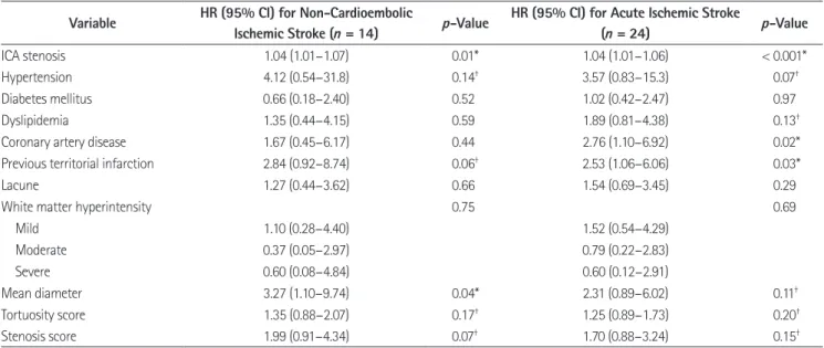
Table 3. Univariate Analysis Result for HRs for Ischemic Strokes (n = 86) Variable HR (95% CI) for Non-Cardioembolic
Ischemic Stroke (n = 14) p-Value HR (95% CI) for Acute Ischemic Stroke
(n = 24) p-Value
ICA stenosis 1.04 (1.01–1.07) 0.01* 1.04 (1.01–1.06) < 0.001*
Hypertension 4.12 (0.54–31.8) 0.14† 3.57 (0.83–15.3) 0.07†
Diabetes mellitus 0.66 (0.18–2.40) 0.52 1.02 (0.42–2.47) 0.97
Dyslipidemia 1.35 (0.44–4.15) 0.59 1.89 (0.81–4.38) 0.13†
Coronary artery disease 1.67 (0.45–6.17) 0.44 2.76 (1.10–6.92) 0.02*
Previous territorial infarction 2.84 (0.92–8.74) 0.06† 2.53 (1.06–6.06) 0.03*
Lacune 1.27 (0.44–3.62) 0.66 1.54 (0.69–3.45) 0.29
White matter hyperintensity 0.75 0.69
Mild 1.10 (0.28–4.40) 1.52 (0.54–4.29)
Moderate 0.37 (0.05–2.97) 0.79 (0.22–2.83)
Severe 0.60 (0.08–4.84) 0.60 (0.12–2.91)
Mean diameter 3.27 (1.10–9.74) 0.04* 2.31 (0.89–6.02) 0.11†
Tortuosity score 1.35 (0.88–2.07) 0.17† 1.25 (0.89–1.73) 0.20†
Stenosis score 1.99 (0.91–4.34) 0.07† 1.70 (0.88–3.24) 0.15†
*p < 0.05.
†p < 0.2.
CI = confidence interval, HR = hazard ratio, ICA = internal carotid artery
on follow-up DWI. Among them, fourteen patients had non- cardioembolic strokes. Hypertension was more frequent in the patients in the stroke group (p = 0.012) than it was in the non- stroke group. The mean diameters were larger in patients with strokes than in those without stroke (p = 0.025). The clinical characteristics of the patient population according to the occur- rence of ischemic stroke and its type are listed in Table 2.
Univariate Analysis of Future Stroke Risk
Univariate analysis (Table 3) revealed that both the degree of ICA stenosis and the mean diameter of the intracranial arteries were significantly associated with future non-cardioembolic ischemic stroke risk (p < 0.05). Hypertension, previous territo- rial infarcts, the tortuosity score, and stenosis score were also marginally associated with future ischemic stroke (p < 0.2). For all ischemic strokes, the degree of ICA stenosis, history of coro- nary artery disease, and previous territorial infarction were sig- nificantly associated with future ischemic stroke risk. Hyper- tension, dyslipidemia, the mean diameter of the intracranial arteries, tortuosity score, and stenosis scores were marginally associated with future ischemic stroke (p < 0.2).
Multivariate Analysis of Future Stroke Risk
The multivariate analysis results are presented in Table 4.
Among the three morphological characteristics, the mean di- ameter of the intracranial arteries was significantly associated with future non-cardioembolic strokes [hazard ratio (HR) 9.45, 95% confidence interval (CI) 1.41–63.55, p = 0.02] (Fig. 2). The stenosis score was marginally associated with future non-cardio-
embolic strokes (HR 2.26, 95% CI 0.84–6.07, p = 0.11). In con- trast, tortuosity was not associated with future non-cardioem- bolic strokes (HR 0.97, 95% CI 0.57–1.63, p = 0.9).
The performance of models 1 (conventional risk factors only) and 2 (conventional risk factors and morphological findings) were significantly different with regard to modeling future non- cardioembolic stroke risk (p = 0.031). Harrell’s C-index of model 2 (0.75, 95% CI 0.571–0.929) was higher than that of model 1 (0.678, 95% CI 0.467–0.889). In contrast, models 1 and 2 did not differ in modeling the future risk of all ischemic strokes (p = 0.367). The C-index of model 1 (0.717, 95% CI 0.560–0.874) was similar to that of model 2 (0.736, 95% CI 0.587–0.885). We did not identify multicollinearity of the included variables in any of the models.
DISCUSSION
The association between imaging characteristics of the intra- cranial arteries, atherosclerotic changes and aging has been previously reported. The presence of a stenotic segment is a well- known manifestation of atherosclerotic changes (1, 19, 20).
Furthermore, tortuous and dilated intracranial arteries, aging and hypertension have also been reported as important athero- sclerosis risk factors (3, 5, 7, 21). We investigated three morpho- logical characteristics of the intracranial arteries. Our result sug- gested that three morphological characteristics of intracranial arteries may be imaging phenotypes and surrogates for athero- sclerotic changes. We think that the prognostic value of a high burden of intracranial atherosclerotic changes may be associat- Table 4. Multivariate Analysis Result for HRs for Ischemic Stroke (n = 86)
Variable
Non-Cardioembolic Stroke All Ischemic Stroke
Model 1 p-Value Model 2 p-Value Model 1 p-Value Model 2 p-Value
HR (95% CI) HR (95% CI) HR (95% CI) HR (95% CI)
Hypertension 3.64 (0.46–28.59) 0.22 3.95 (0.44–35.28) 0.22 2.71 (0.60–12.29) 0.20 2.41 (0.51–11.34) 0.27 Previous territorial infarction 3.49 (1.03–11.87) 0.04 3.55 (1.03–12.20) 0.04 4.14 (1.39–12.31) 0.01 4.04 (1.28–12.77) 0.02 ICA stenosis 1.04 (1.01–1.08) 0.009 1.04 (1.00–1.08) 0.061 1.04 (1.02–1.07) 0.002 1.04 (1.02–1.07) 0.002
Coronary artery disease NA NA 2.54 (0.87–7.43) 0.09 2.24 (0.77–6.48) 0.14
Dyslipidemia NA NA 0.98 (0.33–2.91) 0.97 1.02 (0.33–3.13) 0.98
Mean diameter NA 9.45 (1.41–63.55) 0.02 NA 2.01 (0.66–6.10) 0.22
Tortuosity score NA 0.97 (0.57–1.63) 0.90 NA 0.95 (0.66–1.39) 0.80
Stenosis score NA 2.26 (0.84–6.07) 0.11 NA 1.65 (0.74–3.66) 0.22
Model 1: Analysis with conventional risk factors, Model 2: Analysis with conventional risk factors and morphological characteristics of the intracranial ar- teries.
CI = confidence interval, HR = hazard ratio, ICA = internal carotid artery, NA = not available
ed with future non-cardioembolic stroke. An interesting find- ing is that the dilatation of intracranial artery was more impor- tant than other morphologic characteristics, including stenosis or tortuosity. One study suggested that the non-stenotic intra- cranial arteries of patients with acute ischemic stroke patients showed the tendency of positive remodeling (11). Also, increased intracranial arterial diameter was suggested as a possible mark- er of chronic vascular insufficiency of the brain and was associ- ated with vascular risk factors (3, 4).
In our study, the imaging surrogates of intracranial atheroscle- rosis had added value for future non-cardioembolic stroke risk modeling. Because there is an association between the morpho- logical characteristics of intracranial arteries and some stroke risk factors, one might argue that there was a multicollinearity effect between the risk factors and morphological characteristics. How- ever, our results suggested the lack of multicollinearity between known stenotic risk factors and morphological characteristics.
In addition, the mean diameters of intracranial arteries were independently associated with future non-cardioembolic stroke in model 2. The lack of multicollinearity might be explained by the unique value of imaging surrogates (22). There may be in- dividual variability of imaging phenotypes among patients with the same risk factors and demographics. This was due to differ- ent susceptibility of individual risk factors. For this reason, im- aging surrogates have different clinical significance from the risk factors. Different imaging phenotypes might result from varia- tion in susceptibility to stroke and atherosclerotic risk factors.
The morphologic characteristics of intracranial arteries were thought to be imaging the phenotypes of intracranial arterial atherosclerosis, with additional clinical significance.
Acute ischemic stroke is a collection of heterogeneous diseas- es causing brain tissue ischemia. The Trial of ORG 10172 in Acute Stroke Treatment classification is one example that high- lights the heterogeneous nature of ischemic stroke (23). Cardio- embolic stroke, which accounts for approximately one in four ischemic strokes, has a relatively different pathophysiology and treatment strategy than does other types of ischemic stroke (23-25). Embolism from the heart to the brain results from one of the following mechanisms: structural heart abnormalities (e.g., left ventricular aneurysm), cardiac valvular disease, right- to-left shunts (paradoxical embolism), and rhythm disturbance (6). Dilatation, tortuosity and stenotic segments of the intracra- nial arteries are associated with atherosclerotic changes. How- ever, these characteristics were not closely linked to the mecha- nism of cardioembolic stroke. This finding explains why the morphological characteristics of intracranial arteries do not add value to the prediction of all types of ischemic stroke. One clinical study found that atrial fibrillation and the sudden onset of symptoms were independently associated with cardioembol- ic stroke; in contrast, hypertension, chronic obstructive pulmo- nary disease, diabetes mellitus, hyperlipidemia and age were all
1.0
0.8
0.6
0.4 Event free survival probability
0 500 1000
Time (days)
Non-cardioembolic ischemic stroke
Mean diameter ≤ 3.1
p = 0.00046
Mean diameter > 3.1
A
1.0 0.8 0.6 Event free survival probability 0.4
0 500 1000
Time (days)
Non-cardioembolic ischemic stroke
Tortuosity score ≤ 3
p = 0.158
Tortuosity score > 3
C 1.0
0.8
0.6
Event free survival probability 0.4
0 500 1000
Time (days)
Non-cardioembolic ischemic stroke
Stenosis score = 0 or 1
p = 0.3
Stenosis score = 2 or 3
B
Fig. 2. Survival curves of non-cardioembolic strokes according to three morphological characteristics. Three survival curves for non-car- dioembolic stroke according to the intracranial arterial diameter (A), stenosis score (B) and tortuosity score (C). A mean intracranial arteri- al diameter > 3.1 was associated with significant risk for future non- cardioembolic stroke (p < 0.001). Tortuous intracranial arteries were also associated with a tendency toward a high probability of future non-cardioembolic stroke (p = 0.158).
significantly associated with atherothrombotic infarctions (26).
In our study, hypertension was significantly more frequent in the non-cardioembolic stroke group than it was in the cardio- embolic stroke group.
We performed a systematic, retrospective review to determine future stroke risk based on conventional ischemic stroke risk factors, in addition to the morphologies of intracranial arteries.
We found that the risk of future ischemic strokes is influenced by coronary artery disease, hypertension, stenosis of the proxi- mal ICA, and imaging evidence of prior ischemic strokes. These findings are concordant with those of previous reports (27-29).
This concordance also suggests that our retrospective cohort was relevant to assess future ischemic stroke risk. A history of coro- nary artery disease particularly increases the risk of future isch- emic stroke, although it does not affect the risk of future non- cardioembolic stroke.
In this study, we assessed the added prognostic value of the morphological characteristics of intracranial arteries to predict future ischemic stroke risk. When these morphological charac- teristics were combined with conventional risk factors, they were valuable in the prediction of future non-cardioembolic strokes. Among the three morphological characteristics that we included, the increased mean diameter or dilatation of the in- tracranial arteries was independently associated with future non-cardioembolic stroke risk. Stenosis of the intracranial ar- teries was also marginally associated with future risk of non- cardioembolic stroke.
This study has several limitations. First, the sample size of in- cluded patients was relatively small. Fortunately, however, we reviewed a large number of patients systematically. The prog- nostic values of known ischemic stroke risk factors were con- cordant with a previous large-scale study. Small sample size and small number of event might limit the statistical power of the result. Future study with large subject is required to validate our result. Due to small size of population, subgroup analysis ac- cording to a variety of stroke etiology cannot be done. Second limitation is that our retrospective approach involved a degree of selection bias. Patients who visited another hospital for stroke episode cannot be included. Furthermore, we could not include other known risk factors, such as smoking, heart dis- ease (other than coronary artery disease), alcohol consumption, or family history, because this information was not available in
the medical records. Third limitation, which came using TOF- MRA, is that we could not detect some atherosclerotic changes.
Because TOF-MRA only visualizes the lumen of the vessel, ath- erosclerotic plaques with positive remodeling cannot be detect- ed (10, 11). Instead, we analyzed the diameter, degree of tortu- osity and stenosis, all of which are well demonstrated on TOF- MRA. These characteristics are also associated with atherosclerotic changes. Additionally, the spatial resolution of the analyzed TOF- MRA might be not enough to quantify the degree of intracranial arterial stenosis, and measured value might be overestimating the true stenosis. Instead, we used the number of significantly stenotic segment, which might not be enough to represent the significance of stenosis degree of each segment. Although the number of stenotic segment showed marginal significance, fu- ture study is needed to validate this finding. Lastly, we used im- age-based definitions of acute ischemic stroke, rather than using medical records. Ischemic stroke was defined as only positive on DWI. Therefore, small lacunar infarcts or transient ischemic at- tack may have been missed. Also, because we defined stroke events based on diffusion-restriction lesion, we may miss sub- acute and chronic infarctions, or acute infarct patients those did not underwent DWI.
In conclusion, in combination with conventional stroke risk factors, assessment of the morphological characteristics of in- tracranial arteries adds value to predicting the risk of future non-cardioembolic ischemic stroke.
RefeReNCeS
1. Han J, Qiao H, Li X, Li X, He Q, Wang Y, et al. The three-di- mensional shape analysis of the M1 segment of the middle cerebral artery using MRA at 3T. Neuroradiology 2014;56:
995-1005
2. Diedrich KT, Roberts JA, Schmidt RH, Kang CK, Cho ZH, Parker DL. Validation of an arterial tortuosity measure with application to hypertension collection of clinical hyperten- sive patients. BMC bioinformatics 2011;12 Suppl 10:S15 3. Pico F, Labreuche J, Touboul PJ, Amarenco P. Intracranial ar-
terial dolichoectasia and its relation with atherosclerosis and stroke subtype. Neurology 2003;61:1736-1742 4. Pico F, Labreuche J, Touboul PJ, Leys D, Amarenco P. Intra-
cranial arterial dolichoectasia and small-vessel disease in
stroke patients. Ann Neurol 2005;57:472-479
5. Del Corso L, Moruzzo D, Conte B, Agelli M, Romanelli AM, Pastine F, et al. Tortuosity, kinking, and coiling of the carotid artery: expression of atherosclerosis or aging? Angiology 1998;49:361-371
6. MacDougall NJ, Amarasinghe S, Muir KW. Secondary pre- vention of stroke. Expert Rev Cardiovasc Ther 2009;7:1103- 1115
7. Gutierrez J, Sacco RL, Wright CB. Dolichoectasia-an evolving arterial disease. Nat Rev Neurol 2011;7:41-50
8. Çoban G, Çifçi E, Yildirim E, Ag˘ıldere AM. Predisposing fac- tors in posterior circulation infarcts: a vascular morpho- logical assessment. Neuroradiology 2015;57:483-489 9. Nakamura Y, Hirayama T, Ikeda K. Clinicoradiologic features
of vertebrobasilar dolichoectasia in stroke patients. J Stroke Cerebrovasc Dis 2012;21:5-10
10. Qiao Y, Anwar Z, Intrapiromkul J, Liu L, Zeiler SR, Leigh R, et al. Patterns and implications of intracranial arterial remod- eling in stroke patients. Stroke 2016;47:434-440
11. Lee WJ, Choi HS, Jang J, Sung J, Kim TW, Koo J, et al. Non- stenotic intracranial arteries have atherosclerotic changes in acute ischemic stroke patients: a 3T MRI study. Neuro- radiology 2015;57:1007-1013
12. Morris SA, Orbach DB, Geva T, Singh MN, Gauvreau K, La- cro RV. Increased vertebral artery tortuosity index is associ- ated with adverse outcomes in children and young adults with connective tissue disorders. Circulation 2011;124:
388-396
13. Deguchi I, Ohe Y, Fukuoka T, Dembo T, Nagoya H, Kato Y, et al. Relationship of obesity to recanalization after hyper- acute recombinant tissue-plasminogen activator infusion therapy in patients with middle cerebral artery occlusion.
J Stroke Cerebrovasc Dis 2012;21:161-164
14. Samuels OB, Joseph GJ, Lynn MJ, Smith HA, Chimowitz MI.
A standardized method for measuring intracranial arterial stenosis. AJNR Am J Neuroradiol 2000;21:643-646
15. Kapeller P, Barber R, Vermeulen RJ, Adèr H, Scheltens P, Fre- idl W, et al. Visual rating of age-related white matter chang- es on magnetic resonance imaging: scale comparison, in- terrater agreement, and correlations with quantitative measurements. Stroke 2003;34:441-445
16. North American Symptomatic Carotid Endarterectomy Tri-
al Collaborators; Barnett HJM, Taylor DW, Haynes RB, Sack- ett DL, Peerless SJ, Ferguson GG, et al. Beneficial effect of ca- rotid endarterectomy in symptomatic patients with high- grade carotid stenosis. N Engl J Med 1991;325:445-453 17. Arboix A, Garcia-Eroles L, Massons JB, Oliveres M, Pujades
R, Targa C. Atrial fibrillation and stroke: clinical presenta- tion of cardioembolic versus atherothrombotic infarction.
Int J Cardiol 2000;73:33-42
18. Martí-Vilalta JL, Matías-Guiu J. [Nomenclature of cerebral vascular diseases]. Neurologia 1987;2:166-175
19. Li ML, Xu WH, Song L, Feng F, You H, Ni J, et al. Atheroscle- rosis of middle cerebral artery: evaluation with high-reso- lution MR imaging at 3T. Atherosclerosis 2009;204:447- 452
20. Degnan AJ, Gallagher G, Teng Z, Lu J, Liu Q, Gillard JH. MR angiography and imaging for the evaluation of middle ce- rebral artery atherosclerotic disease. AJNR Am J Neurora- diol 2012;33:1427-1435
21. Ince B, Petty GW, Brown RD Jr, Chu CP, Sicks JD, Whisnant JP. Dolichoectasia of the intracranial arteries in patients with first ischemic stroke: a population-based study. Neurology 1998;50:1694-1698
22. Gillies RJ, Kinahan PE, Hricak H. Radiomics: images are more than pictures, they are data. Radiology 2016;278:563-577 23. Adams HP Jr, Bendixen BH, Kappelle LJ, Biller J, Love BB, Gor- don DL, et al. Classification of subtype of acute ischemic stroke. definitions for use in a multicenter clinical trial.
TOAST. trial of Org 10172 in acute stroke treatment. Stroke 1993;24:35-41
24. Arboix A, Alió J. Acute cardioembolic stroke: an update. Ex- pert Rev Cardiovasc Ther 2011;9:367-379
25. Galanis T, Merli GJ. Direct thrombin and factor Xa inhibition for stroke prevention in patients with atrial fibrillation. Hosp Pract (1995) 2013;41:26-36
26. Arboix A, Oliveres M, Massons J, Pujades R, Garcia-Eroles L.
Early differentiation of cardioembolic from atherothrom- botic cerebral infarction: a multivariate analysis. Eur J Neu- rol 1999;6:677-683
27. Sacco RL, Benjamin EJ, Broderick JP, Dyken M, Easton JD, Feinberg WM, et al. American heart association prevention conference. IV. prevention and rehabilitation of stroke. risk factors. Stroke 1997;28:1507-1517
두개내 동맥의 형태학적 특성이 비심인성 색전 뇌경색 발생 위험에 미치는 추가적 예후 가치
한나혜
1· 장진희
1* · 변호균
1· 이기정
2· 구자성
2· 최현석
1· 정소령
1· 안국진
1· 김범수
1목적: 두개내 동맥의 형태학적 특성이 비심인성 색전 뇌경색 발생 위험에 미치는 추가적 예후 가치를 분석하고자 하였다.
대상과 방법: 후향적 연구를 시행하였으며, 급성 뇌경색이 없으면서 유체 속도 강조 자기공명 혈관조영술(time-of-flight magnetic resonance angiography; 이하 TOF-MRA)을 포함한 3T 자기공명영상을 시행한 86명의 환자가 등록되었다.
환자들은 첫 자기공명영상 시행 이후 > 120일 뒤에 확산강조영상(diffusion-weighted imaging; 이하 DWI)을 포함한 추 적검사를 시행하였다. TOF-MRA에서 두개내 동맥의 확장, 협착, 사행의 세 가지 형태학적 특성을 분석하고 추적 DWI에 서 급성 허혈성 뇌경색 여부를 평가하여 두 가지 예측 모형을 만들었다. 모형 1은 전통적 뇌경색 위험인자를, 모형 2는 이 와 두개내 동맥의 형태학적 특성을 포함하였다. 두 모형 비교에 우도비 검정(likelihood-ratio)을 사용하였고, 모형의 성능 은 Harrell의 일치 지수로 평가하였다.
결과: 14명의 환자가 비심인성 색전 뇌경색으로 진단되었고, 두 모형의 성능은 비심인성 색전 뇌경색 발생 위험 예측에 있 어서 유의한 차이를 보였다(p = 0.031). Harrell의 일치 지수는 모형 2 (0.78 ± 0.05)가 모형 1 (0.72 ± 0.07)보다 높 았다.
결론: 전통적 뇌경색 위험인자와 더불어, 두개내 동맥의 형태학적 특성은 비심인성 색전 뇌경색 발생 위험 예측에 있어서 유용하다.
가톨릭대학교 의과대학 서울성모병원 1영상의학과, 2신경과
28. Harmsen P, Lappas G, Rosengren A, Wilhelmsen L. Long-term risk factors for stroke: twenty-eight years of follow-up of 7457 middle-aged men in Göteborg, Sweden. Stroke 2006;
37:1663-1667
29. Wolf PA, D’Agostino RB, Belanger AJ, Kannel WB. Proba- bility of stroke: a risk profile from the Framingham Study.
Stroke 1991;22:312-318