Table 1. Results of Sonography, Tc-99m DISIDA Scan and MR Cholangiography in 22 Patients with Neonatal Cholestasis Sonography Tc-99m DISIDA scan MR cholangiography No. Sex Age (days) Final Dx
Dx GB; shape/length (cm) TC Dx GB SB HE Dx GB shape EHBD PPH
01 f 62 BA BA E, I / 1.63 + NH low BA E +
02 f 61 BA BA O, S / 3.32 BA high BA O +
03 f 42 BA BA A, I / 1.1 NH low BA A
04 m 58 BA BA A, I / 0.5 + BA low BA A +
05 f 31 BA BA A, I / 1.1 + BA high NH E + +
06 m 57 BA NH E, S / 1.58 NH low NH E + +
07 f 51 BA BA E, I / 1.59 + NH low BA E +
08 f 32 BA NH E, S / 1.65 NH low BA E +
09 f 65 BA BA A, I / 1.2 + NH low BA E +
10 f 63 BA BA A, I / 0.97 BA high BA A +
11 f 90 BA BA A, I / 1.79 + BA low BA A +
12 m 51 BA BA E, I / 1.69 + BA low BA E +
13 f 55 BA NH O, S / 2.12 NH low BA O +
14 m 23 BA BA A, I / 1.09 + NH low BA A +
15 f 55 BA BA E, I / 3.06 + BA high BA E +
16 m 58 NH NH E, S / 2.46 BA high BA E +
17 m 58 NH BA E, I / 1.4 BA high NH E +
18 m 1030 NH NH E, S / 1.62 NH + + low NH E +
19 m 63 NH NH E, S / 2.16 NH + + low BA E
20 m 70 NH NH E, S / 3.3 BA high NH E +
21 m 72 NH NH E, S / 1.6 BA high NH E +
22 f 31 NH NH E, S / 1.58 NH + low NH O +
BA: biliary atresia, NH; neonatal hepatitis, TC: triangular cord sign, GB: gallbladder, SB; small bowel HE: hepatic extraction of radiotracer, EHBD: extrahepatic bile duct, PPH: periportal high signal intensity E: elongated, O: oval, A: atretic, I: irregular margin, S: smooth margin
A B
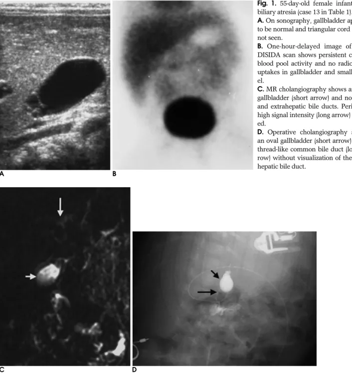
Fig. 1. 55-day-old female infant with biliary atresia (case 13 in Table 1).
A. On sonography, gallbladder appears to be normal and triangular cord sign is not seen.
B. One-hour-delayed image of 99mTc DISIDA scan shows persistent cardiac blood pool activity and no radiotracer uptakes in gallbladder and small bow- el.
C. MR cholangiography shows an oval gallbladder (short arrow) and no intra- and extrahepatic bile ducts. Periportal high signal intensity (long arrow) is not- ed.
D. Operative cholangiography shows an oval gallbladder (short arrow) and a thread-like common bile duct (long ar- row) without visualization of the intra- hepatic bile duct.
C D
A B C Fig. 2. 58-day-old male infant with neonatal hepatitis (case 17 in Table 1).
A. Sonography shows small, contracted gallbladder. There is no triangular cord sign (arrow).
B. One-hour-delayed image of 99mTc DISIDA scan shows normal hepatic extraction and no radiotracer uptakes in gallbladder and small bowel.
C. MR cholangiography shows normal extrahepatic bile duct (arrow).
Table 2. Summary of Diagnostic Results in Each Imaging Modality Compared with Final Diagnosis
Final Diagnosis Sonography 99mTc DISIDA scan MR cholangiography
BA NH BA NH BA NH
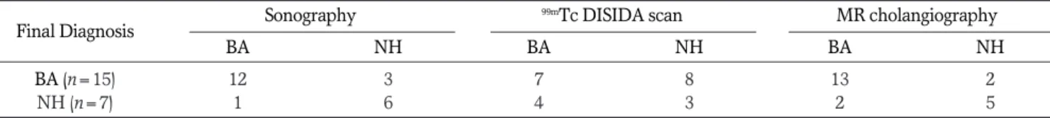
BA (n=15) 12 3 7 8 13 2
NH (n=7) 1 6 4 3 2 5
BA: biliary atresia, NH: neonatal hepatitis
A B C Fig. 3. 31-day-old female infant with biliary atresia (case 5 in Table 1).
A. MR cholangiography shows an elongated gallbladder, periportal high signal intensity (thin arrow), and curvilinear structure (thick arrow) mimicking common bile duct so it is diagnosed as neonatal hepatitis. Extrahepatic bile duct is not visualized on opera- tive cholangiography.
B. Sonography shows an atrophic gallbladder (short arrow) and triangular cord sign (long arrow).
C. One-hour-delayed image of 99mTc DISIDA scan shows high hepatic extraction and no radiotracer uptakes in gallbladder and small bowel.
Table 3. Validity and Reliability of Sonography, 99mTc DISIDA Scan and MR Cholangiography for the Diagnosis of Biliary Atresia
Sonography 99mTc DISIDA scan MR cholangiography
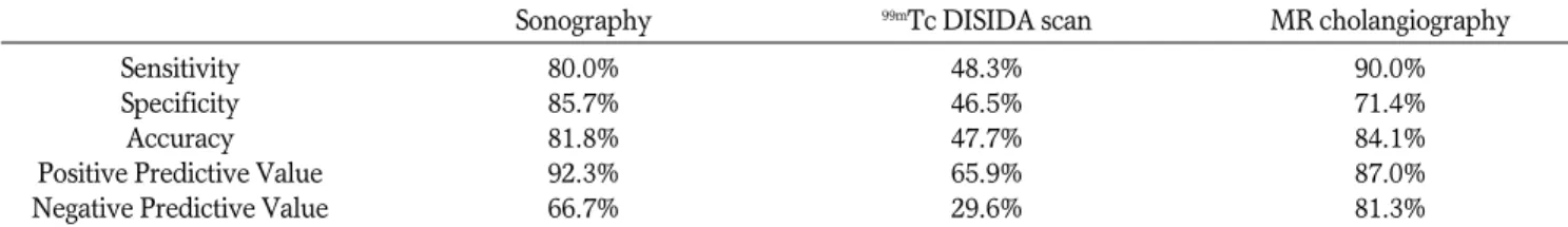
Sensitivity 80.0% 48.3% 90.0%
Specificity 85.7% 46.5% 71.4%
Accuracy 81.8% 47.7% 84.1%
Positive Predictive Value 92.3% 65.9% 87.0%
Negative Predictive Value 66.7% 29.6% 81.3%
A B
Fig. 4. 58-day-old male infant with neonatal hepatitis (case 16 in Table 1).
A. Extrahepatic bile duct cannot be demonstrated on MR cholangiography due to improper selection of the imag- ing plane. Preoperative diagnosis is bil- iary atresia.
B. Sonography shows an elongated gall- bladder. There is no triangular cord sign (not shown).
C. One-hour-delayed image of 99mTc DISIDA scan shows high hepatic ex- traction and no radiotracer uptakes in gallbladder and small bowel.
D. Operative cholangiography shows normal intra- and extrahepatic bile ducts.
C D
1. Kirks DR, Coleman RE, Filston HC, Rosenberg ER, Merten DF. An imaging approach to persistent neonatal jaundice. AJR Am J Roentgenol 1984;142:461-465
2. Shah HA, Spivak W. Neonatal cholestasis. New approaches to di- agnostic evaluation and therapy. Pediatr Clin North Am 1994;41:
943-966
3. Park WH, Choi SO, Lee HJ, Kim SP, Zeon SK, Lee SL. A new diag- nostic approach to biliary atresia with emphasis on the ultrasono- graphic triangular cord sign: comparison of ultrasonography, hepa- tobiliary scintigraphy, and liver needle biopsy in the evaluation of infantile cholestasis. J Pediatr Surg 1997;32:1555-1559
4. Tan Kendrick AP, Phua KB, Ooi BC, Subramaniam R, Tan CE,
Goh ASW. Making the diagnosis of biliary atresia using the trian- gular cord sign and gallbladder length. Pediatr Radiol 2000;30:69-73 5. Park WH, Choi SO, Lee HJ. Technical innovation for noninvasive and early diagnosis of biliary atresia: the ultrasonographic trian- gular cord sign. J Hepatobiliary Pancreat Surg 2001;8:337-341 6. Gerhold JP, Klingensmith WC 3rd, Kuni CC, et al. Diagnosis of bil-
iary atresia with radionuclide hepatobiliary imaging. Radiology 1983;146:499-504
7. Spivak W, Sarkar S, Winter D, Glassman M, Donlon E, Tucker KJ.
Diagnostic utility of hepatobiliary scintigraphy with 99mTc- DISIDA in neonatal cholestasis. J Pediatr 1987;110:855-861
8. Gilmour SM, Hershkop M, Reifen R, Gilday D, Roberts EA.
Outcome of hepatobiliary scanning in neonatal hepatitis syn- drome. J Nucl Med 1997;38:1279-1282
9. Lin WY, Lin CC, Changlai SP, Shen YY, Wang SJ. Comparison technetium of Tc-99m disofenin cholescintigraphy with ultra- sonography in the differentiation of biliary atresia from other forms of neonatal jaundice. Pediatr Surg Int 1997;12:30-33 10. Hay SA, Soliman HE, Sherif HM, Abdelrahman AH, Kabesh AA,
Hamza AF. Neonatal jaundice: the role of laparoscopy. J Pediatr Surg 2000;35:1706-1709
11. Senyuz OF, Yesildag E, Emir H, et al. Diagnostic laparoscopy in prolonged jaundice. J Pediatr Surg 2001;36:463-465
12. Guibaud L, Lachaud A, Touraine R, et al. MR cholangiography in neonates and infants: feasibility and preliminary applications. AJR Am J Roentgenol 1998;170:27-31
13. Miyazaki T, Yamashita Y, Tang Y, Tsuchigame T, Takahashi M, Sera Y. Single-shot MR cholangiopancreatography of neonates, in- fants, and young children. AJR Am J Roentgenol 1998;170:33-37 14. Jaw TS, Kuo YT, Liu GC, Chen SH, Wang CK. MR cholangiogra-
phy in the evaluation of neonatal cholestasis. Radiology 1999;212:
249-256
15. Kim MJ, Park YN, Han SJ, et al. Biliary atresia in neonates and in- fants: triangular area of high signal intensity in the porta hepatis at T2-weighted MR cholangiography with US and histopathologic correlation. Radiology 2000;215:395-401
16. Han SJ, Kim MJ, Han A, et al. Magnetic resonance cholangiogra- phy for the diagnosis of biliary atresia. J Pediatr Surg 2002;37:599- 604
17. Norton KI, Glass RB, Kogan D, Lee JS, Emre S, Shneider BL. MR cholangiography in the evaluation of neonatal cholestasis: initial results. Radiology 2002;222:687-691
18. Avni FE, Segers V, De Maertelaer V, et al. The evaluation by mag- netic resonance imaging of hepatic periportal fibrosis in infants with neonatal cholestasis: preliminary report. J Pediatr Surg 2002;
37:1128-1133
19. Bu LN, Chen HL, Ni YH, et al. Multiple intrahepatic biliary cysts in children with biliary atresia. J Pediatr Surg 2002;37:1183-1187 20. Azar G, Beneck D, Lane B, Markowitz J, Daum F, Kahn E.
Atypical morphologic presentation of biliary atresia and value of serial liver biopsies. J Pediatr Gastroenterol Nutr 2002;34:212-215
Address reprint requests to : Myung-Joon Kim, M.D., Department of Diagnostic Radiology and Research Institute of Radiological Science, Yonsei University College of Medicine, 134 Shinchon-dong, Seodaemun-gu, Seoul 120-752, Korea.
Tel. 82-2-361-5837 Fax. 82-2-393-3035 E-mail: [email protected]
Usefulness of MR Cholangiography in the Evaluation of Neonatal Cholestasis: Comparison with 99mTc DISIDA Scan
1
Jinna Kim, M.D., Myung-Joon Kim, M.D., Choon Sik Yoon, M.D., Jong-Doo Lee, M.D., Si-Yeon Kim, M.D., Seok Joo Han, M.D.
2, Eui Ho Hwang, M.D.
21Department of Diagnostic Radiology and Research Institute of Radiological Science, Yonsei University College of Medicine
2Department of Surgery, Yonsei University College of Medicine
Purpose:
To evaluate the diagnostic validity of MR cholangiography as a second-line imaging tool following sonography in the evaluation of neonatal cholestasis, we compared MR cholangiography with
99mTc DISIDA scan.
Materials and Methods:
We retrospectively evaluated sonography,
99mTc DISIDA scan and MR cholangiogra- phy in twenty-two neonates and infants (age range, 23 103 days; mean age, 57 days) presenting with conju- gated hyperbilirubinemia. Of the 22 patients, 15 were diagnosed as biliary atresia by operative cholangiogra- phy and liver biopsy and six as neonatal hepatitis by imaging finding and clinical data. Remaining one patient was diagnosed as neonatal hepatitis by operative cholangiography and liver biopsy. Two independent ob- servers for each study were assigned to review the images of
99mTc DISIDA scan and MR cholangiography without giving the final diagnosis or other clinical data. Diagnostic accuracy and interobserver variability for each study were evaluated.
Results:
On
99mTc DISIDA scan, biliary atresia was mistaken for neonatal hepatitis in eight patients and vice versa in four patients. On MR cholangiography, it was mistaken biliary atresia as neonatal hepatitis and vice versa in each two patients. Sensitivity, specificity, accuracy, positive and negative predictive values of
99mTc DISIDA scan were 48%, 47%, 48%, 66% and 30%, respectively, and those of MR cholangiography were 90%, 71%, 84%, 87% and 81%, respectively. Interobserver variabilities for
99mTc DISIDA scan and MR cholangiogra- phy were 0.62 and 0.85, respectively.
Conclusion:
In the evaluation of patients with neonatal cholestasis, it would be advisable to use MR cholan- giography, having superior diagnostic accuracy to
99mTc DISIDA scan, as a second-line imaging tool following sonography.
Index words :