Introduction
To overcome the esthetic problems and to comply with clinician's and patient's increased demands for highly esthetic results, ceram- ic abutments started to be developed.
Ceramic abutments for dental implants have been in clinical use since early 1990s.1At first, the abutments were made of the dense- ly sintered high-purity alumina (Al2O3) which in some studies showed good survival rates.2,3
Zirconia abutments are successors to alumina (Al2O3) abut-
ments. Compared with the former, zirconia abutments are radiopaque and demonstrate significantly higher resistance to fracture property.
Some studies had reported that the flexural strength (900 - 1200 MPa) of yttrium-stabilized zirconium oxide is three times that of pure alu- minum oxide,4,5and it's fracture toughness (9 - 10 MPa m1/2) is 2 times as high whereas Young's modulus registers only half of the aluminum oxide values.4,6The clinical outcome and survival rate of zirconia abutments have shown better reliability in vitro and vivo studies and also been comparable to those observed for titanium abutments in all area including molar substitutes.7,8
지르코니아와 레진나노세라믹 임플란트 지대주의 두께에 따른 열순환 후 파절저항
이정원∙차현석∙이주희*
울산대학교 의과대학 서울아산병원 치과보철과
Fracture resistance of zirconia and resin nano ceramic implant abutments according to thickness after thermocycling
Jung-Won Lee, Hyun-Suk Cha, Joo-Hee Lee*
Department of Prosthodontics, College of Medicine, University of Ulsan, Asan Medical Center, Seoul, Republic of Korea
Purpose: The aim of this in vitro study is to investigate load bearing capacity of esthetic abutments according to the type of material and wall thickness. Materials and meth- ods: 70 specimens equally divided into seven groups according to their abutment wall thicknesses. The abutments prepared with titanium 0.5 mm wall thickness were used as a control group (Ti-0.5), whereas zirconia abutments and resin nano ceramic abutments with wall thickness 0.5 mm, 0.8 mm and 1.0 mm were prepared as test groups (Zir-0.5, Zir-0.8, Zir-1.0 and RNC-0.5, RNC-0.8, RNC-1.0). All specimens were tested in a universal testing machine to evaluate their resistance to fracture and all of them underwent thermo-cycling before loading test. Mean fracture values of the groups were measured and statistical analyses were made using two-way ANOVA. Results: Zir-1.0 showed the highest mean strength (2,476.3 ± 342.0 N) and Zir-0.8 (1,518 ± 347.9 N), Ti-0.5 (1,041.8 ± 237.2 N), Zir-0.5 (631.4 ± 149.0 N) were followed. The strengths of RNC groups were significantly lower compared to other two materials (RNC-1.0 427.5 ± 72.1, RNC-0.8 297.9 ± 41.2) and the strengths of all the test groups decreased as the thickness decreases (P < .01). RNC-0.5 (127.4 ± 35.3 N) abutments were weaker than all other groups (P < .05). Conclusion: All tested zirconia abutments have the potential to withstand the physiologic occlusal forces in anterior and posterior regions. In resin nano ceramic abutments, wall thickness more than 0.8 mm showed the possibility of withstanding the occlusal forces in anterior region. (J Korean Acad Prosthodont 2017;55:144-50)
Keywords: Nano ceramics; Dental abutment; Load-bearing capacity; Zirconia; Fracture
c cc
2017 The Korean Academy of Prosthodontics
This is an Open Access article distributed under the terms of the Creative Commons Attribution Non-Commercial License (http://creativecommons.org/licens- es/by-nc/3.0) which permits unrestricted non-commercial use, distribution, and reproduction in any medium, provided the original work is properly cited.
*Corresponding Author: Joo-Hee Lee
Department of Prosthodontics, College of Medicine, University of Ulsan, Asan Medical Center, 88 Olympicro 43 gil, Songpa-gu, Seoul 05505, Republic of Korea
+82 (0)2 3010 5803: e-mail, [email protected]
Article history: Received March 25, 2017 / Last Revision April 6, 2017 / Accepted April 10, 2017
Today, the majority of implant manufacturers offer zirconia abutments which are also available in prefabricated or customized form and can be prepared in the dental laboratory either by the tech- nician or by utilizing computer aided design/computer aided man- ufacturing (CAD/CAM) systems. Often, prefabricated form do not provide refined morphologic enhancement of dental implant esthet- ics that in some clinically challenging cases require quite reduction of the abutment wall for esthetically favorable outcomes. There is rare study which investigated the effect of clinical abutment grind- ing procedures on the resistance of zirconia abutments by means of using rotary instruments.
Adatia et al.9concluded that margin preparation with irrigation up to 1.0 mm did not adversely affect the fracture strength of abutment assemblies and Att et al.10 studied the effect of high speed grinding procedures which concluded that zirconia reduction up to wall thickness of 0.8 mm showed a favorable resistance to frac- ture. Both studies showed favorable fracture resistance after clin- ical reduction of zirconia abutments but the reported studies did not fully provide about this information whether a smaller wall thick- ness of zirconia abutments will lead to a detrimental effect on the stability of the abutment. So there is a need to define a minimal wall thickness that guarantees long-term stability without the effect of chair side modification procedures.
On the other hand, numerous researches about ceramic abutments had been reported, there were no reports about using resin-based mate- rials as an esthetic abutment substitute. It is well known that resin composites have been used as an effective replacement of tooth struc- ture for a long time though it was not considered as an implant sub- stitute because of its relatively low mechanical strengths com- pared to ceramics like zirconia.
But their mechanical strengths had been quite improved with the development of various filler contents and linking methods includ- ing manufacturing systems as well. In company with these devel- opments, a new resin composite block (Lava Ultimate CAD/CAM Restorative 3M ESPE Dental Products, St. Paul, MN, USA) has been proposed as substitutes of tooth - based fixed prosthesis including implant crowns. According to the manufacturers, Lava Ultimate is another new material for CAD/CAM technique. As introduced by its manufacturer, this material is called Resin Nano Ceramic (RNC) that is composed of approximately 80% nano ceramic filler (silica and zirconia) and 20% resin matrix.
From the view of dental materials, this material is one kind of zir- conia reinforced resin composite, which is supposed to combine the properties of composite and ceramic. Like a glass ceramic, the mate- rial has excellent polish retention for lasting esthetics. Different from ceramic, the material is not brittle and shows favorable linking with the resin, easy shade matching, easy milling procedure without fir-
ing, higher fracture toughness and flexural strength than glass ceramics and composites. Due to its recent introduction to the market, few studies have been presented about its properties.11
It was, therefore, of interest to study whether this material could tolerate the occlusal strength as a dental implant abutment.
The aim of this investigation was to determine the minimal wall thickness of zirconia abutment that can withstand physiolog- ic occlusal forces and explore the possibility of the resin-based mate- rials as esthetic implant abutment alternatives as well.
Materials and Methods
Seventy implant abutments and fixtures (TiU, Nobel Biocare Inc., Zürich-Flughafen, Switzerland) with a diameter of 4 mm external hex were used in this study. The implant abutments were divided into 7 groups of 10 specimens each. Implant abutments of 0.5 mm Ti abut- ment wall were used as a control group (Ti-05), whereas abutments of zirconia (Lava Zirconia 3M ESPE, Seefeld, Germany) and resin nano ceramic abutments (Lava Ultimate CAD/CAM Restorative 3M ESPE Dental Products, St. Paul, MN, USA) (Zir-0.5, Zir-0.8, Zir-1.0, RNC-0.5, RNC-0.8, RNC-1.0) were prepared with different wall thicknesses. Group Zir-0.5 received zirconia abutments with a wall thickness of 0.5 mm by CAD/CAM. Group Zir-08 and Zir-1.0 received zirconia abutments with a wall thickness of 0.8 mm and 1.0 mm each. Group RNC-0.5, RNC-0.8, and RNC-1.0 were also prepared resin nano abutments with a wall thickness of 0.5 mm, 0.8 mm, 1.0 mm respectively by CAD/CAM technology.
Regardless of the wall thickness, the abutments of all groups had standard dimensions with a total height 10 mm. The shape of the abut- ment was cylinder with parallel walls. The internal dimension of the abutment was 3.6 mm and the top of the abutment was flat. The abut- ments representing each group (0.5, 0.8, 1.0 mm wall thickness) were fabricated according to the above mentioned thicknesses using a light- cured resin (Visio-FORM, 3M ESPE, Seefeld, Germany). The dimensions of the abutments were controlled using a precise thick- ness-measuring device (Digitmatic Micrometer, Mitsutoyo, Hama- matsu, Japan). Then, the representing sample abutments were scanned using a mechanical scanner (Lava Scan ST Design System, Seefeld, Germany) that operates by surface detection. After that, the zirconia abutments underwent milling and sintering through Lava CAD/CAM system. In case of resin nano ceramic abutments, the same representing samples were scanned and Lava Ultimate blocks were milled via the same CAD/CAM process.
The head of the milled abutment is then bonded to the titanium link (OUR-H & DER-H, Osung MND Co., Ltd., Seoul, Korea) inter- face with bonding material (Nimetic Cem, 3M Deutschland GmbH, Leipzig, Germany) by finger pressure. Specimen preparation and
testing were performed by the same operator and completed in ran- dom sequence to avoid potential errors.
After delivery, all abutments were connected on the implant fixtures using titanium screws and tightened according to the manufacture's recommendation (35 Ncm) using the torque control system (TorqueTite, Nobel Biocare AB, Zürich-Flughafen, Switzerland). After 5 minutes, the above mentioned procedure was repeated to ensure proper tightening of the implant-abutment component.
All the specimens of each group were exposed to thermo-cycling device to simulate 1 year of clinical function. To simulate aging, the specimens underwent 10,000 cycles of thermo-cycling (KR/DTRC- 640, JEIO TECH, Seoul, Korea) between two water baths; 5。C and 55。C for 30 seconds each, with an intermediate pause of 15 seconds.
For the final placement, the implants were fixed with a custom made steel universal chuck which can withstand 1,000 kg weight.
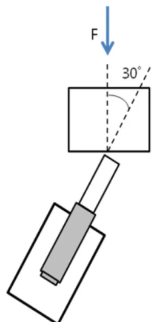
Afterward, they were loaded compressively with a universal test- ing machine (Instron, Canton, MA, USA) with force application at an angle of 30�to the loading axis (Fig. 1). The testing angle was chosen as it represents mechanical loading conditions in the max- illary anterior areas where the esthetic abutments are consid- ered.8,12,13A thin layer of tin foil was placed on the surface of the pis-
ton to prevent surface damage. And the abutments were loaded at the upper part of the specimen until the abutment failures occurred.
The crosshead speed was 1 mm/ min and the applied force was graph- ically recorded on an x-t recorder (Instron Series IX version:
8.07.00, Canton, MA, USA). Fracture of the abutment was accom- panied by an audible pop and failure was also recognized by a devi- ation from graphic linearity. The fracture mode of the abutments was also evaluated visually.
The obtained data was expressed by mean and standard deviation.
The distribution of strength was satisfied with normality assump- tion (result of the Shapiro-Wilk's test, P > .05) and a parametric method one-way analysis of variance (ANOVA) was used. If the global test revealed significant difference, the Tamhane post-hoc comparison method was applied in comparing each pair as the assumption of equal variances was not accepted (result of the Levene's test, P < .001).
Also a two-way ANOVA was performed to assess whether there is an interaction effect between material and thickness. The statistical software SPSS 12.0 (SPSS Inc., Chicago, IL, USA) was used and a type one error rate P > .05 was applied to determine significance of a result.
Results
The final loads at fracture / distortion of all groups are shown in Table 1 and Fig. 2. During the test, two screw loosening were found in group Ti-0.5 and Zir-0.8 which revealed no statistically signifi- cant differences.
Among all the experimental groups, Zir-10 showed the highest mean strength 2,476.3 ± 342.0 N and Ti-0.5 and Zir-0.8 group were followed. All the strengths of Ti and Zir groups were higher than 500 N. The strengths of RNC groups were significantly lower compared to other two materials (RNC-1.0 427.5 ± 72.1, RNC-0.8 297.9 ± 41.2) and the strengths of all the test groups decreased as the thickness decreases (P < .01). And the RNC-0.5 (127.4 ± 35.3 N) abutments were weaker than all other groups (P < .05). The strengths of RNC-0.8 and RNC-1.0 were higher than 290 N.
Results of the two-way ANOVA showed significant interac- tion effect between materials and thicknesses (P < .001): the slope of strength according to wall thickness was significantly higher in Zir groups than RNC groups. Zir groups showed higher mean fracture strength than any resin groups (P < .001).
Fig. 1. Loading of the abutment with a universal testing machine. 30�degree angle was maintained.
Table 1. Fracture/distortion strength of all tested groups (unit: N)
Group Ti-0.5 Zir-0.5 Zir-0.8 Zir-1.0 RNC-0.5 RNC-0.8 RNC-1.0
Mean 1,041.8 631.4 1,518.1 2,476.3 127.4 297.9 427.1
SD 237.2 149 347.9 342 35.3 41.2 72.5
The failure modes were also observed. In the control group (Ti-0.5), failure occurred through the bending of the junction of implant- abutment connection in all samples which showed implant neck dis- tortion. In test groups, all specimens showed abutment fracture before screw bending or fracture. The failure mode of the abutments in test groups showed the homogeneous manner; the total destruction of the abutment body with crack lines. No titanium link distortion was found.
Discussion
The main finding of this study was that all the zirconia abutments tested including 0.5 mm wall thickness have the potential to with- stand the physiologic occlusal forces in anterior and posterior regions of oral cavity.
On investigating the fracture strength of different implant abut- ments, maximum biting forces should be considered. A large number of researches have been focused on the biting forces dur- ing human mastication.14-16And as well known, maximum bite forces vary according to the region of oral cavity. While the great- est bite forces are found in posterior regions especially in the first molar area, one- third or one-fourth of that maximum force is found in the anterior area. Some studies reported that the maximum force levels vary from 216 to 847 N and smaller force levels were reported in anterior area varying from 108 to 299 N.14-16The zirconia abutments investigated in this study exhibited mean fracture strengths of 631.4 at 0.5 mm wall thickness and 1,518.1 and 2,476.3 at 0.8 mm and 1mm wall thickness respectively which all exceeds above mentioned posterior maximum bite force.
In case of resin abutments, 0.5 mm wall thickness showed mean
strength level of 127.4 N which represents unfavorable results even in the anterior area but 0.8 mm and 1.0 mm wall thicknesses revealed strength level of 297.9 N and 427.1 N, which exceed the average strength in anterior region.15,17
The test method used in this study was static loading, so cyclic load- ing with fatigue might yield different results. The lowest mean strength of zirconia 0.5 mm wall thickness in this study was 631.4 N which is higher than results of previous investigations.18,19Data on the fracture stability of zirconia abutments are difficult to compare between studies because of different study designs. However, pre- vious researches have shown that the zirconia abutment was resis- tant enough for normal bite force.18,19In an in vitro study, unprepared titanium-reinforced zirconia showed median fracture load of 294 N and Ti control groups 324 N respectively, after fatigue and static load- ings.18The conclusion of that study was that titanium-reinforced zir- conia abutment performed almost the same manner to titanium abut- ment, so it can be recommended as an aesthetic substitute for sin- gle implant abutment in anterior region.
Currently zirconia implant abutments are designed and sup- plied with different types of implant-abutment connections. The exter- nal and internal connection of zirconia abutments can be accomplished either by the abutment itself (entire ceramic one-piece) or by means of secondary components (two-piece). One-piece abut- ments are made entirely of ceramic, whereas for two-piece abutments, the internal connecting parts are made by titanium insert or separate titanium link.
The influence of the type of connection on the stability of implant- zirconia abutment complex has been studied by Sailer et al.20who concluded that two-piece zirconia abutments with a sec- ondary coupling abutment or metallic insert exhibited significant- ly higher bending moments than one-piece abutments. Another stud- ies reported that abutment fractures occurred at the internal insert- ing part of the zirconia abutment after static fracture loading.9,21For this reason the specimens used in this study were designed as externally connected abutments with connecting titanium link to exclude the possibility of connecting part fracture of one-piece internal type.
Before the loading test, the whole specimens underwent thermo- cycling to simulate artificial aging. Although there are still many con- troversies about exact temperatures and dwelling cycles, no reports have suggested about the necessary number of thermo-cycles to sim- ulate the use-time of a material in vivo. Therefore this study used the most commonly used temperatures 5。C - 55。C and 10,000 cycles to represent around a year of service in the mouth.22
Preparation of specimen and test set-up was basically performed according to the ISO 14801:2007 protocol. This investigation did not include a full veneer crown in the specimens. One study com- paring the effect of adhesively cemented all-ceramic crowns of zir- Fig. 2. Box plots of the results after the load-fracture test in N after thermocycling
(n=10). Zir-1.0 group showed the highest mean strength and RNC-0.5 group showed the lowest mean strength among the test groups.
conia abutments concluded that ceramic restoration did not influ- ence the bending moment of abutments in any of the test groups.20 Another study with ceramic crowns over zirconia abutments had found the zirconia abutment failed in 40% of the specimens prior to either the all-ceramic crown fracturing or the screw component dis- tortion.8An assumption that a crown may act as a stress-shield that allows a larger load to be applied before noticing any abutment frac- ture was extrapolated by the authors. In this study all the tested abut- ments were restored with neither metal crowns nor all-ceramic crowns which allowed not to obscure the cause of failure, that is, abutment related or crown related.10 Also this type of experiment design was already used in other studies.9,16,21It might be needed some new kind of loading protocol or abutment design to ensure not only even distribution of the occlusal force but also force transfer without stress shield .
Generally, the clinical application of prefabricated zirconia abut- ments may need some kind of modifications or grinding procedures.
It should be considered that the mechanical resistance of prepared ones might be different from that of unprepared ones. It is well known that zirconia material is highly susceptible to surface modifications and improper handling techniques.23Although some former stud- ies showed that the prepared zirconia abutments did not yield sta- tistically significant difference compared with unprepared ones, the current literatures does not provide exact information about this issue.9,10 Hence there is still a need of guideline for the preparation of zirconia abutments as well as the minimal wall thickness of abutment that guarantees long-term stability. In this study, the smallest wall thickness of 0.5 mm showed mean fracture strength of 631.4 N which could tolerate posterior bite force of 500 N.
A few of ceramic abutments has been proposed as means of sub- stitute of titanium abutments before the introduction of yttrium-sta- bilized zirconium dioxide (Y-TZP) abutments. However, there are no published clinical trials using resin materials as an implant abutment. The resin blocks used in this study are resin nano ceramic CAD-CAM materials introduced as a substitute for ceram- ics for inlays, onlays, crowns, and implant crowns by the manufacturer.
It was produced as a new kind of composite product with 80% nano ceramic (silica and zirconia) fillers and 20% of resin matrix which underwent heat curing process. As this material contains resin matrix, it is beneficial in tensile strength than ceramic which has mechanical shortcoming of brittleness.
Although there are no reports published about this material as an implant abutment, it has shown favorable fracture resistances in this study at the wall thickness more than 0.8 mm. As well known, to suc- ceed as a dental implant abutment, in many aspects, not only the mechanical strengths like fracture strength but also the bio-physi- ological compatibility including subgingival area should be proved.
Additional further studies about resistance to persistent stress using cyclic loading and screw loosening due to resin nano ceram- ic material will be needed. And longitudinal clinical evaluations includ- ing fracture and discoloration etc. will be also necessary. Although the manufacturer reported comparable biocompatibility and mechan- ical strengths, more in-vitro studies about this material would be need- ed before any clinical appplication.
Conclusion
Within the limitations of this study, all the tested zirconia abut- ments have the potential to withstand the physiologic occlusal forces in anterior and posterior regions. In resin nano ceramic abutments, wall thickness more than 0.8 mm showed the possibility to withstand the occlusal forces in anterior regions. As the results of this study cannot be generalized to other implant system, further studies are needed to verify the minimal wall thickness of implant abutments.
ORCID
Joo-Hee Lee http://orcid.org/0000-0002-7907-3098
References
1. Manicone PF, Rossi Iommetti P, Raffaelli L. An overview of zirconia ceramics: basic properties and clinical applica- tions. J Dent 2007;35:819-26.
2. Henriksson K, Jemt T. Evaluation of custom-made procera ce- ramic abutments for single-implant tooth replacement: a prospective 1-year follow-up study. Int J Prosthodont 2003;16:626- 30.
3. Zembic A, Bösch A, Jung RE, Hämmerle CH, Sailer I. Five- year results of a randomized controlled clinical trial compar- ing zirconia and titanium abutments supporting single-im- plant crowns in canine and posterior regions. Clin Oral Implants Res 2013;24:384-90.
4. Yildirim M, Edelhoff D, Hanisch O, Spiekermann H. Ceramic abutments-a new era in achieving optimal esthetics in im- plant dentistry. Int J Periodontics Restorative Dent 2000;20:81- 91.
5. Lüthy H, Wohlwend A, Pietrobon N, Studer R, Tangorra E, Loeffel O. Flexion resistance of a layered integral ceramic sys- tem and two reinvested integral ceramic systems. Zahntechnik (Zur) 1990;47:497-501.
6. Christel P, Meunier A, Heller M, Torre JP, Peille CN. Mechanical properties and short-term in-vivo evaluation of yttrium-oxide- partially-stabilized zirconia. J Biomed Mater Res 1989;23:45- 61.
7. Glauser R, Sailer I, Wohlwend A, Studer S, Schibli M, Schärer P. Experimental zirconia abutments for implant-supported
single-tooth restorations in esthetically demanding regions: 4- year results of a prospective clinical study. Int J Prosthodont 2004;17:285-90.
8. Yildirim M, Fischer H, Marx R, Edelhoff D. In vivo fracture re- sistance of implant-supported all-ceramic restorations. J Prosthet Dent 2003;90:325-31.
9. Adatia ND, Bayne SC, Cooper LF, Thompson JY. Fracture re- sistance of yttria-stabilized zirconia dental implant abutments.
J Prosthodont 2009;18:17-22.
10. Att W, Yajima ND, Wolkewitz M, Witkowski S, Strub JR.
Influence of preparation and wall thickness on the resistance to fracture of zirconia implant abutments. Clin Implant Dent Relat Res 2012;14:e196-203.
11. Koller M, Arnetzl GV, Holly L, Arnetzl G. Lava ultimate resin nano ceramic for CAD/ CAM: customization case study.
Int J Comput Dent 2012;15:159-64.
12. Dixon DL, Breeding LC, Sadler JP, McKay ML. Comparison of screw loosening, rotation, and deflection among three implant designs. J Prosthet Dent 1995;74:270-8.
13. Strub JR, Gerds T. Fracture strength and failure mode of five different single-tooth implant-abutment combinations. Int J Prosthodont 2003;16:167-71.
14. Waltimo A, Kemppainen P, Könönen M. Maximal contraction force and endurance of human jaw-closing muscles in isometric clenching. Scand J Dent Res 1993;101:416-21.
15. Waltimo A, Könönen M. A novel bite force recorder and maximal isometric bite force values for healthy young adults.
Scand J Dent Res 1993;101:171-5.
16. Gehrke P, Dhom G, Brunner J, Wolf D, Degidi M, Piattelli A.
Zirconium implant abutments: fracture strength and influ- ence of cyclic loading on retaining-screw loosening. Quintessence Int 2006;37:19-26.
17. Helkimo E, Carlsson GE, Helkimo M. Bite force and state of dentition. Acta Odontol Scand 1977;35:297-303.
18. Butz F, Heydecke G, Okutan M, Strub JR. Survival rate, frac- ture strength and failure mode of ceramic implant abutments af- ter chewing simulation. J Oral Rehabil 2005;32:838-43.
19. Zembic A, Sailer I, Jung RE, Hämmerle CH. Randomized-con- trolled clinical trial of customized zirconia and titanium implant abutments for single-tooth implants in canine and posterior re- gions: 3-year results. Clin Oral Implants Res 2009;20:802-8.
20. Sailer I, Sailer T, Stawarczyk B, Jung RE, Hämmerle CH. In vitro study of the influence of the type of connection on the frac- ture load of zirconia abutments with internal and external implant-abutment connections. Int J Oral Maxillofac Implants 2009;24:850-8.
21. Hjerppe J, Lassila LV, Rakkolainen T, Narhi T, Vallittu PK. Load- bearing capacity of custom-made versus prefabricated com- mercially available zirconia abutments. Int J Oral Maxillofac Implants 2011;26:132-8.
22. Gale MS, Darvell BW. Thermal cycling procedures for labo- ratory testing of dental restorations. J Dent 1999;27:89-99.
23. Luthardt RG, Holzhüter MS, Rudolph H, Herold V, Walter MH.
CAD/CAM-machining effects on Y-TZP zirconia. Dent Mater 2004;20:655-62.
지르코니아와 레진나노세라믹 임플란트 지대주의 두께에 따른 열순환 후 파절저항
이정원∙차현석∙이주희*
울산대학교 의과대학 서울아산병원 치과보철과
목적: 이 연구의 목적은 심미적인 임플란트 지대주의 종류와 두께에 따른 파절 강도를 측정하여 구강 내 저작압에 견디는 최소한의 두께를 평가하 기 위함이다.
재료 및 방법: 대조군으로 0.5 mm 두께의 티타늄 임플란트 지대주를(Ti-0.5), 실험군으로 지르코니아 임플란트와 레진 나노 세라믹 지대주를 사용하 여 각각 0.5 mm, 0.8 mm, 1.0 mm 두께로 각 그룹에 10개씩 총 70개의 시편을 제작하였다(그룹Zir-0.5, Zir-0.8, Zir-1.0, RNC-0.5, RNC-0.8, RNC-1.0). 모든 시편은 파절 실험 이전에 열순환을 시행하여 구강 내에서의 사용을 재현한 후, universal testing machine을 이용하여 각 시편의 파절 강도를 측정하여 평균값을 측정하였다. 그룹들의 평균 파절 값을 측정하였으며 이원분산분석을 이용하여 통계학적으로 분석하였다.
결과: Zir-1.0군이 가장 높은 파절 강도 2,476.3 ± 342.0 N를 보였으며 뒤를 이어 Zir-0.8 (1,518 ± 347.9 N), Ti-0.5 (1,041.8 ± 237.2 N), Zir-0.5 (631.4 ± 149.0 N), 의 순이었다. RNC 그룹의 경우에 Ti와 Zir 그룹에 비교하여 유의하게 낮은 파절 강도값을 나타내었으며(RNC-1.0 427.5 ± 72.1, RNC-0.8 297.9 ± 41.2), 모든 실험군에서 지대주 두께가 감소할수록 파절 강도 값도 유의하게 감소했다(P < .01). RNC-0.5 (127.4 ± 35.3 N) 그룹은 다른 모든 군에 비해 유의하게 낮은 값을 보였다(P < .05).
결론: 이번 실험에서 사용된 모든 두께의 지르코니아 지대주는 전치부와 구치부의 교합압을 견딜 수 있는 정도의 파절 강도를 보여주었다. 레진 나 노 세라믹 지대주의 경우 0.8 mm 두께 이상에서 전치부의 교합압을 견딜 수 있는 가능성을 보여주었다. (대한치과보철학회지 2017;55:144-50) 주요단어: 나노세라믹; 지대주; 힘저항능; 지르코니아; 파절
*교신저자: 이주희
05505 서울 송파구 올림픽로 43길 88
울산대학교 의과대학 서울아산병원 치과보철과 02 3010 5803: e-mail, [email protected]
원고접수일: 2017년 3월 25일 / 원고최종수정일: 2017년 4월 6일 / 원고채택일: 2017년 4월 10일
2017 대한치과보철학회
이 글은 크리에이티브 커먼즈 코리아 저작자표시-비영리 3.0 대한민국 라이선스에 따라 이용하실 수 있습니다.
c cc