간 경화를 가진 환자는 간암의 위험 때문에 간 검사의 기회 를 자주 갖게 된다. 간암은 발견 시기에 따라 평균 생존율이 현저한 차이를 보이기 때문에 암의 조기 발견이 중요하다(1).
간암 진단에서 전산화단층촬영(Computed Tomopraphy, CT와 자기공명영상(Magnelic Resonance Image, MRI) 조영 제의 사용 원리는 암이 동맥에 의해 공급을 받고, 정상 간의 주 공급원은 문맥이 이라는 사실에 의존 한다. 간 경화 환자에 서 문맥 고혈압에 의한 혈류 양상의 변화로 간과 간암에서 조 영 증강의 시간 창(time window) 이 소실되고 간의 구조적인 변형 때문에 MRI에서 경화된 간은 비 균질적인 신호 강도를 보인다. 이 때문에 작은 크기의 간암은 간과되는 수가 있으므 로 국소 간 병변의 발견과 특성화에는 특이 MRI 조영제가 필 요하다(2).
간의 특이 조영제인 Ferumoxides (FeridexⓇ, Berlex Laboratories, NJ, U.S.A.)는 철 산화물로 만들어진 초미소립 자 약물이며 세망내피계통(Reticuloendothelial System, RES) 에서 제거되는 MRI 조영제이다(3). MRI에서 철 침전이 많은
조직은 짧은 T2 이완 시간으로 신호 강도가 떨어진다는 특성 에 따라 간의 RES에서 흡수된 Ferumoxides는 간의 신호 강 도를 T2 강조 영상에서 감소시킨다. 대부분의 간암은 RES를 가지지 않기 때문에 T2 강조 영상에서 고신호강도를 보인다.
Ferumoxides의 주입 후의 간암 발견 예민도는 71-86%이다 (4, 5). 경화된 간에서 기능 이상 세망내피세포와 분화된 간암 에서 기능하는 세망내피세포가 제한된 예민도에 기여한다.
Semelka 등 (6)이 Ferumoxides 와 Gd을 순서적으로 주입하 였을 때 간 종양의 발견에 도움이 되었다는 보고를 하였다.
Ferumoxides 가 Gd에 의한 병변 주위 간의 조영 증강 효과를 감소시키므로 Ferumoxides 주입 후에 병변 부위는 더 뚜렷해 진다. 이런 이론적인 배경하에 국소 간암 진단에 대한 Ferumoxides 와 Gd 조합의 효과를 알아보고자 한다.
대상과 방법
29명의 간 경화 환자(남자19명, 여자10명)를 대상으로 하 였다. 나이 평균은 57세(37세-70세)였다. Imaging protocol 의 영상을 모두 얻은 환자 만을 대상으로 하였다. 12명의 환
─ 433 ─ 대한영상의학회지 2004;50:433-436
간암에서 Ferumoxides와 Gadolinium의 이중 조영 자기공명 영상
1최 상 희
목적: 만성 간 질환과 간암이 발생한 환자에서 Gadolinium(Gd) 조영증강 동적 자기공명영상에 서 Ferumoxides의 효과를 알아 보고자 한다.
대상과 방법: 29명의 간 경화 환자를 대상으로 하였다. 12명은 간암이 발생한 환자 였다. 몸무
게 당 0.05 ml의 Ferumoxides 주사 전과 주사 후 30-60분에 gradient echo(GE) T1 in phase 와 opposed -phase, breath-hold turbo spin echo(TSE) T2을 얻었고 20 ml의 Gd을 초당 2 ml의 속도로 주사 후에 4 dynamic in-phase GE series를 얻었다. 간과 간암에 대한 signal to noise ratio(SNR)과 contrast to noise ratio(CNR)을 구하고 paired t-test 를 이용하여 통 계적 분석을 하였다.
결과: Ferumoxides 주사 후 간의 SNR은 8.6±1.20 감소하였다(p<0.001). Gd 주사후 간은 Ferumoxides 주사 후와 비교해 6.09±1.15 증가 하여 Ferumoxides 주사 전과 의미있는 차이 는 없었다(p<0.01). Ferumoxides 주사 후에 간암의 SNR은 의미 있게 변하지 않았으나 배경 간에 대한 간암의 CNR은 7.54±1.61 증가 되었다(p<0.05). Gd은 간암의 CNR을 15.6±3.87 로 더욱 증가 시켰다(p<0.05).
결론: 간암은 Gd으로 조영 증강되고 Ferumoxides는 배경 간의 Gd 조영 증강 효과를 없애기 때문에 Ferumoxides와 Gd의 조합은 간암의 CNR을 높인다.
1성균관대학교 의과대학 삼성서울병원 영상의학과
이 논문은 2003년 11월 12일 접수하여 2004년 4월 2일에 채택되었음.
자에서 13개의 간암이 있었다. 조영제 사용에 대해 환자의 동 의를 얻고 이중 조영 증강 MRI를 시행하였다.
MRI Imaging Protocol
1.5 Tesla Siemens Vision(Siemens Medical Solutions, Inc, Malvern, PA, U.S.A.) 기종의 위상차 배열 코일을 이용하여 영 상을 얻었다. 모든 환자에서 조영 증강 전 GE T1강조 in- phase, opposed phase(TR=150 ms), breath-hold turbo spin echo(TSE) T2영상(TR=3500 ms, TE=138 ms)을 얻고 몸 무게 1 kg 당 0.05 ml의 Ferumoxides 를 5% dextrose 100 ml에 희석하여 30분 동안 주입한 후 30분에서 60분 사이에 일련의 GE 영상을 얻었다. Ferumoxides 주입 후 영상을 얻고 20 ml의 gadolinium-diethylenetri-aminepentaacetate(Gd- DTPA-BMA) (OptimarkⓇ, Mal-linkrodt, St. Louis, MO, U.S.A.)을 초 당 2 ml로 주사한 후에 동맥기, 문맥기, 정맥기, 평형기의 4 dynamic breath-hold GE를 얻고 평형기에서 fat saturated GE를 얻었다.
Image Analysis
내부 장기 대조를 위해 각 간암과 경화된 배경 간, 비장, 허 리근육의 신호 강도를 각각 측정하였다. Pre-Ferumoxides, post-Ferumoxides와 Gd 조영 증강이 최고일 때의 이미지에 서 각 부위 ROI의 mean signal intensity 를 얻었다. ROI 크기 는 0.5 cm2로 하였고 배경 잡음은 촬영상 범위(Field of view) 내의 공기에서 잰 신호 강도를 사용하였다.
■ Liver SNR = (Liver - Muscle) / Noise
■ HCC SNR = (HCC - Muscle) / Noise
■ HCC-to-Liver CNR = (HCC - Liver) / Noise
Statistical Analysis
pre-Ferumoxides, post-Ferumoxides와 post-Gd 신호 강 도의 비교에 paired Student’s t-test를 이용하였고 간암의 경 우는 비 모수 통계를 이용하였다. p-value가 0.05보다 작을 때 통계적으로 의미가 있다고 간주 하였다.
결 과
Ferumoxides 주사 후에 간의 SNR은 T2 강조 영상에서 8.6
±1.20 감소하였다(p<0.001). Gd 조영 증강 효과가 최고 일 때 간의 SNR은 post Ferumoxides 신호 강도에 비교하여 6.09
±1.15 증가하여 (p < 0.001) 간의 신호 강도는 pre- Ferumoxides level 로 되었다(Fig. 1).
종양의 pre Ferumoxides SNR 은 post Ferumoxides 의 SNR과 통계적으로 차이가 없었다(p=0.821). Gd 조영 증강 효과로 종양의 SNR은 10.67±3.39 증가 하였다(p<0.05) (Fig. 1).
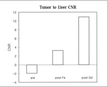
Ferumoxides 주사 후에 간에 대한 병변의 CNR은 7.54±
1.61 증가 하였고(p<0.005), Gd 주사 후에는 15.6±3.87 로 더욱 증가 하였다(p<0.0001) (Fig. 2).
고 찰
간암은 주로 간 경화 환자에서 생기므로 고식적인 방법으로 는 찾기가 어렵다. Teefey 등(7)은 CT, 초음파, MRI의 간암 에 대한 예민도는 유사하였다고 보고하였다. MRI에서 간암과 주변 간의 감별 능력은 종양의 크기와 영상의 대조에 의존한 다. 간암의 위험성이 있는 환자에서 MRI를 이용한 검사는 중 재적 시술로 고칠 수 있는 작은 크기의 간암을 발견하고자 함 이 그 목적이다. 간 경화가 있는 환자에서 작은 간암은 주변의
─ 434 ─
최상희: 간암에서 Ferumoxides와 Gadolinium의 이중 조영 자기공명 영상
Fig. 1. Signal to Noise Ratio (SNR), Liver and Tumor.
The SNR of the liver decreased after Ferumoxides injection due to iron accumulation. At peak Gd effect, the liver SNR re- turned to its baseline pre-Ferumoxides level.
The SNR of the tumor at pre-Ferumoxides was not significant- ly different from that of post Ferumoxides. At peak Gd-en- hanced image, the tumor SNR increased.
Fig. 2. Contrast to Noise Ratio (CNR), Tumor to Liver.
At post- Ferumoxides image, the lesion CNR relative to liver increased (p<0.005). After Gd injection, lesion CNR relative to tumor further increased (p<0.0001).
Tumor to Liver CNR
경화된 실질과 구분이 어려워 찾기가 어려우므로 종양과 주변 간의 대조를 증가 시키는 방법이 필요하다.
Gd은 정상 주변 조직에서 종양의 발견은 효과적이나 간경화 가 있는 간에서 간암의 발견은 제한적이다. 간의 다른 조영제 로 Ferumoxides 는 국소 간 병변을 찾는데 효과적이라고 알 려져 왔다(2). Ferumoxides로 조영 증강시킨 MRI는 조영시 키지 않은 MRI 보다 더 정확하고, 간 전이 암을 보는데도 Computed Tomography of Arterial Portography(CTAP) 만 큼 정확하다고 보고되었다(8). 이는 배경 간을 어둡게 만들어 서 배경 간과 종양의 대조를 증가 시킨다는 이론으로 간 경화 환자에서 구조적인 변화로 인해 철 산화물 입자의 흡수가 불 균질하게 변한다 하더라도 간-종양의 CNR이 증가되어 T2- 강조 영상에서 병변의 발견이 향상된다(9). 이런 이론을 배경 으로 Gd과 Ferumoxides를 함께 이용한 MRI를 비교 했을 때 Gd 주입으로 간암이 더 또렷이 보였다는 보고가 있다(10, 11).
두 조영제는 간암과 주변 간에 약리적, 생리적으로 다른 작 용기전을 가진다. Ferumoxides는 배경 간을 병변보다 더 어둡 게 하고 Gd은 배경 간 보다 병변을 더 밝게 조영 증강 시킨다.
또한 같은 검사에 두 가지 약물을 넣어도 진단적 정보의 손실 이 없다(6)는 이론적 근거 하에 Ferumoxides를 먼저 주입하 고 Gd 을 후에 주입하여 간과 병변의 CNR을 증가 시키는 상 호 상승 효과를 볼 수 있다(Fig. 3).
저자의 연구는 Ward et al(12)의 보고에 비교해 1.5T magnet, 위상차 배열 코일, in-phase Ferumoxides T1 강조 영상을 이용함으로써 이전 연구를 확인하고 더 많은 소견을 찾 을 수 있었다. 또한 in-phase영상으로 지방 침윤에 의한 위음 성을 완전히 배제하고 Ferumoxides의 효과를 알 수 있었다.
이 연구로 두 약물을 주입하였을 때 Ferumoxides는 Gd의 조영 증강 효과를 상쇄시켜 배경 간 SNR이 처음의 in-phase GE 영 상 의 SNR과 유 사 해 진 다 는 것 을 보 여 주 었 고 , Ferumoxides 만을 사용하였을 때 보다 두 가지의 조합시에 간 암과 주변 간의 신호 강도에 의미있는 차이가 있음을 보였다.
이 연구의 모든 병변이 Ferumoxides 조영 전 영상에서 보였 고 표본의 숫자가 적었으나 이론적으로 이 방법의 장점은 분 명히 입증되었다.
요약하면 Ferumoxides와 Gd의 조합에 의한 순서적인 주입
으로 경화된 배경 간의 신호 강도는 Ferumoxides에 의해 감 소되고 간암은 Gd으로 조영 증강되어 간암의 CNR을 증가시 킨다.
결론적으로 Ferumoxides와 Gd의 조합에 의해 증가된 간암 의 CNR으로 간 경화가 있는 환자에서 작은 간암을 조기에 진 단하는데 유용할 수 있다.
참 고 문 헌
1. Nguyen MH, Keeffe EB. Screening for hepatocellular carcinoma. J Clin Gastroenterol 2002;35:S86-91
2. Ros PR, Freeny PC, Harms SH, et al. Hepatic MR Imaging with fer- umoxides: a multicenter clinical trial of the safety and efficacy in the detection of focal hepatic lesions. Radiology 1995;196:481-488 3. Clement O, Siauve N, Cuenod CA, Frija G. Liver imaging with fer-
umoxides (Feridex(r)): Fundamentals, controversies, and practical aspects. Top in Magn Reson Imaging 1998;9;167-182
4. Hori M, Murakami T, Kim T, et al. Detection of hypervascular he- patocellular carcinoma: comparision of SPIO-enhanced MRI with dynamic helical CT. J Comput Assist Tomogr 2002;26:701-710 5. Schultz JF, Bell JD, Goldstein RM, Kuhn JA, McCarty TM. Hepatic
tumor imaging using iron oxide MRI: comparision with computed tomography, clinical impact, and cost analysis. Ann Surg Oncol 1999;6:691-698
6. Semelka RC, Lee JK, Worawattanakul S, Noone TC, Patt RH, Ascher SM. Sequential use of ferumoxide particles and gadolinium chelate for the evaluation of focal liver lesions on MRI. J Magn Reson Imaging 1998;8:670-4
7. Teefey SA, Hildeboldt CC, Dehdashti F, et al. Detection of primary hepatic malignancy in liver transplant candidates: prospective comparision of CT, MR imaging, US, and PET. Radiology 2003;226:
533-542
8. Seneterre E, Taourel P, Bouvier Y, et al. Detection of hepatic metastases: ferumoxides-enhanced MR imaging versus unen- hanced MR imaging and CT during arterial portography. Radiology 1996;200:785-792
9. Elizondo G, Weissleder R, Stark DD, et al. Hepatic cirrhosis and hepatitis: MR imaging enhanced with superparamagnetic iron ox- ide. Radiology 1990;174:797-801
10. Tang Y, Yamashita Y, Arakawa A, et al. Detection of hepatocellu- lar carcinoma arising in cirrhotic livers: comparison of gadolinium and ferumoxides-enhanced MR imaging. AJR Am J Roentgenol 1999;172:1547-1554
─ 435 ─ 대한영상의학회지 2004;50:433-436
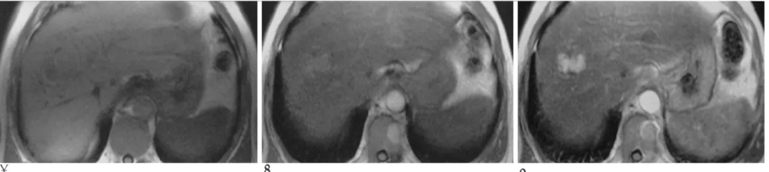
A B C
Fig. 3. Hepatocellular carcinoma.
A. Pre-Ferumoxides T1 in-phase. The tumor shows lower signal intensity than the normal liver.
B. Post-Ferumoxides T2 image. The liver is darkened. Finally tumor-signal is higher than that of liver.
C. Post-Gd T2 image. The tumor shows intense enhancement, consistent with HCC.
11. Kim MJ, Kim JH, Chung JJ, Park MS, Lim JS, Oh YT. Focal hepatic lesions: detection and characterization with combination gadolini- um- and superparamagnetic iron oxide- enhanced MR imaging.
Radiology 2003; 228:719-726
12. Ward J, Guthrie JA, Scott DJ, et al. Hepatocellular carcinoma in the cirrhotic liver: double-contrast MR imaging for diagnosis.
Radiology 2000;216:154-162
─ 436 ─
최상희: 간암에서 Ferumoxides와 Gadolinium의 이중 조영 자기공명 영상
J Korean Radiol Soc 2004;50:433-436
Address reprint requests to : Sang-Hee Choi, M.D., Department of Radiology and Center for Imaging Science, Sungkyunkwan University School of Medicine, 50 Ilwon-dong, Kangnam-gu, Seoul 135-710, Korea.
Tel. 82-2-3410-6436 Fax. 82-2-3410-0049 E-mail: [email protected]
Double Contrast Media enhanced MRI with Ferumoxides- Gadolinium on Hepatocellular Carcinoma
1Sang-Hee Choi, M.D.
1Department of Radiology and Center for Imaging Science, Sungkyunkwan University School of Medicine
Purpose: To evaluate the effects of Ferumoxides on Gadolinium (Gd) enhanced dynamic liver magnetic reso- nance imaging (MRI) in cirrhotic patients and also for the diagnosis of hepatocellular carcinoma (HCC).
Materials and Methods: 29 patients with liver cirrhosis were examined at 1.5T. 12 patients had HCC. The imaging protocol included GE T1 in and opposed phases, and a breath-hold TSE T2 before and 30-60 min fol- lowing 0.05 ml/kg Ferumoxides. Four dynamic in-phase GE series were also acquired after an injection of 20 ml of Gd at 2 ml/sec. SNR and CNR were calculated for liver lesion relative to the muscle and background liv- er respectively. Statistical analysis was performed using the paired t-test.
Results: The SNR of the liver decreased by 8.6±1.20 (p<0.001) after Ferumoxides injection. At the peak of the Gd effect, the liver increased by 6.09±1.15 relative to the post Ferumoxides, but it was not significantly different from the pre-Ferumoxides study (p<0.01). Although there was no significant change in post Ferumoxides SNR of HCC, CNR of HCC relative to the liver increased by 7.54±1.61 (p<0.05). After the Gd injection, CNR of HCC increased by 15.6±3.87 (p<0.05).
Conclusion: The administration of Ferumoxides made HCC CNR increase, and it canceled the effect of Gd en- hancement of the cirrhotic liver. The combination of Ferumoxides and Gd makes HCC CNR increase.
Index words :Abdomen, MR Liver, cirrhosis Liver, MR
Liver neoplasms, MR
Magnetic resonance (MR), contrast media