https://doi.org/10.4093/jkd.2017.18.2.102 ISSN 2233-7431
Abstract
Non-alcoholic fatty liver disease (NAFLD) is one of the most common metabolic liver disorders, and its incidence is expected to increase rapidly in the future as the rate of obesity increases and populations age. The gold standard for diagnosing NAFLD is liver biopsy, which involves sample error, high cost, and can be complicated due to its invasive nature. Therefore, many studies have been reported to establish accurate and convenient models to detect NAFLD using clinical and laboratory parameters. Most were derived from relatively small number of subjects and lack external validation, especially in the Korean population. This article summarizes the established and emerging risk factors for NAFLD and reviews non-invasive diagnostic algorithms for NAFLD including hepatic fibrosis.
Keywords:
Diagnosis, Liver cirrhosis, Non-alcoholic fatty liver disease
임상 및 생화학적 지표에 기반한 비알코올성 지방간의 진단
이용호
연세대학교 의과대학 내과학교실
Diagnosis of Non-Alcoholic Fatty Liver Disease Based on Clinical and Laboratory Data
Yong-ho Lee
Department of Internal Medicine, Yonsei University College of Medicine, Seoul, Korea
Corresponding author: Yong-ho Lee
Department of Internal Medicine, Yonsei University College of Medicine, 50-1 Yonsei-ro, Seodaemun-gu, Seoul 03722, Korea, E-mail: [email protected] Received: May 4, 2017; Accepted: May 16, 2017
This is an Open Access article distributed under the terms of the Creative Commons Attribution Non-Commercial License (http://creativecommons.org/licenses/by- nc/4.0/) which permits unrestricted non-commercial use, distribution, and reproduction in any medium, provided the original work is properly cited.
Copyright ⓒ 2017 Korean Diabetes Association
서론
비알코올성 지방간질환(non-alcoholic fatty liver
disease, NAFLD)은 과량의 알코올이나 지방간을 유발하 는 약물의 복용력이 없음에도 간내 지방이 5% 이상 축적 되는 질환이다[1]. NAFLD는 염증이나 섬유화 반응이 없
는 단순 지방간(simple steatosis), 비알코올 지방간염(non- alcoholic steatohepatitis, NASH), 비알코올 지방간연관 간경변증으로 진행되는 간질환을 포괄하며, 지방간염 및 섬유화의 진행정도가 질환의 예후에 큰 영향을 주기 때문 에 이러한 병의 진행을 판단하기 위한 가장 좋은 방법(gold standard)은 간생검이다. 하지만, 간생검은 침습적이며, 국 소적 평가에 의한 표본 오차의 한계, 고비용 등의 단점을 가 지고 있어 다양한 혈액학적 바이오마커 및 영상학적 도구를 이용하여 비침습적인 방법으로 진단율을 높이려는 노력이 많아지고 있다. 본 글에서는 NAFLD에 대한 위험인자와 혈 액검사 및 임상 데이터에 기반한 NAFLD의 진단 접근법에 대하여 정리해보았다.
본론
1. NAFLD의 위험인자
비만과 인슐린 저항성이 NAFLD를 유발하는 기전은 많 은 연구를 통해 입증되었다[2]. 2012년 미국간학회와 2013 년 대한간학회 가이드라인에서는 입증된 NAFLD의 위험 인자로 비만, 제2형 당뇨병, 이상지질혈증, 대사증후군을, 가능성이 있는 인자로 갑상선기능저하증, 다낭성 난소 증 후군, 수면무호흡증을 제시하고 있다[3,4]. 국내 연구를 통 해 NAFLD의 유병률은 비비만 인구집단에서 10~15%이 지만, 비만 인구에서는 55~70%로 추정된다고 보고하였고 [5], 제2형 당뇨병 환자에서는 70%까지 지방간이 동반되 어 있고 발표한 연구도 있다[6]. 그 외 뇌하수체 기능저하 증, 생식선기능저하증 등의 내분비 질환도 가능성 있는 지 방간의 위험인자로 언급하였다. 비만한 사람은 비만으로 인 해 나타나는 여러 가지 대사적 이상(이상지질혈증, 고혈압, 인슐린저항성 등)을 동반하고 있는 경우가 흔하며, 대사적 으로 건강한 비만한 사람(metabolically healthy obese)도 상당수를 차지한다. 대사적으로 건강한 비만한 사람은 심 혈관계 질환의 위험성이 낮다고 알려져 있지만, NAFLD에 있어서는 비만으로 인한 대사적 이상뿐만 아니라 대사적으
로 건강하더라도 비만 자체가 지방간의 발생의 위험인자임 이 최근 밝혀졌다[7]. 최근에는 사지의 근육량이 적은 근감 소증 환자에서 NAFLD 또는 지방간염의 위험성이 유의하 게 높다고 여러 연구에서 보고되고 있으며[5,8,9], 사지의 피하지방량이 많을수록 지방간의 위험도는 낮다는 보고가 있어[10], body mass index (BMI) 자체보다는 근육과 지 방의 상대적 분포가 지방간의 발생에 더 중요할 것으로 추 정된다. B형 간염 환자에서는 NAFLD가 적게 발생한다는 연구 결과도 있지만[11], 더 많은 연구를 통한 검증이 필요 한 상황이다. 제2형 당뇨병 환자에서의 NAFLD에 대한 대 규모 전향적 연구는 홍콩에서 1,918명을 대상으로 간섬유화 스캔(transient elastrography, fibroscan)이라는 영상 장비 를 이용하여 지방간을 진단한 코호트 연구가 있다[12]. 연 령이나 당뇨병 유병기간과 지방간 유무 간의 유의한 관련 성은 없었으나, 여성, BMI, 중성지방, 공복 혈당, alanine aminotransferase (ALT) 수치와 양의 통계적 유의성을 나 타내었다. 또한, 간 내 섬유화의 정도와 유의한 관련성이 있었던 변수는 당뇨병 유병기간, BMI, 낮은 high density lipoprotein (HDL), 높은 ALT, 알부민뇨로 나타났다. 제2 형 당뇨병 환자에서 NAFLD의 위험인자에 대한 연구 결과 는 많지 않아 향후 연구가 더 필요할 것으로 생각된다.
2. NAFLD 진단 모델: steatosis
현재까지 비알코올 지방간질환에 대한 확립된 선별 검사 법은 없다. aspartate aminotransferase (AST), ALT와 같 은 간기능 검사는 NAFLD 환자에서 정상인 경우가 흔하 기 때문에 선별검사로는 민감도가 떨어진다. 2016년 발표 된 유럽 간학회의 가이드라인에서는 NAFLD가 의심되는 환자에서 당뇨병, 고혈압, 심혈관질환의 과거 병력 및 가족 력, BMI, 허리둘레, AST, ALT, γ-glutamyl-transferase (GGT), 공복혈당, HbA1c, 공복 인슐린, 경구당부하검사 (고위험군에서), 콜레스테롤(total HDL), 중성지방, 요산, 전혈구검사(complete blood count) 등의 검사를 시행할 것 을 권고하고 있다[1].
NAFLD 진단 모델로서 지금까지 가장 많이 검증되고 보 고된 비침습적, 비영상학적 방법은 fatty liver index (FLI), SteatoTest, NAFLD liver fat score로 일반인구에서 타 연 구자들에 의해 검증이 된 바 있다.
1) Fatty liver index
FLI는 지방간 진단 모델로서 쉽게 이용할 수 있다는 장 점이 있어, 현재까지 많은 역학 연구에서 사용되고 있다. 이 탈리아인을 대상으로 초음파를 이용하여 지방간을 진단한 496명 코호트에서 BMI, 허리둘레, triglycerides, GGT 4 가지 변수를 이용하여 계산되며, 0~100 사이의 점수로 환 산하여 30과 60이라는 두 개의 cut-off를 가지고 있다[13].
한국인을 대상으로 한 연구에서도 AUROC (area under receiver operating characteristic curve) 0.86으로 비교적 높은 수치를 나타내었지만 다른 연구에서 충분한 검증이 되 어 있지 않으며, 한국인에 적용 가능한 cut-off는 서양인에 서 제시된 30과 60이 아닌 수치가 조정이 되어야 한다는 한 계점이 있다[14].
FLI = 1 / (1 + exp(–x)) × 100,
x = 0.953 × loge (triglycerides) + 0.139 × BMI + 0.718 × loge (γ-glutamyl-transferase) + 0.053
× (Waist circumference) – 15.745
2) SteatoTest
SteatoTest는 884명 이상의 프랑스 환자 코호트에 서 간생검으로 진단된 중등증~중증의 지방간에 대해 α2-macroglobulin, haptoglobin, apolipoprotein A1, total bilirubin, GGT, 공복혈당, triglycerides, cholesterol, ALT, 나이, 성별, BMI라는 열두 가지 변수를 이용한 미공개 수식 으로, 지방간을 진단하는 모델이다[15]. NAFLD뿐만 아니 라, C형 간염, 알코올성 지방간 환자를 포함하여 모형이 구 축되었다는 특징을 가지고 있으며, 프랑스 코호트에서만 검 증되었으며, 수식이 공개되어 있지 않아 일반적으로 사용하 기 어렵다는 제한점이 있다.
3) NAFLD liver fat score
NAFLD liver fat score는 magnetic resonance spectroscopy로 지방간을 진단한 핀란드 환자 470명을 대상 으로 확립된 모델로, 대사증후군 또는 제2형 당뇨병의 유무, 공복 인슐린, AST, ALT 수치를 이용하였다[16]. 비교적 많 은 연구에서 이용된 진단법이며, 한국인을 대상으로 검증한 연구에서 AUROC 0.77~0.82로 비교적 높은 수치를 나타 내었지만[14], 다른 연구를 통한 충분한 검증이 필요하다.
NAFLD liver fat score
= –2.89 + 1.18 × metabolic syndrome (yes = 1 / no = 0) + 0.45 × diabetes (yes = 2 / no = 0) + 0.15 × (fasting insulin, mU/L) + 0.04 × AST + 0.94 × AST/ALT ratio
4) Hepatic steatosis index (HSI)
건강검진을 통해 초음파로 진단된 한국인 10,724명을 대 상으로 개발된 지방간 진단법으로 ALT, AST, BMI, 성 별, 당뇨병 유무라는 변수로 비교적 간단하게 구성되어 있 다[17]. FLI처럼 30과 36이라는 두 개의 cut-off를 가지고 있어 intermediate 구간에 대한 판단이 쉽지 않다는 제한 점이 있지만, 다른 한국인을 대상으로 한 검증 연구에서도 AUROC 0.85로 비교적 높은 수치를 보였다[14].
HSI = 8 × ALT / AST ratio + BMI (+2, if diabetes; +2, if female)
5) Comprehensive/simple NAFLD score
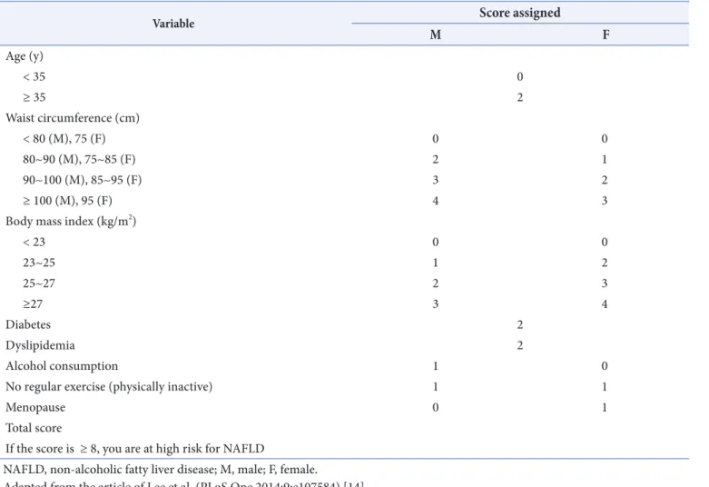
건강검진에서 초음파로 진단된 한국인 15,676명을 대상 으로 개발되어 독립된 검진 코호트 66,868명에서 검증된 지 방간 예측 모델이며, 임상 변수로 구성된 simple score와 임 상 및 생화학적 변수로 구성된 comprehensive score로 구 분되어 있다[14]. Simple 모델(SNS)은 나이, BMI, 허리둘 레, 당뇨병 및 이상지혈증의 유무, 음주, 운동, 폐경 변수로 구성되어 혈액 검사 수치 없이 쉽게 적용가능하다는 장점이 있다(Table 1). Comprehensive 모델(CNS)은 나이, BMI, 허리둘레, 당뇨병 유무, 음주, 운동, 폐경 이외에 공복 혈당, triglyceride, HDL-cholesterol, uric acid, AST, ALT 수치
가 추가되었다. 두 모델 모두 AUROC 0.8~0.9로 비교적 높은 수치를 보였지만, 다른 연구를 통한 검증이 필요하다.
Comprehensive NAFLD score (CNS) NAFLD = 1 / (1 + exp(–x)) × 100 if male,
x = 0.016 × age + 0.182 × BMI + 0.089 × WC + 0.391 × alcohol + 0.124 × exercise + 0.018 × fasting glucose + 0.773 × loge (triglycerides) – 0.014 × HDL cholesterol + 0.145 × uric acid – 0.674 × loge (AST) + 1.632 × loge (ALT) – 21.695.
if female,
x = 0.320 × BMI + 0.044 × WC + 0.533 × diabetes + 0.016 × fasting glucose + 0.951 × loge (triglycerides) – 0.015 × HDL cholesterol + 0.199 × uric acid – 0.645 × loge (AST) + 1.302 × loge (ALT) + 0.255 × menopause – 19.741.
6) Framingham Steatosis Index (FSI)
심혈관질환 관련 유명 코호트인 Framingham 대상자 1,181명에서 복부 computed tomography를 이용하여 지 방간을 진단하였으며, 나이, 성별, BMI, 고혈압, 당뇨병, triglycerides, ALT/AST 변수를 포함하는 모델을 개발하였 다[18]. 독립된 미국 국민건강영양조사 코호트를 이용하여 검증하였을 때 AUROC 0.76으로 비교적 높은 수치를 보였 지만, 다른 연구나 한국인을 대상으로 한 검증이 필요하다.
FSI = −7.981 + 0.011 × age (years) − 0.146
× sex (female = 1, male = 0) + 0.173 × BMI (kg/m2) + 0.007 × triglycerides (mg/dL) + 0.593
× hypertension (yes = 1, no = 0) + 0.789
× diabetes (yes = 1, no = 0) + 1.1 × ALT : AST ratio ≥ 1.33 (yes = 1, no = 0).
Table 1. A simple NAFLD score (SNS)
Variable Score assigned
M F
Age (y)
< 35 0
≥ 35 2
Waist circumference (cm)
< 80 (M), 75 (F) 0 0
80~90 (M), 75~85 (F) 2 1
90~100 (M), 85~95 (F) 3 2
≥ 100 (M), 95 (F) 4 3
Body mass index (kg/m2)
< 23 0 0
23~25 1 2
25~27 2 3
≥27 3 4
Diabetes 2
Dyslipidemia 2
Alcohol consumption 1 0
No regular exercise (physically inactive) 1 1
Menopause 0 1
Total score
If the score is ≥ 8, you are at high risk for NAFLD
NAFLD, non-alcoholic fatty liver disease; M, male; F, female.
Adapted from the article of Lee et al. (PLoS One 2014;9:e107584) [14].
3. NAFLD 진단 모델: steatohepatitis/NASH
간생검이 아닌 임상적, 생화학적, 영상학적 방법을 이용하 여 단순 지방간과 지방간염을 구별하기는 매우 어렵다[1]. 현 재까지 잘 알려진 마커는 세포의 자가사멸이나 파괴될 때 생 성되는 cytokeratin-18 (CK-18) 분절(fragment)이 있지만 [19], 정확도에 대한 논란이 있는 실정이다. 그 외 여러 가 지 생화학적 지표들을 종합하여 만들어진, NASH TestⓇ (Biopredictive, Paris, France), NASH diagnostics 등 많은 진단예측 모델이 개발되었지만, 아직까지 지방간염을 진단하 는데 이러한 비침습적 검사법이 인정되고 있지는 않다[1,4].
4. NAFLD 진단 모델: fibrosis
간 섬유화를 예측하기 위한 임상/생화학적 변수를 이 용한 모델은 중등도 이하의 경미한 섬유화에 대한 정확도 는 낮지만, 진행된 섬유화에 대해서는 높은 정확도를 보이 고 있다. 많은 모델 중에서 NAFLD fibrosis score (NFS) [20]과 fibrosis 4 calculator (FIB-4) [21]는 다양한 인종 의 NAFLD에서 검증되었으며 비교적 정확도가 높은 방 법이다. 이들 모델은 진행된 섬유화에 대한 검증뿐만 아니 라, 전체/심혈관/간관련 사망과 밀접한 연관된다는 역학 연 구 결과도 다수 보고된 바 있다. NFS 모델은 나이, 공복혈 당, BMI, platelet, albumin, AST/ALT로 구성되어 있으 며, FIB-4 모델은 나이, platelet, AST, ALT로 이루어져 비교적 간단하게 계산이 가능하다. 그 외, enhanced liver fibrosis (ELF), BARD, APRI (AST to Platelet Ratio Index) 등 많은 점수화된 공식이 발표되었지만, 충분한 검 증이 더 필요한 상황이다.
NAFLD fibrosis score (NFS)
= (–1.675 + 0.037 × age (years) + 0.094 × BMI (kg/m2) + 1.13 × IFG / diabetes (yes = 1, no = 0) + 0.99
× AST / ALT ratio – 0.013 × platelet count (×109 / l) – 0.66 × albumin [g/dL])
FIB–4 = age (years) × AST [U/L] / (platelets [109 / l]
× (ALT [U/L])1/2)
결론
전 세계적으로뿐만 아니라 국내에서도 비만 및 고령화 사 회가 진행되면서, NAFLD 환자들은 점차적으로 증가하고 있지만, 현재까지 지방간 진단을 위한 명확한 방법이 확립 되어 있지 않은 실정이다. 간생검이나 영상학적인 방법을 이용하지 않고 임상 및 생화학적 지표를 기반으로 진단하고 자 하는 노력이 많이 이루어져 왔다. 본 종설에서 다양한 지 방간 진단 모델을 제시하고 있지만, 각 모델이 개발된 환자 군의 특성에 큰 차이가 있고, 독립된 다른 환자군이나 한국 인을 대상으로 충분한 검증이 이루어지지 않았기 때문에 적 용하는 데에 있어 신중함이 요구된다. 유럽 간학회 가이드 라인에서는 진행된 섬유화를 예측하기 위한 목적으로 이러 한 점수화된 모델을 이용할 수 있다 언급하고 있지만, 그 외 의 경우에 사용하기 위해서는 좀더 많은 연구가 필요할 것 이다.
REFERENCES
1. European Association for the Study of the Liver (EASL);
European Association for the Study of Diabetes (EASD);
European Association for the Study of Obesity (EASO).
EASL-EASD-EASO Clinical Practice Guidelines for the management of non-alcoholic fatty liver disease. J Hepatol 2016;64:1388-402.
2. Yoon HJ, Lee YH, Cha BS. [Causal relationship of non- alcoholic fatty liver disease with obesity and insulin resistance]. J Korean Diabetes 2014;15:76-81. Korean.
3. Korean Association for the Study of the Liver (KASL).
KASL clinical practice guidelines: management of nonalcoholic fatty liver disease. Clin Mol Hepatol 2013;19:325-48.
4. Chalasani N, Younossi Z, Lavine JE, Diehl AM, Brunt EM, Cusi K, Charlton M, Sanyal AJ. The diagnosis and management of non-alcoholic fatty liver disease: practice Guideline by the American Association for the Study of
Liver Diseases, American College of Gastroenterology, and the American Gastroenterological Association.
Hepatology 2012;55:2005-23.
5. Lee YH, Jung KS, Kim SU, Yoon HJ, Yun YJ, Lee BW, Kang ES, Han KH, Lee HC, Cha BS. Sarcopaenia is associated with NAFLD independently of obesity and insulin resistance: Nationwide surveys (KNHANES 2008-2011). J Hepatol 2015;63:486-93.
6. Kim SK, Choi YJ, Huh BW, Park SW, Lee EJ, Cho YW, Huh KB. Nonalcoholic fatty liver disease is associated with increased carotid intima-media thickness only in type 2 diabetic subjects with insulin resistance. J Clin Endocrinol Metab 2014;99:1879-84.
7. Chang Y, Jung HS, Cho J, Zhang Y, Yun KE, Lazo M, Pastor-Barriuso R, Ahn J, Kim CW, Rampal S, Cainzos- Achirica M, Zhao D, Chung EC, Shin H, Guallar E, Ryu S. Metabolically healthy obesity and the development of nonalcoholic fatty liver disease. Am J Gastroenterol 2016;111:1133-40.
8. Hong HC, Hwang SY, Choi HY, Yoo HJ, Seo JA, Kim SG, Kim NH, Baik SH, Choi DS, Choi KM. Relationship between sarcopenia and nonalcoholic fatty liver disease:
the Korean Sarcopenic Obesity Study. Hepatology 2014;59:1772-8.
9. Lee YH, Kim SU, Song K, Park JY, Kim DY, Ahn SH, Lee BW, Kang ES, Cha BS, Han KH. Sarcopenia is associated with significant liver fibrosis independently of obesity and insulin resistance in nonalcoholic fatty liver disease: nationwide surveys (KNHANES 2008-2011).
Hepatology 2016;63:776-86.
10. Subramanian V, Johnston RD, Kaye P, Aithal GP. Regional anthropometric measures associated with the severity of liver injury in patients with non-alcoholic fatty liver disease. Aliment Pharmacol Ther 2013;37:455-63.
11. Joo EJ, Chang Y, Yeom JS, Ryu S. Hepatitis B virus
infection and decreased risk of nonalcoholic fatty liver disease: a cohort study. Hepatology 2017;65:828-35.
12. Kwok R, Choi KC, Wong GL, Zhang Y, Chan HL, Luk AO, Shu SS, Chan AW, Yeung MW, Chan JC, Kong AP, Wong VW. Screening diabetic patients for non-alcoholic fatty liver disease with controlled attenuation parameter and liver stiffness measurements: a prospective cohort study. Gut 2016;65:1359-68.
13. Bedogni G, Bellentani S, Miglioli L, Masutti F, Passalacqua M, Castiglione A, Tiribelli C. The Fatty Liver Index: a simple and accurate predictor of hepatic steatosis in the general population. BMC Gastroenterol 2006;6:33.
14. Lee YH, Bang H, Park YM, Bae JC, Lee BW, Kang ES, Cha BS, Lee HC, Balkau B, Lee WY, Kim DJ. Non- laboratory-based self-assessment screening score for non-alcoholic fatty liver disease: development, validation and comparison with other scores. PLoS One 2014;9:e107584.
15. Poynard T, Ratziu V, Naveau S, Thabut D, Charlotte F, Messous D, Capron D, Abella A, Massard J, Ngo Y, Munteanu M, Mercadier A, Manns M, Albrecht J. The diagnostic value of biomarkers (SteatoTest) for the prediction of liver steatosis. Comp Hepatol 2005;4:10.
16. Kotronen A, Peltonen M, Hakkarainen A, Sevastianova K, Bergholm R, Johansson LM, Lundbom N, Rissanen A, Ridderstråle M, Groop L, Orho-Melander M, Yki- Järvinen H. Prediction of non-alcoholic fatty liver disease and liver fat using metabolic and genetic factors.
Gastroenterology 2009;137:865-72.
17. Lee JH, Kim D, Kim HJ, Lee CH, Yang JI, Kim W, Kim YJ, Yoon JH, Cho SH, Sung MW, Lee HS. Hepatic steatosis index: a simple screening tool reflecting nonalcoholic fatty liver disease. Dig Liver Dis 2010;42:503-8.
18. Long MT, Pedley A, Colantonio LD, Massaro JM, Hoffmann U, Muntner P, Fox CS. Development and
validation of the framingham steatosis index to identify persons with hepatic steatosis. Clin Gastroenterol Hepatol 2016;14:1172-80.e2.
19. Feldstein AE, Wieckowska A, Lopez AR, Liu YC, Zein NN, McCullough AJ. Cytokeratin-18 fragment levels as noninvasive biomarkers for nonalcoholic steatohepatitis:
a multicenter validation study. Hepatology 2009;50:1072- 8.
20. Angulo P, Hui JM, Marchesini G, Bugianesi E, George J, Farrell GC, Enders F, Saksena S, Burt AD, Bida JP, Lindor
K, Sanderson SO, Lenzi M, Adams LA, Kench J, Therneau TM, Day CP. The NAFLD fibrosis score: a noninvasive system that identifies liver fibrosis in patients with NAFLD. Hepatology 2007;45:846-54.
21. Vallet-Pichard A, Mallet V, Nalpas B, Verkarre V, Nalpas A, Dhalluin-Venier V, Fontaine H, Pol S. FIB-4: an inexpensive and accurate marker of fibrosis in HCV infection. comparison with liver biopsy and fibrotest.
Hepatology 2007;46:32-6.