141
©The Korean Society of Food Science and Technology
혈관내피세포에서 꽃송이버섯(Sparassis crispa) 소수성 추출물의
항혈관신생 활성
한장미
1·공소연
1·송재경
1·강예재
2,*·정혜진
1,*
1선문대학교 제약생명공학과, 2선문대학교 수산생명의학과
Antiangiogenic activity of non-aqueous fraction from
Sparassis crispa
extract in human umbilical vein endothelial cells
Jang Mi Han1, So Youn Gong1, Jae Kyung Sohng1, Yue Jai Kang2,*, and Hye Jin Jung1,* 1Department of Pharmaceutical Engineering & Biotechnology, Sun Moon University
2Department of Aquatic Life and Medical Sciences, Sun Moon University
Abstract Sparassis crispa is an edible mushroom that is distributed in Korea, Japan, Europe, and North America. It exerts various biological activities such as immunopotentiation, anti-diabetic, anti-cancer, and anti-inflammatory effects. Recently, we separated the health functional non-aqueous fraction from the chloroform extract of S. crispa (SCF4). In this study, we evaluated the antiangiogenic activity of SCF4 in human umbilical vein endothelial cells (HUVECs). SCF4 effectively inhibited vascular endothelial growth factor (VEGF)-induced cell growth at concentrations (5-25µg/mL) showing no cytotoxic effects. SCF4 inhibited VEGF-induced invasiveness and tube formation ability, which are in vitro angiogenic features of HUVECs, in a dose-dependent manner. In addition, SCF4 markedly suppressed in vivo angiogenesis of chorioallantoic membrane from growing chick embryos without cytotoxicity. Furthermore, SCF4 downregulated the phosphorylation of VEGFR2, AKT, and ERK1/2, which are major angiogenic signal mediators. These results suggest that SCF4 inhibited angiogenesis by suppressing the VEGFR2 signaling pathways without cytotoxicity.
Keywords: Sparassis crispa, non-aqueous fraction, angiogenesis, HUVECs, VEGFR2 signaling
서
론
혈관내피세포(endothelial cells)는 응고, 지혈 및 혈관신생과 같 은 정상적인 생리학적 과정에 핵심적인 역할을 하지만, 암 전이 와 염증과 같은 병태생리학적 과정에도 중요한 역할을 한다 (Rajendran 등, 2013). 정상적으로 조절되지 못한 혈관신생은 당 뇨병성 망막증, 심혈관 질환 및 암을 포함한 다양한 질병을 야기 하는 주요 요인이다(Carmeliet과 Jain, 2000; Carmeliet, 2005). 혈 관신생은 혈관내피세포성장인자(vascular endothelial growth factor, VEGF)의 과다 분비에 의해 유도되는데, VEGF는 혈관내피세포 의 표면에 있는 VEGF 수용체(vascular endothelial growth factor receptor 2, VEGFR2)에 결합하여 다양한 신호들을 전달하여, 혈 관내피세포는 생존, 성장, 이동, 주변 조직으로의 침윤 및 관 형 성과 같은 특징들을 나타낸다(Coultas 등, 2005; Holmes 등,2007). VEGFR2의 하위 신호전달 경로 매개자 중 하나인 extra-cellular signal-regulated protein kinases 1/2 (ERK1/2)는 세포 생 존, 증식 및 분화 조절에 결정적인 역할을 하는 mitogen-activated protein kinases (MAPKs)의 서브 패밀리이고, VEGF 발현 조절에 도 핵심적인 역할을 한다고 보고되어 있다(de Falco, 2014; Du 등, 2011; Shi 등, 2005). 또 다른 신호전달 매개자는 phosphoinositide-3 kinase (PIphosphoinositide-3K)/AKT이며, 이는 생존신호전달에 중요하게 작용하 고 혈관신생 관련 질병의 치료 표적으로 밝혀져 있다(Shi 등, 2005). 따라서, VEGF에 의해 유도된 혈관신생 특징 및 VEGFR2 하위 신호전달 경로를 억제하면 혈관신생 관련 질병들을 치료할 수 있으므로 이를 위한 다양한 연구가 활발히 진행되고 있다 (Gerber 등, 1998; Jing 등, 2012). 천연물은 혈관신생 관련 질병을 포함한 다양한 질병 치료제의 풍부한 공급원이다(Fan 등, 2006). 현재, camptothecin (Camptoth-eca acuminate), paclitaxel (Taxus brevifolia) 및 combretastatin (Combretum caffrum)와 같이 천연물로부터 혈관신생억제제가 분 리되어 사용되고 있으며, 지금까지 많은 연구들에서 다양한 천연 물이 항혈관신생 활성을 갖고 있음이 보고되었다(Avramis 등, 2001; Clements 등, 1999; Han 등, 2016; Han 등, 2017; Vincent 등, 2005). 꽃송이버섯으로 불리는 Sparassis crispa (Wulf.)는 면 역 증강, 혈중 콜레스테롤 저하, 항당뇨병, 항암 및 항염증 효과 와 같은 다양한 생물학적 활성이 입증되었다(Han 등, 2019; Hong 등, 2015; Kim 등, 2012; Kimura, 2013; Yamamoto와 Kimura, 2013). 꽃송이버섯은 다당류, 단백질, 섬유, 렉틴 및 폴리페놀을 비롯한 다양한 화합물을 포함하고 있지만 면역 기능을 향상시키 *Corresponding authors: Hye Jin Jung, Department of
Pharmaceuti-cal Engineering & Biotechnology, Sun Moon University, Asan, Chungnam 31460, Korea
Tel: +82-41-530-2354 Fax: +82-41-530-2939
E-mail: [email protected]
Yue Jai Kang, Department of Aquatic Life and Medical Sciences, Sun Moon University, Asan-si, Chungnam 31460, Korea
Tel: +82-41-530-2264
E-mail: [email protected]
Received January 1, 2019; revised January 31, 2019; accepted February 8, 2019
는 효능 때문에 베타-글루칸(β-glucan)과 같은 다당류에 연구가 집중되고 있다(Hida 등, 2013; Ohno 등, 2000; Yoshida 등, 2012). 현재 꽃송이버섯으로부터 분리된 베타-글루칸의 항혈관신 생 활성 및 전이 억제 효과에 대한 연구가 보고되어 있다(Yama-moto 등, 2009). 하지만 꽃송이버섯의 소수성 추출물에 대한 항 혈관신생 활성은 보고된 바가 없다. 최근에 우리는 꽃송이버섯의 클로로포름(chloroform) 추출물로 부터 소수성 이차대사산물을 함유한 건강기능성 분획물(Sparassis crispa fraction 4, SCF4)을 선택적으로 분리하였다(Han 등, 2019). 본 연구에서는 아직 보고된 바 없는 꽃송이버섯 소수성 추출물 의 혈관신생 억제 효과와 그 작용 메커니즘에 대하여 혈관내피 세포를 이용하여 in vitro 및 in vivo에서 새롭게 밝혀내고자 하였 다. 이러한 연구결과는 앞으로 꽃송이버섯의 항혈관신생활성 작 용기전을 심층적이고 다각도로 규명하기 위한 기초 자료로 활용 될 수 있을 것으로 사료된다.
재료 및 방법
실험재료꽃송이버섯(Sparassis crispa)은 LK Biotech (Cheonan, Korea)에 서 제공받았다. 혈관내피세포성장인자(vascular endothelial growth factor, VEGF)는 Koma Biotech (Seoul, Korea), 단백질 항체는 Cell Signaling Technology (Danvers, MA, USA)에서 구입하여 사 용하였으며, 기타 시약들은 Sigma-Aldrich Co. (St. Louis, MO, USA)에서 구입하여 사용하였다. 꽃송이버섯의 추출 꽃송이버섯 건조 분말 10 g을 메탄올(methanol) 1 L에 첨가하고 혼합물을 실온에서 24시간 동안 교반 후 여과시켰다. 이어서, 꽃 송이버섯의 메탄올 추출물을 증발시키고 90% 메탄올 200 mL에 용해시켰다. 상층액 중 유의한 소수성 물질을 획득하기 위해 헥산 (hexane) 300 mL을 첨가하고, 혼합물을 3시간 동안 교반하였다. 헥 산층을 제거한 후 증류수 200 mL와 클로로포름(chloroform) 300 mL를 시료에 넣고 3시간 동안 교반하였다. 이어서, 수층이 제거 된 샘플에 1% 염화나트륨(sodium chloride) 200 mL을 첨가하고, 혼 합물을 30분 동안 교반하였다. 얻어진 클로로포름층을 분취 고성 능액체크로마토그래피(Thermo UltiMate 3,000, ThermoFisher Scien-tific Inc. Waltham, MA, USA)를 사용하여 7개의 분획으로 분리한 후, 감압 농축 및 진공 건조하여 꽃송이버섯 소수성 추출물(SCF4) 을 수득하였다(Han 등, 2019). 수율은 투입량 대비 수득량을 백분 율로 0.013%를 얻었다. 시료는 dimethylsulfoxide (DMSO)에 녹여 100 mg/mL의 농도로 만들어 냉장보관하고 실험에 사용하였다. 세포 배양
실험에 사용한 혈관내피세포인 human umbilical vein endothelial cells (HUVECs)은 10% 태아소혈청(fetal bovine serum, FBS, Invitrogen, Grand Island, NY, USA)이 포함된 EBM-2 (Lonza, Walkersville, MD, USA) 배지를 사용하여 이산화탄소 세포배양기 (37oC, 5% CO)에서 배양하였고, VEGF 유도 혈관신생 실험에는 FBS를 포함하지 않은 EBM-2 배지를 사용하였다.
혈관내피세포의 생존율 측정
꽃송이버섯 추출물 처리에 의한 세포 독성 평가는 trypan blue assay를 이용하여 측정하였다. HUVEC 세포(3×10⁴ cells/well)를 24-well plate의 각 well에 분주하여 24시간 배양 후, 각 세포에
꽃송이버섯 추출물을 5, 10, 25 µg/mL의 농도로 처리하였다. 이 를 3일간 배양한 다음, 세포를 회수하여 배지와 trypan blue 시약 을 각각 70 µL씩 넣어 염색한 후, hemacytometer를 사용하여 염 색된 죽은 세포와 염색되지 않은 살아있는 세포를 위상차 현미 경(X20; Olympus, Center Valley, PA, USA)을 이용하여 200배로 관찰하여 수를 세어 혈관내피세포에 대한 꽃송이버섯 추출물의 독성 여부를 조사하였다.
혈관내피세포의 증식도 조사
꽃송이버섯 추출물 처리에 의한 세포 증식 억제 활성은 MTT assay를 이용하여 측정하였다. HUVEC 세포(3×10³ cells/well)를 96-well plate의 각 well에 분주하여 24시간 배양 후, 각 세포에 꽃송이버섯 추출물을 다양한 농도 범위인 1-25 µg/mL와 혈관신 생유도인자인 VEGF (30 ng/mL)를 처리하였다. 이를 3일간 배양 한 다음, MTT 용액(2 mg/mL) 50 µL을 첨가하고 3시간 동안 반 응시켰다. 조심스럽게 배지를 제거한 다음, MTT 환원에 의해 생 성된 formazan 침전물을 DMSO에 용해시킨 후 microplate reader 를 사용하여 540 nm에서 흡광도를 측정하였고, 추출물의 농도별 성장억제 정도를 조사하였다.
혈관내피세포의 침윤성 분석
꽃송이버섯 추출물 처리에 의한 혈관내피세포 침투 억제는 invasion assay를 이용하여 관찰하였다. 배양 중인 HUVEC 세포의 FBS 포함 EBM-2 배지를 제거하고 FBS 미포함 EBM-2 배지에서 6시간 동안 배양하였다. Transwell의 upper chamber membrane (8 mm pore) 바닥을 gelatin (1 mg/mL) 10 µL으로 coating하고 건 조시킨 후 upper chamber membrane의 안을 Matrigel (3 mg/mL) 10 µL로 coating하여 건조시켰다. 각 lower chamber well에 FBS 미포함 EBM-2 배지 600 µL와 꽃송이버섯 추출물 5, 10, 25 µg/ mL을 각각 처리하고 혈관신생유도인자인 VEGF (30 ng/mL)를 처 리하였다. 각 transwell의 upper chamber에 HUVEC 세포(7×10⁴
cells/well)을 분주하고 18시간 동안 배양하였다. 침윤된 세포를 고 정시키기 위해 70% methanol을 사용하였다. 세포를 고정시킨 transwell의 upper chamber membrane을 hematoxylin과 eosin을 사 용하여 핵과 세포질을 염색시킨 후 100% ethanol을 사용하여 탈 수시켰다. 각 upper chamber well의 안쪽을 면봉으로 조심스럽게 닦아내고 위상차 현미경(X40; Olympus, Center Valley, PA, USA) 을 이용하여 200배로 세포의 침윤 억제 활성 정도를 관찰하였다. 혈관내피세포의 관 형성도 분석
꽃송이버섯 추출물 처리에 의한 혈관내피세포 관 형성 억제는 tube formation assay를 이용하여 관찰하였다. 배양 중인 HUVECs 의 FBS 포함 EBM-2 배지를 제거하고 FBS 미포함 EBM-2 배지 를 처리한 후 6시간 동안 배양하였다. Angiogenesis kit (Ibidi GmbH, Munich, Germany)의 각 well을 Matrigel (10 µg/µL) 10 µL로 coating한 후, 각 well에 꽃송이버섯 추출물 5, 10, 25 µg/mL 과 혈관신생유도인자인 VEGF (30 ng/mL)와 HUVEC 세포(2×10²
cell/well)을 합쳐 50 µL씩 분주하고 4시간 동안 배양하였다. 위 상차 현미경(X40; Olympus, Center Valley, PA, USA)을 이용하여 200배로 관 형성 억제 활성 정도를 관찰하였다.
단백질 발현 분석
꽃송이버섯 추출물 처리에 의한 혈관신생 신호전달 경로 억제 활성은 western blot analysis를 이용하여 확인하였다. 6-well plate 에 배양된 HUVEC 세포(3×10⁵ cells/well)에 꽃송이버섯 추출물 5,
10, 25 µg/mL을 3시간 동안 반응시킨 후 혈관신생유도인자인 VEGF (30 ng/mL)로 5분간 반응시킨 세포 용해액을 10% sodium dodecyl sulfate-polyacrylamide gel electrophoresis (SDS-PAGE)로 분리하였다. 분리된 단백질을 표준 electroblotting 절차에 따라 polyvinylidene difluoride (PVDF) membrane으로 옮긴 후, 4oC에서 p-VEGFR2, VEGFR2, p-AKT, AKT, p-ERK1/2, ERK1/2, β-actin 에 대한 일차 항체로 면역 표지하였다. 면역 표지는 제조사의 지 침에 따라 enhanced chemiluminescence kit (BioRad, Berkeley, CA, USA)로 검출하였다.
In vivo 혈관형성 분석
꽃송이버섯 추출물 처리에 의한 혈관형성 억제활성은 chorioal-lantoic membrane (CAM) assay를 이용하여 확인하였다. 수정된 유정란을 부화기에서 37oC로 3일 동안 유지시켰다. 피하 주사 바 늘로 약 6 mL의 알부민을 제거하여 CAM과 난황낭을 외막으로 부터 떨어뜨릴 수 있게 하였다. 2일 후, 껍질을 펀칭하여 벗겨 냈 다. 꽃송이버섯 추출물과 대조군(DMSO)을 각각 처리한 coverslips 을 공기 건조시키고 CAM 표면에 적용시켰다. 2일 후, 10% intralipid 1 mL를 chorioallantois에 주입하고 위상차 현미경(X40; Olympus, Center Valley, PA, USA)을 이용하여 200배로 CAM을 관찰하였다.
통계처리
모든 실험은 3회 이상 반복 측정하였으며, 결과의 통계분석은 SPSS 14.0 (SPSS Inc., Chicago, IL, USA)의 ANOVA를 이용하 였다. 처리구간의 유의적 차이(p<0.05)가 관찰된 경우 Tukey의 다 중비교검정법을 사용하여 유의성 검정을 실시하였다.
결과 및 고찰
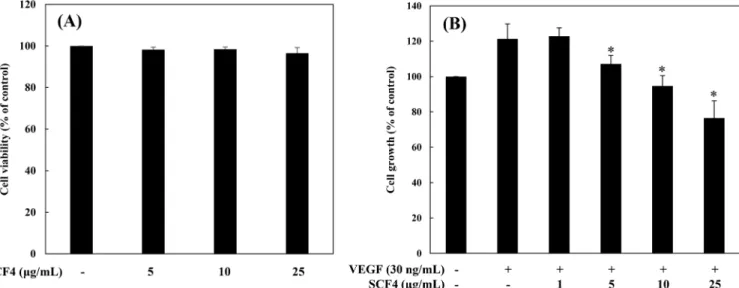
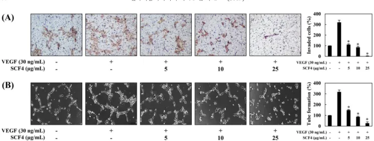
꽃송이버섯 추출물의 혈관내피세포 증식 저해 효과 꽃송이버섯 소수성 추출물(SCF4)의 혈관신생 저해활성 분석에 앞서, 혈관내피세포인 HUVECs에 독성을 나타내는지 여부를 확 인하였다. 세포 독성은 trypan blue assay를 이용하여 분석하였다. HUVEC 세포에 SCF4를 5, 10, 25 µg/mL의 농도로 처리한 결과, 모든 농도에서 SCF4를 처리하지 않은 대조군과 비교했을 때, 독 성이 나타나지 않았다(Fig. 1A). 따라서 이 농도의 SCF4를 처리 하여 혈관신생 저해활성 실험을 진행하게 되었다. 혈관신생의 가장 기본적인 현상은 세포의 활발한 증식이므로 혈관내피세포성장인자(VEGF)를 처리한 혈관내피세포에 SCF4를 농도별로 처리하여 촉진된 세포의 증식을 얼마나 저해하는지 MTT assay로 확인하였다(Hicklin과 Ellis, 2005). 그 결과, 대조군과 비 교하여 VEGF는 혈관내피세포의 증식을 20% 증가시켰으며, SCF4 1 µg/mL에서는 유의적으로 감소하지 않았으나, 5-25 µg/mL에서 는 증가된 세포증식을 농도의존적으로 감소시켰다(*p<0.05) (Fig. 1B). 꽃송이버섯 추출물의 혈관내피세포 침윤성 억제 효과 다음 실험으로, 혈관내피세포의 주요한 혈관신생 특징 중에 하 나인 침윤성에 대한 꽃송이버섯 추출물(SCF4)의 효과를 확인하 였다(Hicklin과 Ellis, 2005). SCF4의 세포 침윤성 억제 활성은 HUVECs이 Matrigel이 코팅된 transwell membrane의 pore를 통해 membrane 아래로 침투하는 능력을 관찰하여 평가하였다. 대조군 와 비교하여 VEGF를 처리한 혈관내피세포들의 침윤성은 현저하 게 증가하였으며, SCF4의 낮은 농도인 5 µg/mL에서는 침윤성이 증가된 세포들을 대조군과 비슷한 수준으로 침윤성을 저해하였 고 SCF4 10, 25 µg/mL의 농도를 처리한 세포들은 VEGF에 의 해 유도된 침투 능력을 농도의존적으로 현저하게 억제하였다 (*p<0.05) (Fig. 2A). 꽃송이버섯 추출물의 혈관내피세포 관 형성 능력 억제 활성 혈관내피세포의 또 다른 주요한 혈관신생 특징인 관 형성 능 력에 대한 꽃송이버섯 추출물(SCF4)의 효과를 확인하였다(Hicklin 과 Ellis, 2005). SCF4의 혈관내피세포 관 형성 억제 활성은 coat-ing된 Matrigel 위에 분주된 HUVEC 세포가 관을 형성하는 것을Fig. 1. The antiproliferative activity of SCF4 on HUVECs. (A) The effect of SCF4 on the viability of HUVECs. Cells were treated with SCF4 (5-25μg/mL) and incubated for 72 h. Cell viability was measured by trypan blue assay. (B) The effect of SCF4 on the growth of HUVECs. Serum-starved HUVECs were stimulated with VEGF (30 ng/mL) in the presence or absence of SCF4 for 72 h. Cell growth was measured using MTT colorimetric assay. The basal levels of the growth of HUVECs that were incubated in serum-free medium without VEGF were normalized to 100%. Each value represents the mean±SD of three different experiments. Values are significantly different from the VEGF control (*p<0.05).
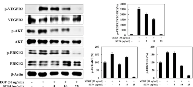
관찰하여 평가하였다. 대조군과 비교하여 혈관신생유도인자인 VEGF를 처리한 세포들은 더 많은 관을 형성하였으며, VEGF와 SCF4 5, 10, 25 µg/mL을 각각 처리한 세포들은 VEGF에 의해 유 도된 튜브 형성 능력을 농도의존적으로 억제하였다(*p<0.05) (Fig. 2B). 이러한 결과는 SCF4가 세포 독성이 없는 농도범위(5-25 µg/ mL)에서 VEGF에 의해 유도된 혈관내피세포의 증식, 침윤성, 관 형성 능력을 억제함으로써 in vitro 혈관신생을 저해함을 입증한다. 꽃송이버섯 추출물의 in vivo 혈관형성 능력 억제 활성 꽃송이버섯 추출물(SCF4)의 혈관신생 억제활성을 in vivo 검증 하기 위해, SCF4가 chorioallantoic membrane (CAM)의 혈관신생 을 저해하는지를 실험하였다. SCF4를 함유한 coverslips을 CAM 표면에 놓고, 혈관신생 유도부위를 현미경으로 관찰하였다. 그 결 과, 대조군 처리시에 혈관신생 억제율은 13% (n=15)인 반면, SCF4 를 처리한 경우에는 기존 혈관에 대한 독성 없이 CAM의 혈관 신생을 강력하게 억제하였다(4 μg/egg에서 88%, n=16) (Fig. 3). 이러한 결과는 SCF4가 혈관내피세포에 대한 독성을 나타내지 않 으면서 in vivo 혈관신생을 유의하게 억제한다는 것을 입증한다. 이상의 실험결과들로 볼 때 꽃송이버섯 소수성 추출물(SCF4)이 in vitro 및 in vivo에서 혈관신생을 효과적으로 저해함을 입증하 여 혈관신생 관련질환의 예방 및 치료를 위한 천연물로 사용될 가능성을 확인하였다. 그러므로, SCF4의 혈관신생 저해 메커니즘 을 규명하기 위한 연구를 이어서 수행하게 되었다. 꽃송이버섯 추출물의 혈관신생 신호전달 경로 억제 활성 VEGFR2 신호 전달은 혈관내피세포의 증식, 이동 및 모세관 형성에 관여하는 다양한 하위 신호 전달 기질의 활성화를 유도 한다고 보고되어 있다(Holmes 등, 2007). VEGFR2의 하위 신호 전달 매개인자로서, PI3K/AKT는 혈관내피세포의 생존 및 증식 신호전달에 중요한 인자로 작용하고 ERK1/2는 혈관내피세포의 침윤성, 관형성과 같은 혈관신생의 주요 표현형에 중요한 인자로 보고되어 있다(de Falco, 2014; Du 등, 2011; Holmes 등, 2007; Shi 등, 2005). 따라서, 꽃송이버섯 추출물(SCF4)의 혈관신생 억 제활성이 VEGFR2 신호전달경로의 억제에 의해 매개된 것인지 확인하기 위해, western blot analysis를 통해 SCF4 처리에 의한 VEGFR2 신호전달경로의 주요 단백질 발현 수준을 확인하였다. 그 결과, SCF4는 전체 단백질 발현 수준에 영향을 주지 않으면 서 VEGF에 의해 유도된 VEGFR2, AKT 및 ERK1/2의 인산화를 효과적으로 억제하였다(Fig. 4). 이러한 결과는 SCF4가 VEGFR2 매개 신호전달 경로를 억제함으로써 항혈관신생 활성을 나타냄 을 시사한다. 우리는 최근 SCF4가 nuclear factor-kappa B (NF-κB) 및 MAPK 신호전달 경로 억제를 통해 lipopolysaccharide (LPS) 자극 에 의한 RAW 264.7 대식세포의 염증 유도 활성을 효과적으로 저해하여 탁월한 항염증 활성을 나타냄을 보고하였다(Han 등, 2019). 현 연구에서는 SCF4가 항염증 활성뿐만 아니라 VEGFR2 매개 신호전달 경로의 저해를 통해 항혈관신생 활성을 가지고 있 Fig. 2. The effect of SCF4 on angiogenesis in vitro. Serum-starved HUVECs were stimulated with VEGF (30 ng/mL) in the presence or absence of SCF4. (A) The inhibitory effect of SCF4 on the VEGF-induced invasion of HUVECs. (B) The inhibitory effect of SCF4 on the VEGF-induced tube-forming ability of HUVECs. The basal levels of invasiveness and tube formation of HUVECs that were incubated in serum-free medium without VEGF were normalized to 100%. Each value represents the mean±SD of three different experiments. Values are significantly different from the VEGF control (*p<0.05).
Fig. 3. The effect of SCF4 on angiogenesis in vivo. Fertilized chick eggs were maintained in a humidified incubator at 37oC. At embryonic day 4.5, coverslips loaded with vehicle alone or SCF4 (4μg/egg) were applied to the CAM surface. After 2 days, the chorioallantois was observed under a microscope. Inhibition ratio was calculated based on the ratio of inhibited eggs relative to the total number of live eggs.
음을 새롭게 입증하였다. 비록 앞선 연구 보고들을 통해 꽃송이 버섯에 함유된 주요 다당류인 베타-글루칸이 항혈관신생 활성을 가지고 있음이 알려져 있다 할지라도(Yamamoto 등, 2009), 꽃송 이버섯 소수성 추출물의 혈관신생 억제활성은 연구되어 보고된 적이 없다. 따라서, 우리의 이번 연구결과는 꽃송이버섯에서 주 요 친수성 유효성분인 베타-글루칸 외에 다른 소수성 유효성분들 이 또한 항혈관신생 활성을 나타냄을 새롭게 밝혀내었다. 흥미롭 게도, 선행 연구 보고에 따르면 꽃송이버섯 유래 베타-글루칸은 in vivo 마우스 실험에서 B16F10 악성흑색종 암세포에 의해 유도 된 혈관신생과 VEGF에 의해 유도된 혈관신생을 억제하였으나, HUVEC 세포의 증식, 이동 및 관 형성 능력을 저해하지 않았다 (Yamamoto 등, 2009). 그러므로, 베타-글루칸의 항혈관신생 활성 은 혈관내피세포에 대한 직접적인 작용에 의해서가 아닌 혈관내 피세포의 기능 조절에 관여하는 다른 생체 내 분자나 신호전달 경로에 영향을 미치는 등의 간접적인 작용기전에 의한 것으로 생 각된다. 그러나, 베타-글루칸과는 달리 SCF4는 HUVEC 세포의 증식, 이동 및 관 형성 능력을 직접적으로 억제하였을 뿐만 아니 라 VEGFR2 매개 신호전달 경로를 직접적으로 저해함으로써 항 혈관신생 활성을 나타내었다. 앞으로의 연구에서는 꽃송이버섯 소수성 추출물(SCF4) 내의 단일 유효성분들을 분리하여 항혈관 신생 작용기전을 보다 다각도로 규명해야 할 것으로 사료된다.
요
약
본 연구에서는 꽃송이버섯 소수성 추출물(SCF4)의 항혈관신생 활성을 혈관내피세포인 HUVECs을 사용하여 확인하였다. 그 결 과, SCF4는 세포 독성을 나타내지 않는 5-25 µg/mL의 농도에서 VEGF에 의해 유도된 혈관내피세포 증식을 유의적으로 감소시켰 을 뿐만 아니라, 혈관내피세포 침윤성과 관 형성 능력을 농도의 존적으로 감소시켜 in vitro 혈관신생을 효과적으로 저해함을 확 인하였다. 또한, SCF4는 독성을 나타내지 않고 CAM의 혈관신생 을 저해함으로써, in vivo 혈관신생을 효과적으로 저해함을 확인 하였다. 마지막으로 SCF4가 혈관신생을 유도하는 주요 신호전달 경로인 VEGFR2, AKT 및 ERK1/2의 전체 단백질 발현 수준의 변화없이 인산화를 저해함을 확인하였다. 따라서, 본 연구는 꽃 송이버섯 소수성 추출물이 혈관신생 저해활성을 나타내고 이러 한 현상은 VEGFR2 신호전달경로의 억제를 통해 진행됨을 입증 하여, 혈관신생 관련 질환 예방 및 치료를 위한 천연물 소재로서 의 적용 가능성을 새롭게 제시하였다.감사의 글
본 논문은 농촌진흥청 연구사업(세부과제번호: PJ01188001)과 한국연구재단 기초연구사업(NRF-2016R1D1A1B03932956)의 지원 에 의해 이루어진 것임.References
Avramis IA, Kwock R, Avramis VI. Taxotere and vincristine inhibit the secretion of the angiogenesis inducing vascular endothelial growth factor (VEGF) by wild-type and drug-resistant human leu-kemia T-cell lines. Anticancer Res. 21: 2281-2286 (2001)
Carmeliet P. Angiogenesis in life, disease and medicine. Nature 438: 932-936 (2005)
Carmeliet P, Jain RK. Angiogenesis in cancer and other diseases. Nature 407: 249-257 (2000)
Clements MK, Jones CB, Cumming M, Daoud SS. Antiangiogenic potential of camptothecin and topotecan. Cancer Chemoth. Pharm. 44: 411-416 (1999)
Coultas L, Chawengsaksophak K, Rossant J. Endothelial cells and VEGF in vascular development. Nature 438: 937-945 (2005) de Falco S. Antiangiogenesis therapy: an update after the first
decade. Korean J. Intern. Med. 29: 1-11 (2014)
Du J, Xu R, Hu Z, Tian Y, Zhu Y, Gu L, Zhou L. PI3K and ERK-induced Rac1 activation mediates hypoxia-ERK-induced HIF-1α expression in MCF-7 breast cancer cells. PLoS One 6: e25213 (2011)
Fan TP, Yeh JC, Leung KW, Yue PY, Wong RN. Angiogenesis: From plants to blood vessels. Trends Pharmacol. Sci. 27: 297-309 (2006)
Gerber HP, Mc Murtrey A, Kowalski J, Yan M, Keyt BA, Dixit V, Ferrara N. Vascular endothelial growth factor regulates endothe-lial cell survival through the phosphatidylinositol 3-kinase/Akt signal transduction pathway. Requirement for Flk-1/KDR activa-tion. J. Biol. Chem. 273: 30336–30343 (1998)
Han JM, Kwon HJ, Jung HJ. Tricin, 4',5,7-trihydroxy-3',5'-dimethox-yflavone, exhibits potent antiangiogenic activity in vitro. Int. J.
Fig. 4. The effect of SCF4 on VEGFR2-dependent signal transduction in HUVECs. Serum-starved HUVECs were pretreated with SCF4 for 3 h at the indicated concentrations and then stimulated with VEGF (30 ng/mL) for 5 min. Protein levels were detected by western blot analysis. The level of β-actin was used as an internal control.
Oncol. 49: 1497-1504 (2016)
Han JM, Lee EK, Gong SY, Sohng JK, Kang YJ, Jung HJ. Sparassis crispa exerts anti-inflammatory activity via suppression of TLR-mediated NF-κB and MAPK signaling pathways in LPS-induced RAW264.7 macrophage cells. J. Ethnopharmacol. 231: 10-18 (2019)
Han JM, Lim HN, Jung HJ. Hovenia dulcis Thunb. and its active compound ampelopsin inhibit angiogenesis through suppression of VEGFR2 signaling and HIF-1α expression. Oncol. Rep. 38: 3430-3438 (2017)
Hicklin DJ, Ellis LM. Role of the vascular endothelial growth factor pathway in tumor growth and angiogenesis. J. Clin. Oncol. 23: 1011-1027 (2005)
Hida TH, Kawaminami H, Ishibashi K, Miura NN, Adachi Y, Ohno N. Oral administration of soluble β-glucan preparation from the cauliflower mushroom, Sparassis crispa (Higher Basidiomycetes) modulated cytokine production in mice. Int. J. Med. Mushrooms 15: 525-538 (2013)
Holmes K, Roberts OL, Thomas AM, Cross MJ. Vascular endothelial growth factor receptor-2: Structure, function, intracellular signal-ling and therapeutic inhibition. Cell. Signal. 19: 2003-2012 (2007)
Hong KB, Hong SY, Joung EY, Kim BH, Bae SH, Park Y, Suh HJ. Hypocholesterolemic effects of the cauliflower culinary-medicinal mushroom, Sparassis crispa (Higher Basidiomycetes), in diet-induced hypercholesterolemic rats. Int. J. Med. Mushrooms 17: 965-975 (2015)
Jing Y, Liu LZ, Jiang Y, Zhu Y, Guo NL, Bamett J, Rojanasakul Y, Agani F, Jiang BH. Cadmium increases HIF-1 and VEGF expres-sion through ROS, ERK, and AKT signaling pathways and induces malignant transformation of human bronchial epithelial cells. Toxicol. Sci. 125: 10-19 (2012)
Kim HH, Lee S, Singh TS, Choi JK, Shin TY, Kim SH. Sparassis crispa suppresses mast cell-mediated allergic inflammation: role of calcium, mitogen-activated protein kinase and nuclear
factor-Kb. Int. J. Mol. Med. 30: 344-350 (2012)
Kimura T. Natural products and biological activity of the pharmaco-logically active cauliflower mushroom Sparassis crispa. Biomed. Res. Int. 2013: 982317 (2013)
Ohno N, Miura NN, Nakajima M, Yadomae T. Antitumor 1,3-beta-glucan from cultured fruit body of Sparassis crispa. Biol. Pharm. Bull. 23: 866-872 (2000)
Rajendran P, Rengarajan T, Thangavel J, Nishigaki Y, Sakthisekaran D, Sethi G. The Vascular Endothelium and Human Diseases. Int. J. Biol. Sci. 9: 1057-1069 (2013)
Shi YH, Wang YX, Bingle L, Gong LH, Heng WJ, Li Y, Fang WG. In vitro study of HIF-1 activation and VEGF release by bFGF in the T47D breast cancer cell line under normotoxic conditions: involvement of PI-3K/Akt and MEK1/ERK pathways. J. Pathol. 205: 530-536 (2005)
Vincent L, Kermani P, Young LM, Cheng J, Zhang F, Shido K, Lam G, Bompais-Vincent H, Zhu Z, Hicklin DJ, Bohlen P, Chaplin DJ, May C, Rafii S. Combretastatin A4 phosphate induces rapid regression of tumor neovessels and growth through interference with vascular endothelial-cadherin signaling. J. Clin. Invest. 115: 2992-3006 (2005)
Yamamoto K., Kimura T. Orally and topically administered Sparassis crispa (Hanabiratake) improved healing of skin wounds in mice with streptozotocin-induced diabetes. Biosci. Biotechnol. Bio-chem. 77: 1303-1305 (2013)
Yamamoto K, Kimura T, Sugitachi A, Matsuura N. Anti-angiogenic and anti-metastatic effects of beta-1,3-D-glucan purified from Hanabiratake, Sparassis crispa. Biol. Pharm. Bull. 32: 259-263 (2009)
Yoshida M, Hida TH, Takeshita K, Tsuboi M, Kanamori M, Akachi N, Miura NN, Adachi Y, Ohno N. Effect of Sasa veitchii extract on immunostimulating activity of β-glucan (SCG) from culinary-medicinal mushroom Sparassis crispa Wulf.:Fr. (higher Basidio-mycetes). Int. J. Med. Mushrooms 14: 537-547 (2012)