INTRODUCTION
Wiskott-Aldrich syndrome (WAS) (Online Mendelian Inheritance in Man [OMIM]301000) is an X-linked reces- sive disorder characterized by thrombocytopenia, eczema, and immunodeficiency. Clinical symptoms include petechi- ae, bloody diarrhea, inability to generate antibodies against polysaccharide antigens, and in some cases, autoimmune manifestations. Affected boys often die because of malignant tumors, particularly lymphoma (1, 2). Treatments for WAS include antimicrobial therapy for infections, intravenous immune globulin, splenectomy for thrombocytopenia, and allogeneic bone marrow transplantation (3-5).
The gene responsible for WAS was isolated, and designat- ed the WAS gene (6). The gene is composed of 12 exons span- ning approximately 9 kilobases. The encoded protein is a 502 amino-acid long intracellular protein, which is broadly expressed in hematopoietic cells (7). The WAS gene was found to be mutated, not only in classic WAS patients, but also in patients with X-linked thrombocytopenia (XLT), which is a clinically mild allelic variant (8, 9). Recent reports have ex- panded the WAS mutation phenotype to include intermit- tent XLT, X-linked neutropenia with or without myelodys- plasia, or WAS/XLT in females with heterozygous mutations
(10, 11).
Many WAS gene mutations have been reported in patients with WAS. However, little is known about Korean WAS pa- tients in terms of their molecular genetic diagnosis. In this report, we describe a Korean family with WAS, which was diagnosed as having exon 2 mutation, based on the WAS gene mutation analysis. This is the first identified case of a hotspot mutation in exon 2 of WAS in Korea.
MATERIALS AND METHODS Subjects
A one-day-old male neonate was admitted to the hospital because of jaundice and petechiae on the body. He was born after 37 weeks gestation, by Cesarean section due to breech presentation and had a birth weight of 2,800 g. Laboratory data showed a hemoglobin of 16.8 g/dL, a white blood cell count of 17,050/ L, a platelet count of 39,000/ L, and a high reticulocyte count (9.1%), but Coombs’ tests were negative.
The level of serum bilirubin (total/direct) was 7.3/0.4 mg/dL, and the result of the liver function test was unremarkable.
Also, immunoglobulin (Ig) M titers to TORCH were nega- Sook-Kyung Park*, Chun-Soo Kim�, Dae-Kyu Song�, Joo-Young Kim�, In-Jang Choi*, Dae-Kwang Kim*,‖,
Departments of Anatomy*, Pediatrics�, Physiology�, Institute for Medical Genetics School of Medicine‖, Keimyung University, Daegu; Department of Anatomy�, Yeungnam University College of Medicine, Hanvit Institute for Medical Genetics¶, Daegu, Korea
Address for correspondence Dae-Kwang Kim, M.D.
Department of Anatomy, Institute for Medical Genetics, School of Medicine, Keimyung University, Hanvit Institute for Medical Genetics, 194 Dongsan-dong, Jung-gu, Daegu 700-712, Korea
Tel : +82.53-250-7475, Fax : +82.53-250-7471 E-mail : [email protected]
*This work was supported by Grant No. R13-2002-028- 03002-0 from the Basic Research Program of KOSEF.
998 J Korean Med Sci 2007; 22: 998-1001
ISSN 1011-8934
DOI: 10.3346/jkms.2007.22.6.998
Copyright � The Korean Academy of Medical Sciences
A Familial Case of Wiskott-Aldrich Syndrome with a Hotspot Mutation in Exon 2 of the WAS Gene
The Wiskott-Aldrich syndrome (WAS) is a severe X-linked disorder characterized classically by thrombocytopenia, immunodeficiency, and eczema. The phenotype observed in this syndrome is caused by mutation in the WAS gene. Peripheral blood DNAs were isolated from an 18-month-old boy with WAS and his mother, maternal uncle, and maternal grandmother. Genetic analysis for the detection of a mutation of WAS gene was performed by polymerase chain reaction-single strand conformational polymorphism analysis (PCR-SSCP) and direct sequencing of the PCR product. In PCR-SSCP, the patient and his maternal uncle had an abnormal shift band, which was not found in normal controls, and his mother and maternal grandmother showed heterozygous bands. In direct sequencing analysis, the patient with WAS had CGC→CAC point mutation in exon 2 that resulted in an amino acid change in codon 86 (Arg86His). The present study identified a gene mutation respon- sible for WAS at a mutation hotspot of the WAS gene in a Korean family.
Key Words : Wiskott-Aldrich Syndrome; Mutation Analysis; Wiskott-Aldrich Syndrome Protein, Neuronal
Received : 7 February 2007 Accepted : 4 May 2007
Exon 2 Mutation of Wiskott-Aldrich Syndrome 999
tive. Serum Ig measurements at day 1 revealed an elevated Ig A level (28.8 mg/dL; normal range, 1.3-3.6) (Ig G: 1,216.8, Ig M<23.8, Ig D<1.5, and Ig E<18.0). The results of periph- eral blood smear were unremarkable, except for a markedly decreased number of platelets. A bone marrow aspiration smear showed slight erythroid and megakaryocytic hypopla- sia. Karyotyping analyses of both the patient and his moth- er were normal. A review of family history revealed his mater- nal uncle was clinically diagnosed as having WAS at five years of age. The patient was treated with platelet transfu- sion, and petechiae on the body disappeared immediately.
However, the patient’s thrombocytopenia persisted. When the patient was 18 months old, genetic counseling was pro- vided at his mother’s request.
Mutation analysis
Genomic DNA was isolated from peripheral blood leuko- cytes by using the Wizard Genomic DNA purification kit, according to the manufacturer’s instructions (Promega, Ma- dison, WI, U.S.A.). In order to examine mutations on the WAS gene, polymerase chain reaction-single strand confor- mational polymorphism (PCR-SSCP) was performed for all 12 coding regions of the WAS gene by using the primer sets as listed in Table 1. The PCR was performed with a thermal
cycler (model 2400, Applied Biosystems, Foster City, CA, U.S.A.) as follows: 32 cycles of 1 min at 94℃for denatura- tion, 1 min at 55-60℃for annealing depending on the primers, and 1 min at 72℃for extension. Final extension was performed at 72℃for 10 min. The PCR products were electrophoresed on an agarose gel and stained with ethidium bromide to confirm the size of the bands. The PCR products were also denatured in formamide loading buffer and elec- trophoresed through 8% and 10% polyacrylamide gels. Sil- ver stain was performed to develop bands (12). To determine the sequences of the DNA samples showing mutant bands, direct sequencing was performed on the ABI Prism 3100 Genetic Analyser (Applied Biosystems).
RESULTS
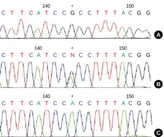
PCR and subsequent SSCP analysis in exon 2 of the WAS gene identified an aberrantly migrating band in the patient’s mother, and in his maternal grandmother, compared to two wild type bands in the controls. The patient and his mater- nal uncle showed two bands, one of which was a lower wild single strand band and the other, an abnormal shifted band (Fig. 1). In the direct sequencing analysis of the PCR prod- ucts for SSCP, the missense mutation was observed in the patient and his maternal uncle. This showed G-to-A transi- tion at complementary nucleotide 257 (c.257G>A) (Fig. 2C), resulting in substitution of His for Arg at codon 86 (Arg86- His) (CGC→CAC). The patient’s mother and his maternal grandmother were heterozygous for the mutation at codon 86 (Fig. 2B).
DISCUSSION
Mutations of the WAS gene result in 3 distinct pheno- types: the classic WAS triad of thrombocytopenia with small platelets, recurrent infections as a result of immunodeficien- cy, and eczema (1); the milder XLT variant, characterized
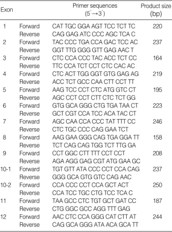
Exon Primer sequences
(5′→ 3′)
Product size (bp) 1 Forward CAT TGC GGA AGT TCC TCT TC 220
Reverse CAG GAG ATC CCC AGC TCA C
2 Forward TAC CCC TGA CCA GAC TCC AC 237 Reverse GGT TTG GGG GTT GAG AAC T
3 Forward CTC CCA CCC TAC ACC TCT CC 164 Reverse TTC CCA TCT CCT CTC CAC AC
4 Forward CTC ACT TGG GGT GTG GAG AG 219 Reverse ACC TCT GCC CAA CTT CCT TT
5 Forward AAG TCC CCT CTC ATG GTC CT 195 Reverse AGC CCT CCT CTT CTC TCT GG
6 Forward GTG GCA GGG CTG TGA TAA CT 223 Reverse GCT CGT CCA TCC ACA TAC CT 7 Forward AGC CAA CCA CCC TAT TTT CC 246
Reverse CTC TGC CCC CAG GAA TCT
8 Forward AAG GAA GGG CAG TGA GGA TT 158 Reverse TCT CAG CAG TGG TCT TTG GA
9 Forward CCT GGC CTT TTT CCT CCT 208 Reverse AGA AGG GAG CGT ATG GAA GC 10-1 Forward TGT GTT ATA CCC CCT CCA CAG 237
Reverse GGG GCA GTG GTC CAG AAC
10-2 Forward CCA CCC CCT CCA GCT ACT 250 Reverse CCA TCC TGC CTG TCC TCA C
11 Forward TAA GCC CTC TGT GCT GAT CC 187 Reverse CTG GGC GCC AGG TTT GAG
12 Forward AAC CTC CCA GGG CAT CTT AT 244 Reverse CAG GCA GGG ATA ACA GCA TT Table 1.PCR primers for amplification of the WAS gene
Fig. 1.Single-strand conformational polymorphism patterns for exon 2 of the WAS gene. Patient’s mother and his maternal grand- mother showed an extra band above upper normal single strand band in lane 5 and 7. In lane 6 and 8, patient and maternal uncle exhibited two bands, a normal lower single strand band and a mobility shift band. In lane 1-4, normal subjects showed two sin- gle strand bands.
1 2 3 4 5 6 7 8
PCR, polymerase chain reaction; WAS, Wiskott-Aldrich syndrome.
1000 S.-K. Park, C.-S. Kim, D.-K. Song, et al.
predominantly by thromobocytopenia with small platelets (8, 9); and finally, congenital neutropenia without the clini- cal findings characteristic for WAS/XLT (10, 11).
As shown in WASPbase (13), a database of mutations, a total of 441 cases consisted of missense mutations in 179 cases (40.6%), deletions in 92 cases (20.9%), nonsense muta- tions in 64 cases (14.5%), splicing defects in 64 cases (14.5
%), and insertions in 42 cases (9.5%). The exon distribu- tion of over 5% frequency of mutation was located in exon 2 (27.0%), exon 10 (16.1%), exon 1 (13.8%), exon 4 (9.8
%), exon 3 (7.5%), and exon 7 (6.4%). Notably, the frequen- cy at codon 86 in exon 2 was 33.6% (40/119 cases). It is well known that the codon 86 of the WAS gene is the most com- mon missense mutation site responsible for WAS/XLT (10, 13). To our knowledge, this is the first report on the hotspot missense mutation in exon 2 of WAS gene in Korea.
There are several reports on WAS mutations in Korean families (Table 2). As shown in Table 2, the predominant mutations were nonsense mutations and missense mutations (14-18). Only one small deletion was reported in exon 10 (15). In this report, we found a missense mutation in exon 2. In Korean patients’ reports, including our results, muta- tions in exon 1, 2, 3, 7, 8, and 10 have been involved in WAS.
Therefore, although it is necessary to accumulate mutation data from a large number of patients with WAS, the distri- bution of mutations may be highly diverse in Korean patients, as had been observed in other reports (9, 10, 13).
A correlation between clinical phenotype and genotype was reported independently by several investigators (19-21), but was not observed by all (22, 23). Imai et al. demonstrat- ed that all WAS patients with missense mutations showed
WASP-positive expression; in contrast, patients with non- sense mutations, large deletions, small deletions, and small insertions were WASP-negative expression. Patients with splicing mutations were either WASP-negative or WASP- positive. Lack of WASP expression was associated with severe clinical symptoms and poor prognosis (24). Jin et al. report- ed 5 mutational hotspots in the WAS gene from 227 WAS/
XLT families with a total of 262 affected members. They also noted that the missense mutation at codon 86 was ob- served most frequently and associated with mild symptoms (25). Two previous reports (24, 25) showed that there is a strong possibility of establishing an association between geno- type and phenotype. In spite of advances in the clinical treat- ment of WAS patients, WAS remains a life-threatening con- dition, resulting in a poor quality of life and a bad long-term prognosis, especially for those who lack an HLA-matched sibling. The discovery of the WAS gene, and the identifica- tion of the molecular basis of WAS, have made it possible to provide genetic counseling for at-risk families. Therefore, this report on the hotspot missense mutation in exon 2 of WAS gene, will help this patient’s parent and his maternal uncle to understand WAS and plan both genetic counseling and family regulation.
REFERENCES
1. Sullivan KE, Mullen CA, Blaese RM, Winkelstein JA. A multiinsti- tutional survey of the Wiskott-Aldrich syndrome. J Pediat 1994; 125:
876-85.
2. Filipovich AH, Mathur A, Kamat D, Kersey JH, Shapiro RS. Lym- phoproliferative disorders and other tumors complicating immun- odeficiencies. Immunodeficiency 1994; 5: 91-112.
3. Mullen CA, Anderson KD, Blaese RM. Splenectomy and/or bone marrow transplantation in the management of the Wiskott-Aldrich syndrome: long-term follow-up of 62 cases. Blood 1993; 82: 2961-6.
4. Litzman J, Jones A, Hann I, Chapel H, Strobel S, Morgan G. Intra- venous immunoglobulin, splenectomy, and antibiotic prophylaxis in Wiskott-Aldrich syndrome. Arch Dis Child 1996; 75: 436-9.
5. Filipovich AH, Stone JV, Tomany SC, Ireland M, Kollman C, Pelz CJ, Casper JT, Cowan MJ, Edwards JR, Fasth A, Gale RP, Junker A, Kamani NR, Loechelt BJ, Pietryga DW, Ringden O, Vowels M, Exon Type of
mutation
Nucleotide No.
Nucleotide mutation
Amino acid
effect Year
1 Nonsense 71 C→T Arg 13 Stop 2003
2 Missense 208 G→A Gly 70 Arg 2005
3 Missense 336 T→C Leu 101 Pro 2004
7 Nonsense 665 C→T Arg 211 Stop 2003 8 Nonsense 756 G→A Trp 252 Stop 2006 10 Deletion 1326-1329 G del Frameshift/444 Stop 2004 Table 2.Published mutations of the WAS gene in Korean fami- lies
Fig. 2.Sequencing identification of the missense mutation in exon 2 of the WAS gene. The asterisk (*) indicated a single base ‘‘G’’
to ‘‘A’’ substitution in the affected patient (C) compared with a nor- mal control (A), causing replacement of arginine by histidine at the amino acid residue 86 (Arg86His). WAS exon 2 sequence in female heterozygotes (B).
A
B
C C T T C A T C C G C C T T T A C G G
140 * 150
C T T C A T C C N C C T T T A C G G
140 * 150
C T T C A T C C A C C T T T A C G G
140 * 150 WAS, Wiskott-Aldrich syndrome.
Exon 2 Mutation of Wiskott-Aldrich Syndrome 1001
Hegland J, Williams AV, Klein JP, Sobocinski KA, Rowlings PA, Horowitz MM. Impact of donor type on outcome of bone marrow transplantation for Wiskott-Aldrich syndrome: collaborative study of the International Bone Marrow Transplant Registry and the Na- tional Marrow Donor Program. Blood 2001; 97: 1598-603.
6. Derry JM, Ochs HD, Francke U. Isolation of a novel gene mutated in Wiskott-Aldrich syndrome. Cell 1994; 78: 635-44.
7. Stewart DM, Treiber-Held S, Kurman CC, Facchetti F, Notarangelo LD, Nelson DL. Studies of the expression of the Wiskott-Aldrich syndrome protein. J Clin Invest 1996; 97: 2627-34.
8. Villa A, Notarangelo L, Macchi P, Mantuano E, Cavagni G, Brugnoni D, Strina D, Patrosso MC, Ramenghi U, Sacco MG, Ugazio A, Vez- zoni P. X-linked thrombocytopenia and Wiskott-Aldrich syndrome are allelic diseases with mutations in the WASP gene. Nat Genet 1995;
9: 414-7.
9. Schwarz K. WASPbase: a database of WAS- and XLT-causing muta- tions. Immunol Today 1996; 17: 496-502.
10. Imai K, Nonoyama S, Ochs HD. WASP (Wiskott-Aldrich syndrome protein) gene mutations and phenotype. Curr Opin Allergy Clin Im- munol 2003; 3: 427-36.
11. Devriendt K, Kim AS, Mathijs G, Frints SG, Schwartz M, Van Den Oord JJ, Verhoef GE, Boogaerts MA, Fryns JP, You D, Rosen MK, Vandenberghe P. Constitutively activating mutation in WASP causes X-linked severe congenital neutropenia. Nat Genet 2001; 27: 313-7.
12. Ha TW, Han KH, Son DG, Kim SP, Kim DK. Analysis of loss of heterozygosity in Korean patients with keratoacanthoma. J Korean Med Sci 2005; 20: 340-3.
13. Imai K. WASPbase. http:// homepage.mac.com/kohsukeimai/wasp/
WASPbase.html.
14. Jo EK, Futatani T, Kanegane H, Kubota T, Lee YH, Jung JA, Song CH, Park JK, Nonoyama S, Miyawaki T. Mutational analysis of the WASP gene in 2 Korean families with Wiskott-Aldrich syndrome. Int J Hematol 2003; 78: 40-4.
15. Kim MK, Kim ES, Kim DS, Choi IH, Moon T, Yoon CN, Shin JS.
Two novel mutations of Wiskott-Aldrich syndrome: the molecular prediction of interaction between the mutated WASP L101P with WASP-interacting protein by molecular modeling. Biochim Biophys Acta 2004; 1690: 134-40.
16. Hwang DJ, Yang JW, Kim SY, Yi HK, Lee DY, Hwang PH. Diag- nostic Approach of Wiskott-Aldrich Syndrome. Korean J Pediatr 2004; 47: 726-34.
17. Baek HJ, Choi SH, Sohn KR, Kook H, Kim SJ, Song ES, Han DK, Hwang TJ. Mutation Analysis in X-linked Recessive Congenital Im- munodeficiency Syndromes. Chonnam Med J 2005; 41: 48-61.
18. Kim HJ, Yoo EH, Ki CS, Yoo GH, Koo HH, Kim JW, Kim SH. A novel mutation W252X in the WAS gene in a Korean patient with Wiskott-Aldrich syndrome. Int J Hematol 2006; 83: 426-8.
19. Wengler GS, Notarangelo LD, Berardelli S, Pollonni G, Mella P, Fasth A, Ugazio AG, Parolini O. High prevalence of nonsense, frame shift, and splice-site mutations in 16 patients with full-blown Wiskott- Aldrich syndrome. Blood 1995; 86: 3648-54.
20. Zhu Q, Watanabe C, Liu T, Hollenbaugh D, Blaese RM, Kanner SB, Aruffo A, Ochs HD. Wiskott-Aldrich syndrome/X-linked thrombo- cytopenia: WASP gene mutations, protein expression, and pheno- type. Blood 1997; 90: 2680-9.
21. Lemahieu V, Gastier JM, Francke U. Novel mutations in the Wiskott- Aldrich syndrome protein gene and their effects on transcriptional, translational, and clinical phenotypes. Hum Mutat 1999; 14: 54-66.
22. Greer WL, Shehabeldin A, Schulman J, Junker A, Siminovitch KA.
Identification of WASP mutations, mutation hotspots and genotype- phenotype disparities in 24 patients with the Wiskott-Aldrich syn- drome. Hum Genet 1996; 98: 685-90.
23. Schindelhauer D, Weiss M, Hellebrand H, Golla A, Hergersberg M, Seger R, Belohradsky BH, Meindl A. Wiskott-Aldrich syndrome:
no strict genotype-phenotype correlations but clustering of missense mutations in the amino-terminal part of the WASP gene product.
Hum Genet 1996; 98: 68-76.
24. Imai K, Morio T, Zhu Y, Jin Y, Itoh S, Kajiwara M, Yata J, Mizutani S, Ochs HD, Nonoyama S. Clinical course of patients with WASP gene mutations. Blood 2004; 103: 456-64.
25. Jin Y, Mazza C, Christie JR, Giliani S, Fiorini M, Mella P, Gandelli- ni F, Stewart DM, Zhu Q, Nelson DL, Notarangelo LD, Ochs HD.
Mutations of the Wiskott-Aldrich Syndrome Protein (WASP): hot- spots, effect on transcription, and translation and phenotype/geno- type correlation. Blood 2004; 104: 4010-9.