INTRODUCTION
Perinatal hypoxic-ischemic encephalopathy (HIE) is com- mon, and reported to affect two to four of every 1,000 full- term neonates, and considerably more preterm babies (1).
Approximately 15 to 20% of term asphyxiated newborns with HIE die during the newborn period and 25% of the survivors exhibit permanent neuropsychological deficits such as mental retardation, cerebral palsy, seizures, and learning disabilities (1). Despite the fact that perinatal asphyxia close- ly corresponds to experimental models of cerebral hypoxia- ischemia (HI), where successful neuroprotective interven- tions have not been introduced, to date, no reliable treatment strategies have been developed to mitigate neurological injury and the resulting impairments in the clinical setting.
The protective effect of exogenously administered erythro- poietin (EPO) has received a great deal of attention for vari- ous brain damage in adult animals, and promising data are
emerging with perinatal animal models also. The neuropro- tective effects exerted by EPO are mediated by various direct and indirect mechanisms (2-4). For evaluation of the effica- cy of a pharmacological treatment of brain injury, it is very important to assess not only the histopathological evidence, but also the improvement of neurobehavioral performance.
Recent studies have suggested that use of EPO has a neuro- protective effect in the neonatal HI rat model, based on neu- ropathological and biochemical data (5-7). However, there has been limited emphasis on the effectiveness of EPO on behavioral alterations after EPO treatment in a postnatal day (P) 7 HI rat model (8-10).
The subventricular zone (SVZ), found along the ependy- mal layer of the lateral ventricle, is known to generate neu- ral progenitor cells throughout postnatal life (11), and neu- rogenesis at this cerebral region might be capable of replac- ing neurons after ischemic brain injury (12, 13). Recent experimental data indicate that diverse forms of ischemic
484
Sung Shin Kim*, Kyung-Hoon Lee�, Dong Kyung Sung�, Jae Won Shim�, Myo Jing Kim, Ga Won Jeon, Yun Sil Chang, and Won Soon Park
Department of Pediatrics*, Soonchunhyang University College of Medicine, Bucheon Hospital, Buchoen;
Department of Molecular Cell Biology�, Sungkyunkwan University School of Medicine/Clinical Trial Center, Clinical Research Institute, Samsung Medical Center;
Samsung Biomedical Research Institute�; Department of Pediatrics�, Kangbuk Samsung Hospital, Sungkyunkwan University School of Medicine, Seoul;
Department of Pediatrics, Samsung Medical Center, Sungkyunkwan University School of Medicine, Seoul, Korea
Address for correspondence Yun Sil Chang, M.D.
Department of Pediatrics, Samsung Medical Center, Sungkyunkwan University School of Medicine, 50 Irwon-dong, Gangnam-gu, Seoul 135-710, Korea Tel : +82.2-3410-3539, Fax : +82.2-3410-0043 E-mail : [email protected]
*This work was supported by the Korea Research Foundation Grant funded by the Korean Government (MOEHRD) (KRF-2005-041-E00346) and IN-SUNG Foundation of Medical Research (C-A5-841-1).
DOI: 10.3346/jkms.2008.23.3.484
Erythropoietin Attenuates Brain Injury, Subventricular Zone Expansion, and Sensorimotor Deficits in Hypoxic-Ischemic Neonatal Rats
The aim of this study was to investigate the effect of erythropoietin (EPO) on histo- logical brain injury, subventricular zone (SVZ) expansion, and sensorimotor func- tion deficits induced by hypoxia-ischemia (HI) in newborn rat pups. Seven-day-old male rat pups were divided into six groups: normoxia control, normoxia EPO, hypox- ia control, hypoxia EPO, HI control, and HI EPO group. Sham surgery or HI was performed in all animals. HI was induced by ligation of the right common carotid artery followed by 90 min of hypoxia with 8% oxygen. Recombinant human EPO 3 U/g or saline was administered intraperitoneally, immediately, at 24- and 48-hr after insult. At two weeks after insult, animals were challenged with cylinder-rearing test for evaluating forelimb asymmetry to determine sensorimotor function. All animals were then sacrificed for volumetric analysis of the cerebral hemispheres and the SVZ. The saline-treated HI rats showed marked asymmetry by preferential use of the non-impaired, ipsilateral paw in the cylinder-rearing test. Volumetric analysis of brains revealed significantly decreased preserved ipsilateral hemispheric volume and increased ipsilateral SVZ volume compared with the sham-operated animals.
Treatment of EPO significantly improved forelimb asymmetry and preserved ipsi- lateral hemispheric volume along with decreased expansion of ipsilateral SVZ fol- lowing HI compared to the saline-treated HI rats. These results support the use of EPO as a candidate drug for treatment of neonatal hypoxic-ischemic brain damage.
Key Words : Hypoxia-Ischemia, Brain; Animals, Newborn; Subventricular Zone; Function
Received : 19 April 2007 Accepted : 1 November 2007
injury stimulate neural precursor proliferation; however, most of these investigations have focused on neuronal pro- duction in the adult rodent brain (12, 13). A recent report from Plane et al. (14) and Chang et al. (15) showed that HI or transient middle cerebral artery occlusion in a neonatal rodent model resulted in augmentation of the SVZ size simi- lar to results from the adult model, and that the expansion of the SVZ was directly proportional to the severity of the injury. Yang et al. (16) and Ong et al. (17) also recently report- ed that hypoxia-ischemia injuries resulted in expansion of the SVZ in neonatal rats.
However, there have been conflicting reports on the find- ing of the effects of EPO on the changes in the SVZ. Wang et al. (18) reported that EPO enhanced neurogenesis and expansion of the SVZ after stroke in the adult rodent; Chang et al. (15) reported that EPO markedly preserved hemispher- ic volume and decreased the expansion of SVZ in a neonatal rat stroke model. However, the effect of EPO on changes in the neonatal SVZ after HI in the newborn rat has not been previously studied.
Therefore, herein, the therapeutic potential effects of EPO in P7 HI rats were investigated by evaluating histopatholo- gy, using volumetric analysis of brain volume; in addition, the neurofunctional outcome was assessed by the examina- tion of sensorimotor function using the cylinder-rearing test.
Furthermore, we also evaluated whether EPO administra- tion affects the expansion of the SVZ in response to a hypox- ic-ischemic insult to the neonatal brain, by a volumetric anal- ysis of SVZ of the rat brain.
MATERIALS AND METHODS Hypoxia-ischemia
The experimental protocols were reviewed and approved by the Institutional Animal Care and Use Committee (IAC- UC) of the Samsung Biomedical Research Institute (SBRI), Seoul, Korea. This study also followed the institutional and National Institutes of Health guidelines for laboratory ani- mal care. Sprague-Dawley rats (Daihan Biolink Co., Seoul, Korea) with dated pregnancies were maintained at the same center and housed in individual cages with free access to water and laboratory chow. The offspring delivered sponta- neously were reared with their dams. To exclude gender effects, male P7 rat pups weighing 12-15 g were used for the experiments (19).
The P7 male rat pups (n=43) were randomly divided into six groups: 1) normoxia control (n=5), normoxia plus saline;
2) normoxia EPO (n=5), normoxia plus EPO; 3) hypoxia control (n=5), hypoxia plus saline; 4) hypoxia EPO (n=5), hypoxia plus EPO; 5) HI control (n=11), HI plus saline; and 6) HI EPO (n=12), HI plus EPO. Cerebral hypoxia-ischemia was induced by a modification of the method reported origi-
nally by Rice et al. (20) as previously described (21, 22). Rat pups were anesthetized in a small jar containing cotton soaked with methoxyflurane, and deep anesthesia was maintained during the surgical procedure by placing a small plastic tube containing cotton soaked with methoxyflurane over the nose.
The neck was incised in the midline, and the right common carotid artery was permanently ligated with 4-0 silk. The total time for surgery in each animal never exceeded three minutes.
Originally described Rice-Vannuci rat hypoxic-ischemic model produces large infarcts (20) and depletes dorsal SVZ (23). Therefore, we reduced the severity of the insult by reduc- ing the hypoxia exposure time to 90 min and found that it produced a moderate level of injury (21, 22). Following a two- hr recovery and feeding period, the animals were exposed to 90 min of hypoxia (8% O2and 92% N2) by placing them in airtight containers partially submerged in a 37℃water bath to maintain a constant thermal environment. Animals in the groups of normoxia control, normoxia EPO, hypoxia control, and hypoxia EPO received a sham-operation. The sham-operated pups underwent the same surgical procedure with exposure of the carotid artery without ligation. Pups received either 0.9% saline with the same volume as the EPO or recombinant human EPO (Epokine prefilled�: erythro- poietin alpha, CJ Corp., Seoul, Korea) (a kind gift from CJ Corp.) at a dose of 3 units per gram of body weight injected intraperitoneally three times: upon retrieval from the hypox- ia chambers or the sham operation, 24 and 48 hr after insult.
After the injection, the pups were returned to their dams for 14 days until sacrifice.
Cylinder-rearing test
It has been shown that episodes of unilateral cerebral HI in P7 rats result in neuronal loss in the cortex, basal ganglia, and hippocampus (24). As in humans, rats that experience cerebral HI have motor and cognitive deficits. In a cylinder- rearing test of paw placement while exploring a transparent cylinder, rats that underwent unilateral HI on P7 have been shown to exhibit an ipsilateral paw weight-bearing preference as early as on P21 (25). This cylinder-rearing test has been shown to reveal lateralizing sensorimotor deficits in neona- tal HI (25).
Forelimb use bias was analyzed by the movements of each rat during exploratory activity in a transparent polymethyl- methacrylate (Atoglas, Atofina Chemicals Inc., Philadelphia, PA, U.S.A.) cylinder that measured 20 cm in diameter and 30 cm in height, at 14 days after surgery (P21). The size of the cylinder allowed free movements but was small enough to encourage rearing and wall exploration. Its height pre- vented the rat from reaching the top edge. The cylinder was sufficiently heavy that it did not move when the animal sup- ported its weight against the wall. Animals were handled for about 10 min per day three days before testing. Each animal was individually placed in the cylinder and observed for three
minutes. The initial forepaw placement of each weight-bear- ing contact with the wall during a full rear was recorded, or
‘‘both’’ were recorded for simultaneous contact with both forepaws (26). Results were expressed as the percentage of use of the non-impaired forelimb (right, ipsilateral) relative to the total number of forepaw movements at initiation.
Investigators blinded to the treatment group were assigned to the scoring. All animals were recorded twice and the aver- age scores were used for data analysis.
Histopathological evaluation
For the histopathological examination, after cylinder-rearing testing, animals were anesthetized with 10 mg/kg ketamine (Yuhan Corp., Gunpo, Korea), and euthanized at P21 by transcardiac perfusion with 0.1 M phosphate-buffered saline (PBS, pH 7.4). Their brains were rapidly removed, cryopro- tected, embedded in optimal cutting temperature (OCT) com- pound (Tissue-Tek�, Sakura Finetek, Torrance, CA, U.S.A.), and quickly frozen. The 10 μm thick serial sections were made using a cryostat (Thermo Electron Corp., Waltham, MA, U.S.A.) and were mounted in the slide with intersec- tion intervals of 80 μm. The mounted sections were air-dried, stained with hematoxylin-eosin, dehydrated in graded ethanol solutions, cleared in Citrosolv (Fischer Scientific, Pittsburg, PA, U.S.A.), and cover-slipped in Permount (Fisher Scientific).
Stereologic volumetric analyses of bilateral hemisphere and SVZ
All volumetric quantifications were performed with an Olympus BX 40 photomicroscope (Olympus Optical Co., Ltd., Tokyo, Japan) equipped with a high-resolution charge- coupled device (CCD) camera (Olympus Optical Co. Ltd.), a motorized XYZ axis computer-controlled stage, and the Stereoinvestigator software package (Version 6.52, Micro- Bright Field, Inc, Williston, VT, U.S.A.). When calculating the volume, the cross-sectional areas of the region of interest (ROI) in each section were traced on the computer screen at low power using a 1.5×/4× lens and then, the volume was calculated using the Stereoinvestigator software according to the Cavalieri’s principle (27). The sections encompassed the whole striatum rostrally from the genu of the corpus callosum and caudally to the rostral part of the hippocam- pus corresponding to Plates 7-30 of the Structure of the rat brain (28). For the ROI, the right and left hemispheres and the SVZ were traced. Morphologic criteria were used con- sistently in all animals to determine the boundaries of the SVZ. The dorsolateral striatal extension of the SVZ, which resembles the dark band and thin long triangle in the sec- tions corresponding to Plates 10-23 of Structure of the rat brain (28), was outlined. Briefly, the superior-medial bound- ary of SVZ was defined by the corpus callosum, the lateral boundary by the striatum, and the inferior boundary by the
lateral ventricular margin.
Using this sampling strategy, approximately ten histologi- cal sections per brain were evaluated for hemispheric mea- surements, and approximately five sections for SVZ measure- ment per brain were analyzed. Size alterations of the SVZ were assessed by calculating the percent SVZ volume in the affect- ed versus control hemisphere for each animal. For anatomi- cal evaluation of cerebral injury, the ratio of the ipsilateral remaining cerebral hemisphere volume to the volume of cor- responding contralateral cerebral hemisphere was expressed as a percentage. Quantification was conducted by an exam- iner blinded to the treatment group.
Data analyses
Data were presented as mean±SD. For statistical analy- sis, nonparametric methods were used. Significance was set at p<0.05. The Kruskal-Wallis test was applied for compar- isons of multiple groups followed by the Wilcoxon rank sum test including the Bonferroni correction for comparison bet- ween two groups. Evaluation of the relationship between the percentage of preserved hemispheric volume, and expansion of the SVZ as well as the percentage of non-impaired paw initiation was performed by linear regression. All statistical analyses were done using SAS�Enterprise Guide, version of 3.0.2 (SAS Institute, Cary, NC, U.S.A.).
% Hemispheric volume (Ipsi/contra)
150 140 130 120 110 100 90 80 70 60
50 Sham Hypoxia-Ischemia Hypoxia-Ischemia
control EPO
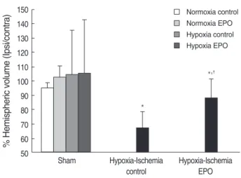
Fig. 1. Stereologic volumetric quantification for histological brain injury at 2 week after insult (P21). Measuring volume of contralat- eral hemisphere in all animals did not show any significant differ- ences among the 6 experimental groups (data not shown). Rema- ining tissue volume in the ipsilateral hemisphere was shown as a ratio of intact ipsilateral over the contralateral hemispheric volume.
Cerebral hypoxia-ischemia significantly decreased percentage ipsilateral hemispheric volume of the P21 rats. EPO significantly ameliorated this ipsilateral hemispheric volume loss following hypoxic-ischemic insult. Data shown as mean±SD.
*p<0.05, compared with sham-operated animals (Sham); �p<0.05, compared with hypoxia-ischemia control.
*
*,�
Normoxia control Normoxia EPO Hypoxia control Hypoxia EPO
RESULTS Brain injury
There were no significant differences in the whole brain volume of animals in the sham-operated groups, whether they were exposed to hypoxia, or administered with EPO, or not. The contralateral hemispheric volumes of the animals were not statistically different among the six experimental groups (data not shown). For anatomical assessment of cere- bral injury, the ratio of the ipsilateral remaining cerebral hemispheric volume to the volume of corresponding con- tralateral cerebral hemisphere was expressed as a percentage
(Fig. 1). For the saline treated animals after HI, there was a significant decrease in the percentage of preserved ipsilateral hemispheric volume compared with the sham-operated ani- mals (p<0.05). Animals that received EPO following HI showed a significant reduction of ipsilateral hemispheric volume loss compared with the saline treated HI animals (p<0.05).
Subventricular zone
From the rostral portion of the hematoxylin and eosin- stained brain sections corresponding to Plate 10 of the Struc- ture of the rat brain (28), the SVZ was observed as a densely
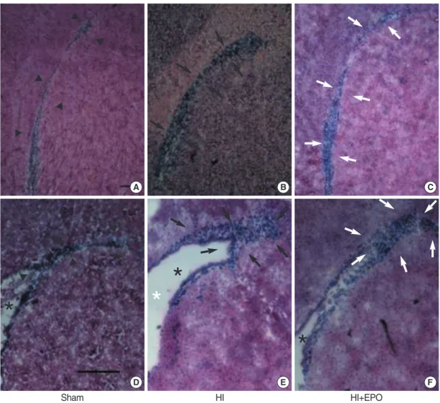
Fig. 2. Morphological changes of subventricular zone (SVZ) of the ipsilateral brain of rats at 2 weeks after insult (P21). Hematoxylin-eosin stained brain sections revealed SVZ as densely stained dark band (arrowheads) rostrally (A) and long triangular shape caudally (D) in sham-operated animals (Sham). Shapes of ipsilateral SVZ in sham operated animals (Sham) were similar, whether they were exposed with hypoxia or treated with EPO or not (data not shown). SVZ of ipsilateral brain of the rats subjected to HI expands significantly (black arrows) rostrally (B) and caudally (E), and shaped more triangular caudally (E). EPO treatment significantly decreased these expansions of SVZ (white arrows) rostrally (C) and caudally (F). Upper row represents sections including rostral part of striatal SVZ corresponding to Plate 11 of Structure of the rat brain. Lower row represents sections including mid-striatal SVZ corresponding to Plate 16 of Structure of the rat brain. Black stars indicate lateral ventricle. White star indicates widening of lateral ventricle due to striatal atrophy following hypoxic-ischemic insult. Each bar represents 0.5 mm.
A B C
D E F
*
* *
*
Sham HI HI+EPO
stained band like structure between the corpus callosum and the subcallosal striatum (Fig. 2A). Moving caudally to the caudal sections corresponding to Plate 23 of the Structure of the rat brain (28), the SVZ widened dorsolaterally and show- ed a long triangular shape with a dorsolateral tail beneath the corpus callosum (Fig. 2D). In the sham-operated ani- mals treated with saline versus those treated with EPO, or exposed with normoxia versus those exposed with hypoxia, there were no differences in the morphology of the SVZ.
Whereas the contralateral SVZ did not show any changes in morphology, the ipsilateral SVZ in the saline-treated HI ani-
mals revealed marked morphological alterations in more ros- tral sections with significant expansion rostrally and lateral- ly (Fig. 2B). In addition, in the more caudal sections, a more triangular shape was noted, caused by ipsilateral atrophy of striatum and corresponding widening of the lateral ventri- cle (Fig. 2E). EPO administration following HI significant- ly ameliorated these morphological changes of SVZ rostral- ly (Fig. 2C) and caudally (Fig. 2F). When we measured the SVZ volume on the contralateral side to operation, there were no statistical differences among the six experimental groups (data not shown). We calculated the percentage vol-
% SVZ volume (Ipsi/contra)
300
250
200
150
100
50
Sham Hypoxia-Ischemia Hypoxia-Ischemia
control EPO
Fig. 3. Stereologic volumetric quantitification of subventricular zone (SVZ) at 2 weeks (P21) after insult in P7 rats (A). Eythropoietin (EPO) or hypoxia-exposure did not affect the ipsilateral SVZ volume in sham-operated animals (Sham), and there were no significant differences in contralateral SVZ volume in the animals among 6 groups (data not shown). Changes of ipsilateral SVZ volume was shown as a per- centage of ipsilateral SVZ volume over the contralateral SVZ volume. Percentage SVZ volume was significantly increased in the saline treated hypoxic-ischemic rat brain compared to that of sham operated animals. EPO treatment significatly decreased this ipsilateral SVZ volume expansion. Data shown as mean±SD. *p<0.05 compared with sham-operated animals (Sham); �p<0.05 compared with hypoxia- ischemia control. Relationship between the percentage preserved ipsilateral hemispheric volume and ipsilateral percent expansion of SVZ volume was evaluated by regression analysis and showed a direct inverse relationship (r2=0.2701, p<0.05) (B).
*
�
Normoxia control Normoxia EPO Hypoxia control Hypoxia EPO
A
% SVZ volume (Ipsi/contra)
350 300 250 200 150 100 50 0
40 60 80 100 120
% Hemispheric volume (ipsi/contra) B
R2=0.2701
% Non-impaired paw initation
110 100 90 80 70 60 50 40 30 20 10
Sham Hypoxia-Ischemia Hypoxia-Ischemia
control EPO
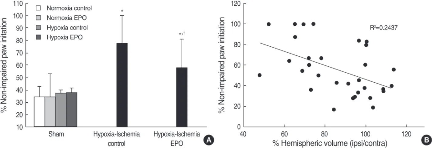
Fig. 4. Percentage of non-impaired (ipsilateral) forepaw initiation at weight-bearing when P21 rats (at 2 weeks after insult) underwent cylin- der-rearing test in 6 different groups (A). Saline-treated hypoxia-ischemia control animals showed a significantly asymmetric preferential use of non-impaired forelimb compared with sham operated animals. EPO treatment significantly ameliorated this sensorimotor fucntion- al deficit following HI insult. Data shown as mean±SD. *p<0.05 compared with sham-operated animals (Sham); �p<0.05 compared with hypoxia-ischemia control. Forelimb use asymmetry was directly correlated with the degree of histological brain injury shown by regression analysis of percentage of non-impaired limb initiation in the cylinder-rearing test and percentage preserved hemispheric volume (r2=0.2437, p<0.05) (B).
*
*,�
Normoxia control Normoxia EPO Hypoxia control Hypoxia EPO
A
% Non-impaired paw initiation
120 100 80 60 40 20 0
40 60 80 100 120
% Hemispheric volume (ipsi/contra) B
R2=0.2437
ume ratio of the SVZ ipsilateral over contralateral to the insult to measure the degree of volume changes in the ipsi- lateral SVZ (Fig. 3A). Saline-treated HI animals showed a significantly increased percentage volume of the ipsilateral SVZ compared to the sham-operated animals (p<0.05). EPO treatment significantly attenuated this ipsilateral SVZ expan- sion following HI (p<0.05). The relationship between the degree of SVZ enlargement and the degree of brain tissue injury was evaluated by comparison between the ratio of percentage of the hemispheric volume and the percentage SVZ volume, which showed an inverted linear relationship with coefficient (r2) of 0.2701 (p<0.05) (Fig. 3B). Therefore, the ipsilateral SVZ size was larger than its counterpart in the contralateral hemisphere, despite the fact that the ipsi- lateral hemisphere itself was smaller in size than the con- tralateral hemisphere.
Cylinder-rearing test
The percentage of non-impaired forepaw initiation dur- ing weight-bearing episodes with the cylinder-rearing test was calculated in P21 rats (Fig. 4A) to evaluate the lateral- izing sensorimotor deficit (25). Sham-operated animals show- ed symmetrical use of paws bilaterally when rearing in the cylinder, and the percentage of ipsilateral and contralateral paw initiation was the same at about 30%. HI animals treat- ed with saline showed significant asymmetry due to the pref- erential use of the non-impaired, ipsilateral paw when rear- ing, compared with the sham-operated animals (p<0.01).
EPO treatment significantly improved this unimpaired fore- limb bias for wall movements compared with saline-treated HI animals (p<0.05) (Fig. 4A). When the percentage of hemispheric volume ipsilateral versus contralateral and the percentage of the non-impaired paw initiation with the cylin- der test was compared, an inverted linear relationship with the coefficient (r2) of 0.2437 was observed (p<0.05) (Fig. 4B).
DISCUSSION
Our results demonstrate that multiple doses of EPO fol- lowed by HI decreased histopathological brain injury along with simultaneous expansion of the ipsilateral SVZ and im- proved lateralizing sensorimotor deficits in a neonatal rat model. This study describes for the first time the detailed morphological effect of EPO on alterations of the SVZ in developing rats subjected to unilateral cerebral HI using irreversible right common carotid artery ligation and hypox- emia.
Perinatal HI insults occur during a period in CNS devel- opment when there is extensive proliferation, migration, and cellular differentiation. The SVZ adjacent to the lateral ventricle is a forebrain region in mammalian animals where neurogenesis persists postnatally (29, 30). The SVZ contains
a population of stem cells and more mature progenitors of neurons, astrocytes, and oligodendrocytes, and normally the SVZ neuroblasts migrate tangentially along the rostral migra- tory stream to reach the olfactory bulb where they mature into local interneurons (29, 31). However, after injury, the cells of the SVZ proliferate and some of these cells migrate toward the damaged areas in the adult animal models (12, 13, 32). Previous studies have provided evidence that a severe HI insult in the neonatal rat brain depleted the precursors within the first 48 hr of recovery, resulting in SVZ regression (23, 33). Recently, however, Plane et al. (14), Yang et al. (16), and Ong et al. (17) using a neonatal HI model, and Chang et al. (15) using a neonatal stroke model reported that insults caused the expansion of the SVZ. These contradictory findings in neonatal animals may be due to the difference in exposure duration of hypoxia, species-related differences in the suscep- tibility and response of HI, and different maturational stages among animals of the same species. Furthermore, there are conflicting reports regarding the effect of EPO in stroke induced SVZ expansion. In an adult stroke model, Wang et al. (18) found that treatment with EPO significantly improv- ed functional recovery, along with increases in density of cere- bral vessels, increased numbers of bromodeoxyuridine (BrdU)- positive cells in the ipsilateral SVZ, but did not decrease the brain infarct volume significantly. Contrary to this finding, Chang et al. (15) reported that EPO treatment preserved injured hemispheric volume and decreased the expansion of SVZ in a neonatal stroke model. These contradictory results might have resulted from the differences in infarct volume reduction by EPO treatment in these two studies. Here, we showed that a moderate HI brain injury during the neona- tal period expands the SVZ, which is consistent with previ- ous studies (13, 14, 16, 17), and that the expansion of the SVZ was directly correlated with the intensity of the injury similar to the results reported by Plane et al. (14). Further- more, our results showed that EPO decreased the SVZ expan- sion induced by HI injury very similar to the findings report- ed by Chang et al. (15). Although we did not evaluate the proliferation or differentiation of precursor cells in the SVZ in this study, morphological SVZ expansion is directly relat- ed with the proliferation of precursor cells comprising the SVZ (12-14). It has been reported that stem or precursor cells in neurogenic areas of the adult brain respond to injury (34). Therefore, we cautiously assume that the ameliorated brain injury as a result of EPO administration might influ- ence the reactive expansion of SVZ. However, caution is needed in interpreting the effect of EPO on neonatal SVZ in this study, because we investigated only the morphologic change of SVZ at one time point, not the serial investigation of proliferation of precursor cells in SVZ. Demonstrating func- tional improvement accompanied by histological improve- ment following brain injury strengthens the promise of the neuroprotective therapy. Several studies have reported that EPO administration improved neurobehavioral dysfunction
caused by HI injury in newborn rats. Kumral et al. (8) demon- strated that EPO administered immediately after a HI insult in P7 rats produced long-lasting improvements in the cog- nitive function of the rats using the Morris water maze test at two weeks and again nineteen weeks after injury. Span- dou et al. (9) demonstrated that EPO administration before HI insult significantly reduced the severity of brain damage and improved the short functional recovery at 24 hr after insult in neonatal rats. Spandou et al. (10) also demonstrat- ed that a single dose of EPO administered immediately after HI insult, in the neonatal period, significantly reduced the severity of brain damage and prevented long-term sensori- motor deficits. The test was done at 42 days postnatally in animals using a battery of behavioral tests including Rota- Rod treadmill, grip traction test, foot fault test, postural reflex test, and limb placing test. In our study, the cylinder- rearing test was used to determine sensorimotor ability at fourteen days after HI insult (P21). Unilateral damage to the forelimb region of the rat brain sensorimotor cortex caus- ed chronic deficits in sensorimotor function and use of the contralateral forelimb was easily quantified. In each of the models of unilateral injury, the rats relied on the non-impair- ed forelimb for exploratory movements along the walls of the cylinder rearing. The cylinder-rearing test has been used in established models of unilateral stroke, Parkinson’s dis- ease, and spinal cord injury (35). The test also proved useful for the evaluation of the neuroprotective effect of previous forced limb use after 6-hydroxydopamine administration (36). Measurement of asymmetry is well suited for evaluat- ing functional consequences after unilateral brain injury. It is a simple, sensitive, and reliable tests that allows the observ- er to study behavior under unforced conditions. In order to obtain reproducible results, it is critical to reduce stressful stimuli. Gustavsson et al. (26) reported that evaluation of sensorimotor function in freely moving animals was achiev- able with the cylinder rearing test ant that this approach is advantageous compared with tests that require handling, such as the grip-traction test, foot-fault test, postural reflex test, and the limb-placing test. In the neonatal stroke model, the cylinder-rearing test has been proven to be useful for senso- rimotor deficits in P24 rats (15). Grow et al. (25) reported that both normal and HI rats explore the walls of a cylinder as early as P21. In the present study, HI animals showed significant asymmetry due to the preference of use of the non-impaired, ipsilateral paw when rearing, compared to the sham-operated animals. EPO treatment significantly improved this asymmetry following HI and this functional improvement was positively correlated with the degree of tissue preservation in the injured hemisphere. This is the first study, to our knowledge, to demonstrate that EPO treat- ment improves sensorimotor function in P21 neonatal rats after HI insult. Thus, the improvement of functional deficits and the neuroprotection observed by histopathology demon- strated in this model may be helpful for the consideration of
potential therapeutic strategies.
In summary, administration of multiple doses of EPO in P7 HI rats demonstrated a neuroprotective effect when given after the end of a hypoxic exposure by showing not only histopathological improvement evidenced by decreased brain injury and reduced reactive SVZ expansion, but also sensorimotor functional improvement. This neuroprotective effect of EPO suggests consideration of possible neonatal interventions in humans with perinatal hypoxic-ischemic encephalopathy.
REFERENCES
1. Vannucci RC. Hypoxic-ischemic encephalopathy. Am J Perinatol 2000; 17: 113-20.
2. Agnello D, Bigini P, Villa P, Mennini T, Cerami A, Brines ML, Ghezzi P. Erythropoietin exerts an anti-inflammatory effect on the CNS in a model of experimental autoimmune encephalomyelitis.
Brain Res 2002; 952: 128-34.
3. Digicaylioglu M, Lipton SA. Erythropoietin-mediated neuroprotec- tion involves cross-talk between Jak2 and NF-kappaB signalling cascades. Nature 2001; 412: 641-7.
4. Shingo T, Sorokan ST, Shimazaki T, Weiss S. Erythropoietin regu- lates the in vitro and in vivo production of neuronal progenitors by mammalian forebrain neural stem cells. J Neurosci 2001; 21: 9733- 43.
5. Aydin A, Genc K, Akhisaroglu M, Yorukoglu K, Gokmen N, Gonul- lu E. Erythropoietin exerts neuroprotective effect in neonatal rat model of hypoxic-ischemic brain injury. Brain Dev 2003; 25: 494-8.
6. Kumral A, Ozer E, Yilmaz O, Akhisaroglu M, Gokmen N, Duman N, Ulukus C, Genc S, Ozkan H. Neuroprotective effect of erythro- poietin on hypoxic-ischemic brain injury in neonatal rats. Biol Neonate 2003; 83: 224-8.
7. Sun Y, Zhou C, Polk P, Nanda A, Zhang JH. Mechanisms of ery- thropoietin-induced brain protection in neonatal hypoxia-ischemia rat model. J Cereb Blood Flow Metab 2004; 24: 259-70.
8. Kumral A, Uysal N, Tugyan K, Sonmez A, Yilmaz O, Gokmen N, Kiray M, Genc S, Duman N, Koroglu TF, Ozkan H, Genc K. Ery- thropoietin improves long-term spatial memory deficits and brain injury following neonatal hypoxia-ischemia in rats. Behav Brain Res 2004; 153: 77-86.
9. Spandou E, Soubasi V, Papoutsopoulou S, Karkavelas G, Sime- onidou C, Kaiki-Astara A, Guiba-Tziampiri O. Erythropoietin pre- vents hypoxia/ischemia-induced DNA fragmentation in an experi- mental model of perinatal asphyxia. Neurosci Lett 2004; 366: 24-8.
10. Spandou E, Papadopoulou Z, Soubasi V, Karkavelas G, Simeonidou C, Pazaiti A, Guiba-Tziampiri O. Erythropoietin prevents long-term sensorimotor deficits and brain injury following neonatal hypoxia- ischemia in rats. Brain Res 2005; 1045: 22-30.
11. Lois C, Garcia-Verdugo JM, Alvarez-Buylla A. Chain migration of neuronal precursors. Science 1996; 271: 978-81.
12. Arvidsson A, Collin T, Kirik D, Kokaia Z, Lindvall O. Neuronal replacement from endogenous precursors in the adult brain after
stroke. Nat Med 2002; 8: 963-70.
13. Parent JM, Vexler ZS, Gong C, Derugin N, Ferriero DM. Rat fore- brain neurogenesis and striatal neuron replacement after focal stroke.
Ann Neurol 2002; 52: 802-13.
14. Plane JM, Liu R, Wang TW, Silverstein FS, Parent JM. Neonatal hypoxic-ischemic injury increases forebrain subventricular zone neurogenesis in the mouse. Neurobiol Dis 2004; 16: 585-95.
15. Chang YS, Mu D, Wendland M, Sheldon RA, Vexler ZS, McQuillen PS, Ferriero DM. Erythropoietin improves functional and histologi- cal outcome in neonatal stroke. Pediatr Res 2005; 58: 106-11.
16. Yang Z, Levison SW. Hypoxia/ischemia expands the regenerative capacity of progenitors in the perinatal subventricular zone. Neuro- science 2006; 139: 555-64.
17. Ong J, Plane JM, Parent JM, Silverstein FS. Hypoxic-ischemic injury stimulates subventricular zone proliferation and neurogenesis in the neonatal rat. Pediatr Res 2005; 58: 600-6.
18. Wang L, Zhang Z, Wang Y, Zhang R, Chopp M. Treatment of stroke with erythropoietin enhances neurogenesis and angiogenesis and improves neurological function in rats. Stroke 2004; 35: 1732-7.
19. Zhu C, Xu F, Wang X, Shibata M, Uchiyama Y, Blomgren K, Hag- berg H. Different apoptotic mechanisms are activated in male and female brains after neonatal hypoxia-ischaemia. J Neurochem 2006;
96: 1016-27.
20. Rice JE 3rd, Vannucci RC, Brierley JB. The influence of immaturity on hypoxic-ischemic brain damage in the rat. Ann Neurol 1981; 9:
131-41.
21. Park WS, Sung DK, Kang S, Koo SH, Kim YJ, Lee JH, Chang YS, Lee M. Neuroprotective effect of cycloheximide on hypoxic-ischemic brain injury in neonatal rats. J Korean Med Sci 2006; 21: 337-41.
22. Park WS, Sung DK, Kang S, Koo SH, Kim YJ, Lee JH, Chang YS, Lee M. Therapeutic window for cycloheximide treatment after hypox- ic-ischemic brain injury in neonatal rats. J Korean Med Sci 2006;
21: 490-4.
23. Levison SW, Rothstein RP, Romanko MJ, Snyder MJ, Meyers RL, Vannucci SJ. Hypoxia/ischemia depletes the rat perinatal subven- tricular zone of oligodendrocyte progenitors and neural stem cells.
Dev Neurosci 2001; 23: 234-47.
24. Vannucci RC, Vannucci SJ. A model of perinatal hypoxic-ischemic brain damage. Ann N Y Acad Sci 1997; 835: 234-49.
25. Grow JL, Liu YQ, Barks JD. Can lateralizing sensorimotor deficits be identified after neonatal cerebral hypoxia-ischemia in rats? Dev Neurosci 2003; 25: 394-402.
26. Gustavsson M, Anderson MF, Mallard C, Hagberg H. Hypoxic pre- conditioning confers long-term reduction of brain injury and improve- ment of neurological ability in immature rats. Pediatr Res 2005; 57:
305-9.
27. Mandarim-de-Lacerda CA. Stereological tools in biomedical research.
An Acad Bras Cienc 2003; 75: 469-86.
28. Swanson L. Brain maps III: structure of the rat brain: an atlas with printed and electronic templates for data, models, and schematics. 3 ed. San Diego, CA, USA: Elsivier Academic Press, 2004.
29. Lois C, Alvarez-Buylla A. Proliferating subventricular zone cells in the adult mammalian forebrain can differentiate into neurons and glia. Proc Natl Acad Sci USA 1993; 90: 2074-7.
30. Goldman JE. Lineage, migration, and fate determination of postna- tal subventricular zone cells in the mammalian CNS. J Neurooncol 1995; 24: 61-4.
31. Luskin MB. Restricted proliferation and migration of postnatally generated neurons derived from the forebrain subventricular zone.
Neuron 1993; 11: 173-89.
32. Zhang RL, Zhang ZG, Zhang L, Chopp M. Proliferation and differ- entiation of progenitor cells in the cortex and the subventricular zone in the adult rat after focal cerebral ischemia. Neuroscience 2001;
105: 33-41.
33. Skoff RP, Bessert DA, Barks JD, Song D, Cerghet M, Silverstein FS. Hypoxic-ischemic injury results in acute disruption of myelin gene expression and death of oligodendroglial precursors in neona- tal mice. Int J Dev Neurosci 2001; 19: 197-208.
34. Lie DC, Song H, Colamarino SA, Ming GL, Gage FH. Neurogene- sis in the adult brain: new strategies for central nervous system dis- eases. Annu Rev Pharmacol Toxicol 2004; 44: 399-421.
35. Schallert T, Fleming SM, Leasure JL, Tillerson JL, Bland ST. CNS plasticity and assessment of forelimb sensorimotor outcome in uni- lateral rat models of stroke, cortical ablation, parkinsonism and spinal cord injury. Neuropharmacology 2000; 39: 777-87.
36. Cohen AD, Tillerson JL, Smith AD, Schallert T, Zigmond MJ. Neu- roprotective effects of prior limb use in 6-hydroxydopamine-treated rats: possible role of GDNF. J Neurochem 2003; 85: 299-305.