Introduction
Total knee arthroplasty (TKA) has been well established as a successful treatment for end-stage knee osteoarthritis. Mostly, the patients are evaluated for radiographic evidence of osteoarthritis,
such as joint space narrowing and lower extremity malalignment to determine the necessity of TKA. For patients complaining of bilateral knee pain, either unilateral TKA on the more painful knee or simultaneous or consecutive bilateral TKA is performed according to the severity of radiographic osteoarthritis. Usually, radiographic evaluation of the former patients reveals severe os- teoarthritis on one knee and mild osteoarthritic changes in the contralateral knee. Most of these patients are advanced in age and have some cartilage damage in the contralateral knee joint; there- fore, they undergo unilateral TKA on the severe osteoarthritic knee and conservative treatment for the contralateral knee with- out further evaluation. Previous studies concerning the prognosis in the contralateral knee after primary unilateral TKA focused mainly on the observation or conservative treatment and showed that radiographic changes and symptomatic severity are associ- ated with subsequent progression to arthroplasty in the contra-
Clinical Results of Contralateral Arthroscopic
Meniscectomy Performed with Unilateral Total Knee Arthroplasty: Minimum 3-year Follow-up
Sang Jin Lee, MD 1 , Bum-Sik Lee, MD 2 , Jong-Min Kim, MD 3 , and Seong-Il Bin, MD 3
1
Department of Orthopedic Surgery, Haeundae Paik Hospital, Inje University College of Medicine, Busan;
2Department of Orthopedic Surgery, Incheon St. Mary’s Hospital, The Catholic University of Korea College of Medicine, Incheon;
3Department of Orthopedic Surgery, Asan Medical Center, University of Ulsan College of Medicine, Seoul, Korea
Purpose: We assessed the clinical outcome of contralateral arthroscopic meniscectomy performed with unilateral total knee arthroplasty (TKA).
Materials and Methods: From May 1999 to June 2006, 23 patients underwent unilateral total knee arthroplasty and contralateral arthroscopic meniscectomy at the same time. All patients were women and followed for at least 36 months, except 2 patients who died. For clinical assessment, range of motion of the knee joint, Hospital for Special Surgery (HSS) knee score and the Lysholm knee score were evaluated preoperatively and at the last follow-up. At arthroscopy, meniscal pathology and cartilage changes were recorded and classified according to the Outerbridge scale. Progression of osteoarthritis in the contralateral knee to subsequent TKA was also assessed.
Results: The mean age of the 21 patients was 67.1 years and the mean follow-up period was 5.7 years. All of the patients were diagnosed with osteoarthritis and had Outerbridge grade 3 or 4 cartilage changes. Eight of the 21 patients had subsequent TKA at an average of 3.1 years after the index operation. The other 13 patients had no further surgery and clinical results including the HSS knee score and the Lysholm score were improved from 74.5 and 60.6 preoperatively to 90.8 and 82.4 postoperatively, respectively (p<0.001).
Conclusions: Contralateral arthroscopic meniscectomy performed simultaneously with unilateral TKA produces relatively good results regardless of the presence of cartilage degeneration.
Keywords: Knee, Meniscus, Contralateral meniscectomy, Arthroplasty pISSN 2234-0726 · eISSN 2234-2451
Knee Surgery & Related Research
Received May 8, 2014; Revised (1st) September 1, 2014;
(2nd) October 15, 2014; Accepted October 28, 2014 Correspondence to: Seong-Il Bin, MD
Department of Orthopedic Surgery, Asan Medical Center, University of Ulsan College of Medicine, 88 Olympic-ro 43-gil, Songpa-gu, Seoul 138- 736, Korea
Tel: +82-2-3010-3528, Fax: +82-2-488-7877 E-mail: [email protected]
76
This is an Open Access article distributed under the terms of the Creative Commons Attribution Non-Commercial License (http://creativecommons.org/licenses/by-nc/4.0/) which permits unrestricted non-commercial use, distribution, and reproduction in any medium, provided the original work is properly cited.
Copyright © 2015 KOREAN KNEE SOCIETY www.jksrr.org
lateral knee
1-3). However, if contralateral knee pain is severe from the beginning or has been exacerbated with time, meniscal lesion or cartilage damage might exist despite mild radiographic find- ings. If a meniscal tear is confirmed with magnetic resonance im- aging and the symptoms are mainly from mechanical pain rather than osteoarthritic pain, surgical treatment such as meniscetomy might be considered. But, it is known that the prognosis of men- iscectomy is closely associated with the cartilage status. In addi- tion, considering that the loading of the contralateral knee joint generally increases in the early stage of rehabilitation after TKA, it is not clear whether the meniscectomy of the contralateral knee would be more beneficial than the conservative treatment. Until recently, there are few studies concerning the prognosis of men- iscectomy of the contralateral knee after primary unilateral TKA for osteoarthritis.
In this study, we assessed the clinical outcomes of arthroscopic meniscectomy as well as the probability of a subsequent contra- lateral TKA when one knee was replaced due to osteoarthritis and the contralateral knee was treated with arthroscopic menis- cectomy simultaneously under the hypothesis that contralateral arthroscopic meniscectomy performed with unilateral TKA would be effective regardless of cartilage degeneration.
Materials and Methods
Between 1999 and 2006, 729 primary TKAs were performed by the same surgeon at one institution. Of these, 23 patients who had both knee pain and underwent additional arthroscopic men- iscectomy in the contralateral knee during TKA were included in this study. All of the patients were female and diagnosed with osteoarthritis of both knees. Institutional Review Board approval was obtained for this study.
Primary TKAs were performed using LPS or LPS-flex implant (NexGen; Zimmer Inc., Warsaw, IN, USA). Indications for sur- gery were varus thrust in walking and obliteration of the joint space on standing radiography. Arthroscopic meniscectomy were performed by the same surgeon based on the following indica- tions: 1) mechanical meniscal pain was more predominant than the other osteoarthritic symptoms; 2) the meniscal signs such as McMurray test and joint line tenderness were clear; and 3) varus thrust in walking was not present
4,5). Meniscectomy was not considered in cases where obliteration of the joint space was ob- served on standing radiography or the mechanical axis lay across the medial half of the medial tibial plateau of the whole lower limb on radiography. No additional procedures such as synovec- tomy, chondroplasty, and microfracture were performed.
Clinical assessment was performed using the medical records and telephone interviews. The range of motion (ROM), Hospital for Special Surgery (HSS) knee score, and the Lysholm knee score were evaluated preoperatively and at the last follow-up. At ar- throscopy, all findings concerning meniscal pathology, ligament status, and cartilage changes were recorded on a special form.
Cartilage changes were classified according to the Outerbridge scale
6). Also, we evaluated whether a contralateral TKA was sub- sequently performed and assessed the mean time from the initial meniscectomy to the subsequent TKA.
Statistical analysis was done using SPSS ver. 17.0 (SPSS Inc., Chicago, IL, USA). Preoperative ROM, knee scores and ar- throscopic findings were compared between the two groups by the use of an independent t-test or a Fisher’s exact test. Clinical results of group II were assessed using paired t-tests. Statistical significance was set at p<0.05.
Results
All patients were followed for at least 36 months, except 2 pa- tients who died within 12 months after the index surgery. The mean age of the 21 patients at operation was 67.1 years (range, 56 to 71 years) and the mean follow-up period was 5.7 years (range, 3.1 to 10.1 years).
At arthroscopic operation, all of the patients had Outerbridge grade III or IV cartilage changes on the medial femoral condyle or the medial tibial plateau. Eight patients had a horizontal tear of the posterior horn of the medial meniscus. Ten patients had a radial tear of the body or posterior horn of the medial meniscus.
The other three patients had a complex tear of the medial menis- cus, a complex tear of the lateral meniscus and a flap tear of the medial meniscus with a horizontal tear of the lateral meniscus, respectively. Partial meniscectomy of the medial meniscus was performed in 17 patients, subtotal meniscectomy of the medial meniscus in 2, partial meniscectomy of the lateral meniscus in 1, and subtotal meniscectomy of the medial meniscus and partial meniscectomy of the lateral meniscus in 1 (Table 1).
Of the 20 patients who had partial or subtotal meniscectomy
of the medial meniscus, 8 had a subsequent TKA surgery at an
average of 3.1 years (range, 2.0 to 6.6 years) after initial opera-
tion (group I). Including two patients who had subtotal men-
iscectomy, 13 of the 21 patients had no further surgery (group
II). The mean age of group I was 65.0 years (range, 60 to 71
years). In group II, the mean age was 63.5 years (range, 56 to 69
years) and the mean follow-up period was 5.0 years (range, 3.1
to 9.1 years). No significant difference was found between the
Ta bl e 1. C linic al D at a o f P at ien ts w ith C on tra lat era l A rthr os co pic M eni sce ct om y P er fo rm ed w ith U ni lat era l T ot al K ne e A rthr op las ty (TK A) N o. Ag e (y r) M eni sc us t ea r Ar thr os co pic m eni sce ct om y
Tim e t o su bs eq uen t TK A (y r)
Fo llo w-u p (y r)
Ar thr os co pic c ar til ag e f in din g (O ut erb ridg e g rade) Pr eo pera tiv e M edi al pa te lla La tera l pa te lla Tr oc hl ea MFC MTP LFC LT P
ROM (
o)
HSS sc Ly sho lm ore sc ore 1 69 LM co m plex Pa rti al N/A 3.1 4 4 4 1 3 2 4 135 62 57 2 64 MM h or izo nt al Pa rti al N/A 3.1 2 2 4 3 4 1 3 130 78 61 3 68 MM h or izo nt al Pa rti al N/A 3.8 2 2 3 1 3 1 2 130 73 58 4 64 MM radi al Pa rti al N/A 4.0 1 1 0 4 4 1 1 140 75 59 5 60 MM radi al Pa rti al N/A 4.1 3 1 3 3 4 1 2 140 82 65 6 57 MM f lap & LM MM s ub to ta l & N/A 4.1 1 1 2 3 4 0 1 125 54 52 h or izon ta l L M pa rti al 7 62 MM co m plex Pa rti al N/A 5.0 1 1 1 4 1 4 1 137 77 59 8 64 MM radi al Pa rti al N/A 5.2 2 2 3 4 4 1 2 120 69 57 9 65 MM radi al Pa rti al N/A 5.4 2 2 2 3 4 1 2 130 91 69 10 63 MM radi al Pa rti al N/A 5.6 3 3 3 4 4 3 3 140 83 65 11 59 MM h or izo nt al Pa rti al N/A 6.2 1 3 2 4 3 1 2 130 73 59 12 69 MM radi al Pa rti al N/A 6.3 2 2 4 3 4 2 2 140 77 58 13 56 MM h or izo nt al Su bt ot al N/A 9.1 2 2 3 4 4 1 2 120 74 69 14 65 MM radi al Pa rti al 2.7 10.1 1 2 0 4 0 4 1 140 77 57 15 63 MM h or izo nt al Su bt ot al 3.7 9.9 4 4 4 4 4 1 2 130 88 65 16 67 MM radi al Pa rti al 1.9 8.6 2 2 3 4 4 2 2 140 77 58 17 60 MM radi al Pa rti al 1.6 4.0 3 3 3 4 4 2 2 135 81 61 18 60 MM h or izo nt al Pa rti al 6.6 7.4 2 4 2 2 3 3 4 140 79 59 19 66 MM h or izo nt al Pa rti al 3.0 6.2 3 3 4 4 4 1 1 125 77 52 20 64 MM radi al Pa rti al 2.0 4.6 2 2 3 4 4 1 2 140 93 69 21 71 MM h or izo nt al Pa rti al 3.1 3.7 2 2 2 4 3 1 2 140 58 57 MFC: m edi al f em ora l co nd yle , MTP : m edi al t ib ia l p lat ea u, LFC: l at era l f em ora l co nd yle , L TP : l at era l t ib ia l p lat ea u, R O M: ra ng e o f m ot io n, HSS: H os pi ta l f or S pe cia l S ur ger y, LM: l at era l m eni sc us, MM: m edi al m eni sc us, N/A: n ot a pp lic ab le.
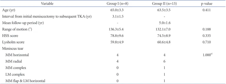
groups in age at index operation (p=0.411), preoperative ROM (136.3±5.4, 132.1.±7.0; p=0.188), preoperative HSS knee score (78.8±9.6, 74.5.±8.9; p=0.335), and the Lysholm knee score (59.8±4.9, 60.6.±4.8; p=0.710) (Table 2). The distribution of meniscal tear type was not significantly different between groups (p=1.000). In group II, the mean preoperative and postoperative ROM (132.1±7.0, 129.6.±11.0) showed no significant difference (p=0.566) and the HSS knee score (74.5.±8.9, 90.8.±5.4) and the Lysholm knee score (60.6±4.8, 82.4±4.6) were improved signifi- cantly (p<0.001, respectively) (Table 3).
Discussion
The major finding of this study is that contralateral meniscec- tomy simultaneously performed with unilateral TKA showed relatively good results regardless of cartilage injury.
There are several studies on the progression of contralateral knee osteoarthritis after primary unilateral knee arthroplasty.
Ritter et al.
1)reported that the probability of undergoing a subse-
quent arthroplasty by 7 years after index surgery in contralateral normal knees and contralateral knees with osteoarthritis was 5%
and 21%, respectively. They also reported that the probability of needing a TKA in patients with contralateral knee osteoarthritis was increased to 37% at 10 years. In the present study, 8 of the 21 patients (38%) had a subsequent TKA surgery by 6.6 years after initial operation and this finding is similar to that of the study of Ritter et al.
1). Mont et al.
2)also reviewed the history of the contra- lateral knee in patients who underwent unilateral primary total knee arthroplasty. According to the study, 93% of the patients who had moderate or severe symptoms and severe radiographic arthritis of the contralateral side with Ahlback grading scale later underwent subsequent TKA. In contrast, patients who initially presented with mild symptoms or no symptom had only 9% in- cidence of subsequent TKA. In addition, McMahon and Block
3)reported that the baseline Kellgren-Lawrence (K-L) grade of the contralateral knee at the time of index surgery was a strong predictor of eventual contralateral TKA
7). In the study, the mean survival time to contralateral arthroplasty was similar between patients with K-L grade II and those with grade III, 131.7 months and 127.6 months, respectively. However, patients with K-L grade IV had a mean survival time of only 80.45 months, whereas no patients with grade 0 or I progressed to subsequent TKA. Sayeed et al.
8)also described that the 10-year probability of having a con- tralateral TKA after index TKA was 36% and the probability in- creased to 70% when grade 4 of K-L grade radiographic changes were present. To sum up, two previous studies mainly used radio- graphic findings such as K-L grade or Ahlback scale to evaluate Table 2. Demographics and Preoperative Data of Group I and Group II
Variable Group I (n=8) Group II (n=13) p-value
Age (yr) 65.0±3.3 63.5±3.5 0.411
Interval from initial meniscectomy to subsequent TKA (yr) 3.1±1.5 -
Mean follow-up period (yr) - 5.0±1.6
Range of motion (
o) 136.3±5.4 132.1±7.0 0.188
HSS score 78.8±9.6 74.5±8.9 0.335
Lysholm score 59.8±4.9 60.6±4.8 0.710
Meniscus tear
MM horizontal 4 4 1.000
a)MM radial 4 6
MM complex 0 1
LM complex 0 1
MM flap & LM horizontal 0 1
Values are presented as mean±standard deviation or number.
TKA: total knee arthroplasty, HSS: Hospital for Special Surgery, MM: medial meniscus, LM: lateral meniscus.
a)