This is an Open Access ar ticle distributed under the terms of the Creative Commons Attribution Non-Commercial License (http:
//creativecommons.org/licenses/by-nc/4.0/) which permits unrestricted non-commercial use, distribution, and reproduction in any medium, provided the original work is properly cited.
© 2021 THE KOREAN SOCIETY OF MYCOLOGY.
Accepted: September 17, 2021 Revised: September 15, 2021 Received: July 02, 2021
https://doi.org/10.4489/KJM.20210037 Kor. J. Mycol. 2021 September , 49(3): 399-403
OPEN ACCESS pISSN : 0253-651X eISSN : 2383-5249
RESEARCH NOTE
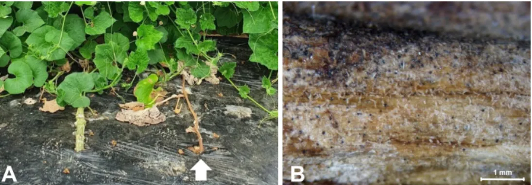
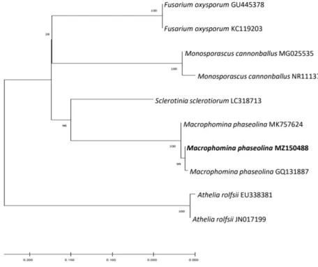
First Report of Macrophomina phaseolina Causing Charcoal Rot in Bottle Gourd in Korea
Sang Gyu Kim
*, Tae Bok Kim, and Oak Jin Lee
Vegetable Research Division, National Institute of Horticultural and Herbal Science, Rural Development Administration, Wanju 55365, Korea
*