Protective Effect of Dried Mackerel Extract on Lipopolysaccharide-induced Inflammation
Kwang-Hyuk Kim
1, Myoung Won Choi
1, Hyang Mi Choi
2and Sun-Young Lim
2*
1
Department Microbiology, Kosin University College of Medicine, Busan 602-703, Korea
2
Division of Marine Environment & Bioscience, Korea Maritime and Ocean University, Busan 606-080, Korea
Received August 2, 2013 /Revised September 9, 2013 /Accepted September 9, 2013The effect of dried mackerel extract on the production of nitric oxide (NO) and cytokines, including interleukin-6 (IL-6), tumor necrosis factor-α (TNF-α), and interferon-γ (IFN-γ), was investigated. All ex- tracts and fractions from dried mackerel significantly reduced NO production induced by lip- opolysaccharide (LPS). Among the extracts, acetone+methylene chloride (A+M), n-hexane, and 85%
aqueous methanol (MeOH) showed the strongest inhibitory effects. The 85% aqueous MeOH fraction at a concentration of 10 μg significantly decreased LPS-induced IL-6 and TNF-α production after 6 hr of incubation. In the case of LPS-induced IFN-γ production, the 85% aqueous MeOH fraction de- creased the production of IFN-γ afer 6, 24, and 72 hr of incubation in a dose-dependent manner. The results show that an 85% aqueous MeOH fraction inhibits the production of NO and proinflammatory cytokines (IL-6, TNF-α, IFN-γ), suggesting that this fraction acts as a potent immunomodulator.
Key words : Interferon-γ, interleukin-6, mackerel extract, nitric oxide, tumor necrosis factor-α
*Corresponding author
*Tel : +82-51-410-4757, Fax : +82-51-404-4750
*E-mail : [email protected]
This is an Open-Access article distributed under the terms of the Creative Commons Attribution Non-Commercial License (http://creativecommons.org/licenses/by-nc/3.0) which permits unrestricted non-commercial use, distribution, and reproduction in any medium, provided the original work is properly cited.
Journal of Life Science 2013 Vol. 23. No. 9. 1140~1146 DOI : http://dx.doi.org/10.5352/JLS.2013.23.9.1140
서 론
선천적 면역은 박테리아나 바이러스 감염에 대해 방어할 수 있는 다양한 작용기전을 통해 활성화되는 고전적 숙주방어 형태이다. 그러나 지속적이며 과도한 면역반응은 도리어 조직 손상을 촉진하고 그 결과 일부에서는 패혈증과 만성염증을 유발하게 된다[3]. 염증과정 중 염증유도 사이토카인 및 nitric oxide (NO)는 inducible NO synthase (iNOS)에 의해 다량 생 성된다[9]. 특히 면역과 염증에 관련된 여러 cytokine 중 tumor necrosis factor-alpha (TNF-α), iterleukin-6 (IL-6), inter- leukin-1β (IL-1β)는 대식세포에서 생산되는 대표적인 pro-in- flammatory 사이토카인으로 각종 염증질환의 발생과 진행에 중요한 작용을 하는 것으로 보고되고 있다[8]. 내독소로 잘 알 려진 LPS (lipopolysaccharide)는 그람음성균의 세포외막에 존재하여, RAW 264.7 세포와 같은 대식세포 또는 단핵세포에 서 TNF-α, IL-6, IL-1β와 같은 pro-inflammatory cytokine 등 염증 매개물질의 생산을 자극한다[1, 18, 25]. Con-A는 작두콩 에서 분리된 렉틴으로 동물의 적혈구를 응집하는 작용뿐만 아니라 T 림프구를 자극하는 역할을 하며 IL-2와 같은 사이토 카인을 생성한다[22]. 이러한 염증반응은 박테리아의 제거에 유리하게 작용하지만, 통제범위를 넘어선 과도한 염증반응은
TNF-α, IL-1 및 IL-6 등의 염증매개 사이토카인을 다량 생산하 게 되고, 이러한 염증 매개물질들은 조직을 손상시킨다[20].
고등어(Scomberjaponicus)는 농어목 고등어과에 속하는 해 산어류로 n-3계열지방산인 eicosapentaenoic acid (EPA, 20:
5n-3)와 docosahexaenoic acid (DHA, 22:6n-3) 같은 다가불포 화지방산(polyunsaturated fatty acid, PUFA)을 많이 함유하 고 있을 뿐만 아니라 단백질, 지방, 칼슘, 인, 나트륨, 칼륨 등의 영양소들도 함유하고 있다. 특히 어류의 근육은 EPA와 DHA 같은 n-3계열 PUFA를 많이 함유하고 있으며[24], 고등어와 같은 회유어종의 근육에서는 비회유어종보다 DHA 함량이 높 게 나타난다[19, 21]. Wu [29] 등은 해양동물에서 EPA를 추출 하여 마우스에 급여하였을 때 EPA가 prostaglandin E
2(PGE
2) 의 생성을 억제한다고 보고하였다. Weldon [28] 등은 EPA와 DHA 투여는 염증을 유발하는 유전자 발현 및 pro-in- flammatory 사이토카인의 생성을 억제한다고 보고하였고 DHA에 의한 저해효과가 더 우수하였다고 보고하였다. 본 연 구의 목적은 건조 고등어 추출물에 의한 항염증 활성을 검증 하는 것으로 특히 고등어 85% aqueous methanol 분획물을 중심으로 비장세포에서 T 세포 증식자극 물질로서의 Con와 B 세포 자극물질로서 LPS를 이용하여 NO 생성 및 면역과정의 생물학적 작용과 대사적 변화를 유도하는 IL-6, IFN-γ 및 TNF- α 같은 염증유발 사이토카인의 생성을 측정하였다.
재료 및 방법
재료 및 시약
본 실험에 사용된 고등어는 부산 자갈치 수산시장에서 구입
하여 적당한 크기로 토막 내어 저온진공 건조기를 이용하여
40℃에서 40 torr의 압력으로 30시간 건조하였다. 사이토카인
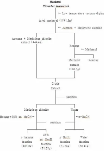
Fig. 1. The procedure for solvent extract and fraction of mackerel.
생성을 위한 실험동물은 수컷 Balb/c 마우스로서 생후 8주 내외, 체중 25 g 내외의 것을 대한바이오링크(음성군,충청북 도)로부터 구입하여 실험에 사용하였다. Lipopolysaccharide (LPS)는 Escherichia coli (serotype 026:B6)에서 분리정제된 제 품(Sigma-Aldrich Corp. Saint Louis, MO, USA)과 concana- valin A (Con-A)는 Pharmacia사(Pharmacia Fire Chemicals, Uppsala, Sweden) 제품을 사용하였다. IL-6, TNF-α, IFN-γ를 측정하기 위한 시약은 Mouse IL-6, TNF-α, IFN-γ ELISA kit (Biolegend, San Diego, USA)를 사용하였다.
건조고등어로부터 유기용매를 이용한 추출물 및 분획물 제조 건조된 고등어는 실험 사용 전까지 -70℃의 deep freezer (NF-400SF, NIHON FREEZER, Tokyo, Japan)에 냉동 보관하 였다가 유기용매 추출을 위하여acetone:methylene chloride를 1:1 비율로 혼합하여 시료가 충분히 잠기도록 하여 24시간 방 치한 후 추출하였고 이 과정을 2회 반복하여 여액을 얻었다.
또한 acetone:methylene chloride (1:1) 혼합용매로 추출되지 않은 성분을 methanol (MeOH)로 추출하고자 남은 잔사에 acetone:methylene chloride (1:1)과 동량의 MeOH로 위와 동 일한 방법으로 2회 반복하여 여액을 얻었다. 두 용매로부터 최대로 수득한 추출물을 혼합하여 다시 용매극성에 따라 순차 적으로 분획하여 n-hexane, 85% aqueous MeOH (85% aq.
MeOH), n-butanol (n-BuOH), water 분획물을 얻었다. 실험에 는 각각의 추출물들을 dimethyl sulfoxide (DMSO)에 녹여 세 포배지로 필요한 농도로 희석하여 실험에 사용하였다(Fig. 1).
Nitric oxide 측정
RAW 264.7 세포를 96-well cell culture plate에 well 당 2×10
4cells/ml가 되도록 분주하여 37℃, 5% CO
2incubator에 서 24시간 동안 배양하였다. 배양액을 10% FBS가 함유된 MEM 배지로 교체한 후 준비된 시료를 1시간 동안 전처리하 고, nitric oxide 생성을 유도하기 위해 LPS (1 μg/ml = 1 ppm) 를 처리한 후, 48시간 동안 37℃, 5% CO
2incubator에서 배양 하였다. 이후 생성된 NO의 양은 Griess 시약(0.1% N-(1-naph- tyl)ethylenediamine : 1% sulfanilamide = 1:1)을 이용하여 microplate reader (VICTOR3, Perkin Elmer, Waltham, MA, USA)로 570 nm에서 흡광도를 측정하였다. sodium nitrate를 사용하여 측정된 흡광도로 표준곡선을 작성하여 nitric oxide 의 농도별 흡광도를 얻었으며, 표준곡선을 실험결과에 적용하 여 생성된 nitric oxide의 함량을 정량하였다[23].
비장세포 배양 상층액 분리 및 IL-6, TNF-α 및 IFN-γ 측정 수컷 Balb/c 마우스를 희생시킨 후 비장을 분리하고, RPMI 1640 배지로 균질화한 후 상층액이 2×10
6/ml가 되도록 10%
FBS가 함유된 RPMI 1640 (Gibco, Buffalo, NY, USA) 배지로 조절한다. 24 wells tissue culture plate (Costar, Corning, NY,
USA)에 1 ml씩 분주한 후 자극제(LPS 2 μg나 Con-A 2 μg)와 건조 고등어의 85% aq. MeOH 분획물을 농도별(1, 3, 10 μg/
ml)로 작용시켜 37℃, 5% CO
2배양기에 배양하였다. 대조군은
0.01% DMSO가 되도록 사용하였으며 배양시간은 상기의 조
건에 6, 24, 48 및 72시간으로 하였다. 배양이 끝난 후 전량
배양액을 수거한 다음 300× g에서 10분간, 10,000× g에서 30분
간 원침시킨 후 그 상층액을 수거하여 -70℃에 보관하였다
[10]. 미리 96 wells microplate에 mouse IL-6, TNF-α 및 IFN-γ
에 대한 capture 항체를 coating buffer에 희석하여 100 μl씩을
분주한 후 4℃에서 하룻밤 방치하였다. 다음날 plate를 세척용
완충액으로 4번 세척한 후 assay diluent 200 μl를 분주한 후
실온에서 1시간 동안 방치하였다. 세척용 완충액으로 4번 세
척한 후 plate의 각 well에 시료 100 μl을 적하하여 실온에서
2시간 동안 방치하였다. 세척용 완충액으로 4번 세척한 후 de-
tection 항체 100 μl을 분주한 후 실온에서 1시간 동안 방치하
였다. 세척용 완충액으로 4번 세척한 후 avidin-horseradish
peroxidase액 100 μl을 적하하여 다시 실온에서 30분 동안 방
치하였다. 세척용 완충액으로 5번 세척한 후 tetrame-
thylbenzidine이 포함된 기질액 100 μl을 적하하여 실온에서
15분 동안 방치한 후 stop액 100 μl씩을 가하여 반응을 정지시
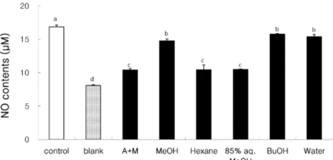
Fig. 2. Effect of extracts and fractions (0.05 mg/ml) from dried mackerel on the production of nitric oxide (NO). The val- ues were expressed as the mean ± SD and different su- perscripts indicate significant differences among treat- ments (
p
<0.05). Control, sample was treated with LPS and phosphatate buffered saline; Blank, sample was treated with phosphate buffered saline. A+M, acetone with methylene chloride extract; MeOH, methanol ex- tract; Hexane,n
-hexane fraction; 85% aq. MeOH, 85%aqueous methanol fraction; BuOH,
n
-butanol fraction;Water, water fraction
켰다. Optical density는 ELISA (Bio-Rad, Model 550,
Hercules, CA, USA)를 이용하여 450 nm에서 측정하였다[17].
통계분석
실험결과는 Mean ± SD (Standard diviation)으로 나타내었 고 분석된 실험 data는 대조군과 각 시료로부터 얻은 실험자료 로부터 ANOVA를 실시하여 유의성이 있을 경우에 post-hoc test로 Duncan's multiple range test를 실시하여 95% 수준에 서 유의성을 검증하였다.
결과 및 고찰
건조 고등어 추출물 및 분획물의 nitric oxide 생성 억제 효과
혈관내피로부터 유래된 nitric oxide는 강력한 혈관확장제 로 작용함으로써 혈관 항상성을 유지시키는 반면 염증상태에 서의 과도한 nitric oxide의 생성은 혈관투과성, 부종 등의 염 증반응을 촉진시킬 뿐만 아니라 염증매개체의 생합성을 촉진 하여 급성과 만성염증에 대한 반응으로 조직의 손상을 일으킬 수 있다[26]. Nitric oxide의 생성량은 RAW 264.7 세포의 배양 액 중에 LPS 자극으로 유도된 nitric oxide의 함량을 측정하는 것으로 세포의 생존율에 영향을 미치지 않는 농도에서 건조 고등어 추출물 및 분획물을 처리하여 배양한 후, 세포배양액 에 Griess 시약을 반응시켜 확인하였다. 시료는 0.05 mg/ml의 농도로 처리하였으며, 대조군으로는 시료 대신 PBS를 사용하 여 LPS를 처리한 control과 시료 및 LPS를 처리하지 않은 blank를 사용하였다. Fig. 2에서 보는 바와 같이 LPS를 처리한 control은 nitric oxide의 생성량이 높게 나타났으며, LPS를 처 리하지 않은 blank는 nitric oxide의 생성량이 상대적으로 매 우 낮게 나타났다. 염증유발에 중요한 역할을 하는 것으로 알 려진 nitric oxide 생성에 대한 건조 고등어 추출물 및 분획물 들의 억제효과를 확인한 결과, acetone:methylene chloride (A+M) 및 methanol (MeOH) 추출물 및 모든 분획물들은 ni- tric oxide 생성을 유의적으로 감소시켰으며(p<0.05), MeOH 추출물과 비교했을 때 A+M 추출물에 의한 nitric oxide 생성 억제효과가 높았다. 또한 각 분획물도 control보다 낮은 nitric oxide 생성량을 나타내었으며, 특히 85% aq. MeOH 및 n-hex- ane 분획물에 의한 억제효과가 높았다. Calder [4]은 참치와 같은 등 푸른 생선에 다량 함유된 EPA와 DHA는 PUFA에 의해 합성된 eicosanoids에 의해 항염증 효과를 나타낸다고 보고하였다. 또한 DHA [14]와 EPA [15]는 LPS로 유도된 ni- tric oxide 생성을 저해한다고 보고되었으며, 특히 DHA는 LPS로 유도된 nuclear transcription factor-kappa B (NF-κB) 활성을 저해함으로써 pro-inflammatory 사이토카인들의 생성 을 저해하는 것으로 보고되었다[7]. NF-κB는 사이토카인, 성 장인자 및 세포부착분자 등에 관련된 여러 유전자들의 발현에
중요한 역할을 하는 전사인자로서 활성화된 NF-κB는 iNOS, IL-6및 TNF-α 등 여러 염증매개물질들의 전사를 촉진한다고 알려져 있다[13].
건조 고등어 추출물 및 분획물의 IL-6, TNF-α 및 IFN-γ 생성 억제 효과
비장세포의 증식반응이 일어나면 증식하는 세포는 여러 종 류의 면역반응을 매개하는 여러 가지 사이토카인을 분비한다.
선행된 항암 및 항산화 실험[12]에서 건조 고등어 분획물들 중 85% aq. MeOH 분획물에 의한 효과가 가장 높았고 nitric oxide 생성 억제능 또한 높았다. 따라서 건조 고등어 85% aq.
MeOH 분획물에 의한 비장세포의 사이토카인(IL-6, TNF-α, and IFN-γ) 생성에 미치는 영향을 6, 24, 48 및 72시간 배양한 후 살펴보았다. 본 실험에서 사용된 비장세포 자극제로는 B 세포 자극물질인 LPS와 T 세포 증식 자극 물질인 Con-A가 사용되었다. IL-6, TNF-α 같은 pro-inflammatory 사이토카인 은 염증반응을 매개하는 물질로 초기염증반응에 관여하고 있 는 것으로 알려져 있다[27]. IL-6는 단핵세포와 대식세포에 의 해 분비되는데 분화된 B 세포가 형질세포로 분화되도록 촉진 시키고 항체의 분비를 자극하고 염증부위에서 항상 높은 수치 를 나타내는 것으로 알려져 있다[6]. Table 1은 IL-6의 생성량 을 나타낸 것으로 비장세포를 활성화시키는 LPS 자극에 의해 IL-6는 분비되며 배양시간이 증가할수록 생성량도 증가하였 다. 건조 고등어 85% aq. MeOH 분획물을 농도별(1, 3 및 10 μ g)로 처리했을 때 농도가 증가할수록 IL-6의 생성량은 감소하 였다. 또한 반응시간별로 살펴보면 건조 고등어 85% aq.
MeOH 분획물 첨가 후 배양시간이 길어질수록 IL-6의 생성량
이 증가하였고 모든 첨가농도의 경우 6시간 배양 후 가장 낮은
Table 1. Effect of 85% aqueous methanol fraction from dried mackerel on the production of LPS induced interleukin-6 at different times in mouse spleen cells
Samples (μg/ml) Concentrations (pg/ml)
6 hr 24 hr 48 hr 72 hr
LPSLPS+85% aq. MeOH 1 LPS+85% aq. MeOH 3 LPS+85% aq. MeOH 10
39.8±4.0a 11.4±16.1b
2.8±4.0b 0.0±0.0b
126.4±14.1 122.2±20.1 127.8±20.1 76.7±36.2
154.8±2.0a 161.9±28.1a 176.1±0.0a 105.1±12.1b
235.8±76.3 255.7±40.2 173.3±12.1 210.2±0.0 The values were expressed as the mean ± SD and different superscripts in a column indicate significant differences among treatments (
p
<0.05). LPS, lipopolysaccharide; 85% aq. MeOH, 85% aqueous methanol fraction from dried mackerel.Table 2. Effect of 85% aqueous methanol fraction from dried mackerel on the production of Con-A induced interleukin-6 at different times in mouse spleen cells
Samples (μg/ml) Concentrations (pg/ml)
6 hr 24 hr 48 hr 72 hr
Con-A
Con-A +85% aq. MeOH 1 Con-A +85% aq. MeOH 3 Con-A +85% aq. MeOH 10
0.0±0.0 0.0±0.0 0.0±0.0 0.0±0.0
108.0±40.2 110.1±20.1 59.7±36.2 93.8±28.1
190.3±4.0 255.7±32.1 176.1±0.0 247.2±36.2
292.6±52.2 267.1±24.1 335.2±24.1 292.6±12.1 The values were expressed as the mean ± SD and different superscripts in a column indicate significant differences among treatments (
p
<0.05). Con-A, concanavalin A; 85% aq. MeOH, 85% aqueous methanol fraction from dried mackerel.Table 3. Effect of 85% aqueous methanol fraction from dried mackerel on the production of LPS induced tumor necrosis factor-α at different times in mouse spleen cells
Samples (μg/ml) Concentrations (pg/ml)
6 hr 24 hr 48 hr 72 hr
LPS
LPS+85% aq. MeOH 1 LPS+85% aq. MeOH 3 LPS+85% aq. MeOH 10
211.2±10.3a 112.4±16.2b 119.3±13.0b 89.5±9.7b
228.2±48.1 160.6±26.0 142.2±19.5 123.9±6.5
242.7±41.2 169.7±19.5 149.1±29.2 174.3±0.0
199.5±16.2 206.4±19.5 142.2±26.0 206.4±0.0 The values were expressed as the mean ± SD and different superscripts in a column indicate significant differences among treatments (
p
<0.05). LPS, lipopolysaccharide; 85% aq. MeOH, 85% aqueous methanol fraction from dried mackerel.IL-6 생성량을 확인할 수가 있었다(p<0.05). 이상의 결과는 건 조 고등어 85% aq. MeOH 분획물에 의한 IL-6 생성의 감소효 과는 6시간 배양 후 가장 효과적이며 24 및 72시간 배양조건에 서는 효과가 없는 것으로 설명할 수 있다. LPS 자극 때와는 다르게 T 세포 자극물질인 Con-A에 대해서는 초기(6시간)에 는 생성되지 않았으나 배양시간과 더불어 IL-6 생성량이 증가 하였다(Table 2). Con-A와 함께 건조 고등어 85% aq. MeOH 분획물을 처리했을 때 초기에는 IL-6이 생성되지 않았으나 24 시간 배양 후 모든 첨가농도에서 Con-A만 처리했을 경우보다 IL-6의 생성량이 감소하는 경향을 나타내었으나 유의적 차이 는 없었다. 이상의 결과로부터 건조 고등어 85% aq. MeOH 분획물은 마우스 비장세포에서 Con-A보다 LPS에 의해 자극 된 IL-6 생성을 감소시키는데 더 효과적인 것으로 여겨진다.
TNF-α의 경우(Table 3과 4), 앞서의 IL-6과 유사한 경향을 나타 내어 건조 고등어 85% aq. MeOH 분획물은 모든 첨가농도에
서 6시간 배양 후 LPS로 유도된 TNF-α의 생성량을 유의적으
로 감소시켰다(Table 3, p<0.05). 반응시간별로 살펴보면 건조
고등어 85% aq. MeOH 분획물은 첨가 후 배양시간이 길어질
수록 TNF-α의 생성량이 증가하는 경향을 나타내었다(Table
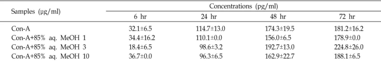
3). Table 4는 Con-A에 의해 유도된 TNF-α의 생성량 변화추이
를 나타낸 것으로 배양시간이 증가할수록 TNF-α의 생성량이
증가하였다. 그러나 건조 고등어 85% aq. MeOH 분획물을 농
도별(1, 3 및 10 μg)로 처리했을 때 Con-A만 처리한 대조군과
비교했을 때 TNF-α의 생성량은 유의적 차이가 없었으나 24시
간 배양조건에서는 농도 의존적으로 감소하는 경향을 나타내
었다. 따라서 IL-6와 유사하게 건조 고등어 85% aq. MeOH
분획물은 LPS에서 유도되는 TNF-α의 생성량을 감소시키는데
더 효과적인 것으로 여겨진다. Table 5와 6은 LPS와 Con-A로
각각 자극하여 IFN-γ의 생성변화를 나타낸 결과로 LPS와
Con-A에 의해 배양시간과 더불어 IFN-γ의 생성량이 증가함
Table 4. Effect of 85% aqueous methanol fraction from dried mackerel on the production of Con-A induced tumor necrosis factor-α at different times in mouse spleen cells
Samples (μg/ml) Concentrations (pg/ml)
6 hr 24 hr 48 hr 72 hr
Con-A
Con-A+85% aq. MeOH 1 Con-A+85% aq. MeOH 3 Con-A+85% aq. MeOH 10
32.1±6.5 34.4±16.2 18.4±6.5 36.7±0.0
114.7±13.0 110.1±0.0
98.6±3.2 96.3±6.5
174.3±19.5 156.0±6.5 192.7±13.0 162.9±22.7
181.2±16.2 178.9±0.0 224.8±26.0 188.1±6.5 The values were expressed as the mean ± SD and different superscripts in a column indicate significant differences among treatments (
p
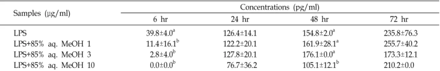
<0.05). Con-A, concanavalin A; 85% aq. MeOH, 85% aqueous methanol fraction from dried mackerel.Table 5. Effect of 85% aqueous methanol fraction from dried mackerel on the production of LPS induced interferon-γ at different times in mouse spleen cells
Samples (μg/ml) Concentrations (pg/ml)
6 hr 24 hr 48 hr 72 hr
LPS
LPS+85% aq. MeOH 1 LPS+85% aq. MeOH 3 LPS+85% aq. MeOH 10
0.0±0.0c 83.3±0.0b 0.0±0.0c 208.3±59.0a
1055.6±157.2a 1250.0±117.8a 1166.7±0.0a
791.7±58.9b
1027.8±39.3 1708.3±530.4 2083.3±471.4 1166.7±117.9
2833.3±0.0a 2166.7±117.9b 1708.3±58.9c 1541.7±58.9c The values were expressed as the mean ± SD and different superscripts in a column indicate significant differences among treatments (
p
<0.05). LPS, lipopolysaccharide; 85% aq. MeOH, 85% aqueous methanol fraction from dried mackerelTable 6. Effect of 85% aqueous methanol fraction from dried mackerel on the production of Con-A induced interferon-γ at different times in mouse spleen cells
Samples (μg/ml) Concentrations (pg/ml)
6 hr 24 hr 48 hr 72 hr
Con-A
Con-A+85% aq. MeOH 1 Con-A+85% aq. MeOH 3 Con-A+85% aq. MeOH 10
125.0±56.0a 83.3±0.0b 83.3±117.9b 83.3±117.9b
2000.0±235.7a 1416.7±353.6b 1791.7±294.6ab 1333.3±0.0b
3916.7±707.1 3750.0±1060.7 3833.3±353.6 4083.3±707.1
4333.3±942.8 4833.3±1414.2 4541.7±294.6 4125.0±176.8 The values were expressed as the mean ± SD and different superscripts in a column indicate significant differences among treatments (
p
<0.05). Con-A, concanavalin A;85% aq. MeOH, 85% aqueous methanol fraction from dried mackerel.을 확인할 수가 있었다. 그러나 IFN-γ의 특성상 초기(6시간) 배양조건에서 LPS 단독으로는 IFN-γ의 생성을 유도하지 않지 만 배양시간 경과와 더불어 크게 증가하였으며 메탄올 추출물 소량(1 μg첨가농도) 첨가는 배양시간이 경과되면서 LPS 단독 군에 비하여 증가하는 경향을 보였다. 이것은 세포성 면역증 강 효과라고 볼 수 있다. 건조 고등어 85% aq. MeOH 분획물 농도별로 살펴보면 농도가 증가할수록 LPS에 의해 자극된 IFN-γ의 생성을 감소시키는 경향을 나타내었다. 첨가농도별 로 살펴보면 건조 고등어 85% aq. MeOH 분획물(1 및 3 μg 첨가농도)을 LPS와 함께 처리했을 때 24 및 72시간 배양했을 때 IFN-γ의 생성량이 유의적으로 감소하였다(Table 5). Con-A 의 경우 건조 고등어 85% aq. MeOH 분획물과 함께 처리했을 때 모든 첨가농도에서 Con-A만 처리한 대조군과 비교했을 때 생성량이 감소되는 것을 확인하였다(Table 6). 특히 건조 고등 어 85% aq. MeOH 분획물을 6 및 24시간 배양했을 때 유의적
으로 Con-A에 의해 자극된 IFN-γ의 생성량을 감소시켰다.
해양동물을 이용한 항염증 효과에 관한 연구는 다양하게
진행되어 있지 않다. 제한된 연구들 중 고등어와 함께 대표적
인 등 푸른 생선인 참치의 항암활성 및 면역학적 연구에서
참치 에탄올 추출물은 in vitro 인체 장암세포 및 백혈병성 임
파모세포의 증식을 억제하였으며 S-180 세포를 접종한 후 참
치 추출물을 투여한 생쥐에서 용혈반형성 세포수와 혈청 단백
질 중 immunoglobin의 상대적 비율이 현저히 증가함을 관찰
하여 생체 내에서 면역 증강효과를 나타내는 성분이 존재함을
보고하였다[11]. 또 다른 연구로 해양동물 중의 하나인 군소
(Aplysiakurodai)를 이용한 연구로 군소로부터 추출한 다당분
획물의 면역조절 효과를 검토한 결과 T cell line의 증식능에
대하여 농도가 증가함에 따라 증식능이 증가하는 효과와
RAW 264.7 cell line에 대하여 IL-12의 경우 47% 이상 증가하
였음을 보고하였다. 본 연구자들도 참치 추출물 및 분획물에
의한 nitric oxide 및 pro-inflammatory 사이토카인의 생성 억 제효과를 검토한 결과 참치 분획물들 중 85% aq. MeOH 분획 물이 nitric oxide 생성과 LPS로 자극된 IL-6과 TNF-α의 생성 량을 유의적으로 감소시키므로써 항염증 효과를 나타내었다 고 보고하였다[16]. 따라서 고등어 및 참치를 포함하는 등 푸른 생선은 대표적인 DHA 및 EPA의 공급원으로 인체 연구에 의 하면 이들 n-3계 PUFA들이 IL-1, IL-6 및 TNF-α와 같은 pro-inflammatory 사이토카인의 생성을 억제한다고 보고되었 다[2, 5]. 지금까지 고등어에 대한 면역학적 연구는 미미하였으 나 본 연구에서는 대표적인 서민 생선이며 DHA 및 EPA의 공급원인 고등어를 두 가지 자극물질, 즉 T 세포 증식자극 물 질로서의 Con와 B 세포 자극물질로서 LPS를 이용하여 NO 생성 및 면역과정의 대사적 변화를 유도하는 IL-6, IFN-γ 및 TNF-α 같은 염증유발 사이토카인의 생성에 대해 연구한 것으 로 고등어의 식품영양학적 가치 측면에서 새로운 시도였다고 여겨진다. 따라서 이상의 결과로부터 건조 고등어 85% aq.
MeOH 분획물은 nitric oxide 생성과 IL-6와 TNF-α와 같은 pro-inflammatory 사이토카인의 생성을 감소시켜 염증반응을 예방할 것으로 기대되며 향후 건조 고등어 85% aq. MeOH 분획물을 더욱 정제하여 면역조절물질의 구조를 동정할 필요 가 있다.
감사의 글
이 논문은 해양수산부의 지원으로 수행한 해양에너지전문 인력양성사업과 2013년도 정부(교육부)의 재원으로 한국연구 재단의 지원을 받아 수행된 기초연구사업(NRF- 2013R1A1A2004694)의 연구결과입니다.
References
1. Axtelle, T. and Pribble, J. 2001. IC14, a CD14 specific mono- clonal antibody, is a potential treatment for patients with severe sepsis.
J Endotoxin Res
7, 310-314.2. Bouwens, M., van de Rest, O., Dellsscharft, N., Bromhaar, M. G., de Groot, L. C., Geleijinse, J. M., Muller, M. and Afman, L. A. 2009. Fish-oil supplementation induces antiin- flammatory gene expression profiles in human blood mono- nuclear cells.
Am J Clin Nutr
90, 415-424.3. Brown, K. L., Cosseau, C., Gardy, J. L. and Hancock, R. E.
2007. Complexities of targeting innate immunity to treat infection.
Trends Immunol
28, 260-266.4. Calder, P. C. 2008. The relationship between the fatty acid composition of immune cells and their function.
Prostagglan- dins Leukot Essent Fatty Acids
79, 101-108.5. Caughey, G. E., Mantzioris, E., Gibson, R. A., Cleland, L.
G. and James, M. J. 1996. The effect on human tumor ne- crosis factor and a interleukin 1β production of diets en- riched in n-3 fatty acid from vegetable oil or fish oil.
Am
J Clin Nutr
63, 116-122.6. Delgado, A. V., McManus, A. T. and Chambers, J. P. 2003.
Production of tumor necrosis factor-alpha, interleukin 1-be- ta, interleukin-2, and interleukin-6 by rat leukocyte sub- population after exposure to substance P.
Neuropeptides
37, 355-361.7. De Smedt-Peyrusse, V., Sargueil, F., Moranis, A., Harizi, H., Mongrand, S. and Laye, S. 2008. Docosahexaenoic acid pre- vents lipopolysaccharide-induced cytokine production in microglial cells by inhibiting lipopolysaccharide receptor presentation but not its membrane subdomain localization.
J Neurochem
105, 296-307.8. Feldmann, M., Brennan, F. M. and Maini, R. N. 1996. Role of cytokines in rheumatoid arthritis.
Annu Rev Immunol
14, 397-440.9. Green, L. C., Wagner, D. A., Logowski, G. J., Skipper, P.
L., Wishnok, J. S. and Tannenbaum, S. R. 1982. Analysis of nitrate, nitrite and [15N] nitrate in biological fluids.
Anal Biochem
126, 131-138.10. Hwang, S. A., Dasgupta, A. and Actor, J. K. 2004. Cytokine production by non-adherent mouse splenocyte cultures to
Echinacea
extracts.Clin Chim Acta
343, 161-166.11. Hwang, W. I., Baik, N. G., Hwang, Y. K. and Lee, S. D.
1992. Antitumor and immunological effects of tuna extracts
. J Korean Soc Food Nutr
21, 353-366.12. Jang, J. R., Choi, H. J., Kim, K. K. and Lim, S. Y. 2008. Effect of extracts from dried mackerel on antioxidant activity and inhibition of growth of human cancer cell lines.
J Life Sci
18, 680-685.13. Kaltschmidt, B., Sparna, T. and Kaltschnidt, D. 1999.
Activation of NF-kappa B by reactive oxygen intermediates in the nervous system.
Antioxid Redox Signal
1, 129-144.14. Khair-El-Din, T., Sicher, S. C., Vazquez, M. A., Chung, G.
W., Stallworth, K. A., Kitamura, K., Miller, R. T. and Lu, C. Y. 1996. Transcription of the murine iNOS gene is in- hibited by docosahexaenoic acid, a major constituent of fetal and neonatal sera as well as fish oils.
J Exp Med
183, 1241-1246.15. Kim, J., Lee, C. M., Jeong, H. J., Kim, D. W. and Lee, K.
Y. 2012. Elevated anti-inflammatory effects of eicosapentae- noic acid based self-aggregated glycol chitosan nano- particles.
J Nanosci Nanotechnol
12, 2672-2678.16. Kim, K. H., Choi, M. W., Choi, H. M. and Lim, S. Y. 2013.
Effect of tuna extracts on production of nitric oxide and in- flammatory cytokines.
J Korean Food Sci
45, 385-390.17. Kim, K. H., Kim, S. H. and Park, K. Y. 2001. Effects of kimchi extracts on production of nitric oxide by activated macro- phages, transforming growth factor-beta 1 of tumor cells and interleukin-6 in splenocytes.
J Food Sci
6, 126-132.18. Lazarov, S., Balutsov, M. and Ianev, E. 2000. The role of bacterial endotoxins, receptors and cytokines in the patho- genesis of septic (endotoxin) shock.
Vutr Boles
32, 33-40.19. Medina, I., Auboug, S. P. and Martin, R. P. 1995. Composi- tion of phospholipids of white muscle of six tuna species.
Lipids
30, 1127-1135.20. Miyake, K. 2004. Innate recognition of lipopolysaccharide
초록:Lipopolysaccharide (LPS)에 의해 유도된 염증에 대한 건조 고등어 추출물의 항염증 효과 김광혁
1․최명원
1․최향미
2․임선영
2*
(
1고신대학교 의과대학 미생물학 교실,
2한국해양대학교 해양환경생명과학부)
본 연구에서는 건조 고등어 추출물 및 분획물들에 의한 nitric oxide 생성에 미치는 영향을 살펴보았고 건조 고등어 85% aq. MeOH 분획물을 중심으로 면역과정의 생물학적 작용과 대사적 변화를 유도하는 IL-6, IFN-γ 및 TNF-α 같은 pro-inflammatory 사이토카인의 생성을 측정하여 건조 고등어 추출물에 의한 항염증 효과에 대하여 검토하였다. 건조 고등어 추출물과 각 분획물은 control보다 낮은 nitric oxide 생성량을 나타내었으며, 특히 85%
aq. MeOH 및 n-hexane 분획물에 의한 저해효과가 높았다. 건조 고등어 분획물은 Con-A 보다는 LPS에 의해 자극 된 IL-6, TNF-α 및 IFN-γ의 생성을 감소시키는데 더 효과적이었다. IL-6 및 TNF-α의 생성은 배양시간 6시간 후 건조 고등어 85% aq. MeOH 분획물의 모든 첨가농도(1, 3 및 10 μg)에서 감소되었다(p<0.05). IFN-γ의 경우, 건조 고등어 85% aq. MeOH 분획물을 LPS와 함께 처리했을 때 특히 72시간 배양 시 IFN-γ의 생성량이 농도 의존적으 로 감소하였고 Con-A에 의해 자극된 IFN-γ의 생성량은 6 및 24시간 배양 후 유의적으로 감소하였다(p<0.05). 이상 의 결과로부터 건조 고등어 85% aq. MeOH 분획물은 nitric oxide 생성과 pro-inflammatory 사이토카인(IL-6, TNF-α, and IFN-γ)을 감소시켜 염증반응을 예방할 것으로 기대된다.
by Toll-like receptor 4-MD-2.
Trends Microbiol
12, 186-192.21. Murase, T. and Saito, H. 1996. The docosahexaenoic acid content in the lipid of albacore Thunnusalalunga caught in the two separate localities.
Fish Sci
62, 634-638.22. Palacios, R. 1982. Concanavalin A triggers T lymphocytes by directly interactingwith their receptors for activation.
J Immunol
361, 98-106.23. Posadas, I., Terencio, M. C., Guilln, I., Ferrndiz, M. L., Coloma, J., Pay, M. and Alcaraz, M. J. 2000. Co-regulation between cyclo-oxygenase-2 and inducible nitric oxide syn- thase expression in the time course of murine inflammation.
Naunyn-Schmiedebergs Arch Pharmacol
361, 98-106.24. Ruxton, C. H., Reed, S. C., Simpson, M. J. A. and Millington, K. J. 2004. The health benefits of omega- polyunsaturated fatty acids.
J Hum Nutr Diet
17, 449-459.25. Scott, M. G. and Hancock, R. E. 2000. Cationic antimicrobial peptides and their multifunctional role in the immune
system.
Crit Rev Immunol
20, 407-431.26. Stokes, K. Y., Cooper D., Tailor, A. and Granger, D. N. 2002.
Hypercholesterolemia promotes inflammation and micro- vascular dysfunction: role of niric oxide and superoxide.
Free Radical Biol Med
33, 1026-1036.27. Tizard, I. R. 1986.
Immunology
, pp. 423-441, 2nd eds., Saunders College Publishing: An introduction inflammation, NY, USA.28. Weldon, S. M., Mullen, A. C., Loscher, C. E., Hurley, L. A.
and Roche, H. M. 2007. Docosahexaenoic acid induces an anti-inflammatory profile in lipopolysaccharide-stimulated human THP-1 macrophages more effectively than eicosa- pentaemoic acid.
J Nutr Biochem
18, 250-258.29. Wu, D., Meydani, S. N., Meydani, M., Hayek, M. G., Huth, P. and Nicolosi, R. J. 1996. Immunologic effects of marine- and plant-derived n-3 polyunsaturated fatty acids in nonhu- man primates.