J. Fish Pathol., 32(2) : 059~067 http://dx.doi.org/10.7847/jfp.2019.32.2.059
서 론
바이러스성출혈성패혈증(viral hemorrhagic sep- ticemia, VHS)은 유럽에서 양식되고 있는 연어과 어류와 동아시아에서 양식되고 있는 넙치(Paral-
ichthys olivaceus)에서 발생하는 질병으로 알려져 있다(Wolf, 1988; Isshiki et at., 2001; Skall et al., 2005; Kim et al., 2009; OIE, 2019). 원인 병원체인 바이러스성출혈성패혈증바이러스(VHS virus, VHSV) 는 북미, 유럽 및 북반구 전역에 걸쳐 약 80여종의 담수 및 해수 어류에서 검출되었다(Skall et al., 2005; Egusa et al., 2006; OIE, 2019). 국내에서는 2001년 양식산 넙치에서 VHSV가 처음으로 검출
VHS (viral hemorrhagic septicemia)의 원인병원체인 VHSV (genotype IVa)에 대한 단클론 항체 개발
공경희 ・ 오명주 ・ 장민석*・ 김춘섭**・ 김위식†
전남대학교 수산생명의학과, *국립수산과학원 남해수산연구소, **㈜엔바이로젠
Development of monoclonal antibodies against viral hemorrhagic septicemia virus (VHSV, genotype IVa), the causative agent of VHS
Kyoung-Hui Kong, Myung-Joo Oh, Min-Seok Jang
*, Choon-Sup Kim
**and Wi-Sik Kim
†Department of Aqualife Medicine, College of Fisheries and Ocean Science, Chonnam National University, Yeosu 59626, Korea
*
South Sea Fisheries Research Institute, National Institute of Fisheries Science (NIFS), Yeosu 59780, Korea
**
Enbiogene, Yeosu 59771, Korea
We developed and subsequently characterized mouse antibodies (MAbs) against viral hemorrhagic septicemia virus (VHSV, genotype IVa), the causative agent of VHS. Five hybridoma clones secreting MAbs against VHSV were established. The MAbs recognized the glycoprotein (MAbs 2C10, 18H4, 23H6, and 30B7) and nucleocapsid protein (15E10) of VHSV by western blot analysis. All five MAbs reacted with VHSV-infected cells and tissue homogenates of VHSV-infected olive flounder (Paralich- thys olivaceus) by western blot analysis. Whereas, no reactivity was observed in normal cells and tissue homogenates of normal olive flounder. Moreover, these MAbs reacted with VHSV, but did not react with other fish viruses (infectious hematopoietic necrosis virus, hirame rhabdovirus, spring viraemia of carp virus, infectious pancreatic necrosis virus, marine birnavirus, and nervous necrosis virus) by enzyme linked immunosorbent assay (ELISA). These results indicate that the MAbs are specific to VHSV and can be of value in VHSV detection.
Key words: Olive flounder, viral hemorrhagic septicemia virus, genotype Ⅳa, VHSV, monoclonal
antibody†Corresponding author: Wi-Sik Kim
Tel: +82-61-659-7177, Fax: +82-61-659-7177 E-mail: [email protected]
되었고(Kim et al., 2003), 그 후 다양한 지역에서 사육중인 양식산 넙치뿐만 아니라 자연산 어류(12 종)와 이매패류(2종)에서도 VHSV가 검출되었다 (Kim et al., 2009; Kim et al., 2012; Jang et al., 2018).
현재까지 연어과 어류에서는 VHSV의 감염이 보 고된 바 없다 (Jang et al., 2018).
VHSV는 Rhabdoviridae 과 Novirhabdovirus 속에 속하는 탄환형의 바이러스로서 6개의 gene (3’-N- P-M-G-NV-L-5’)으로 구성된 약 11.1 kbp의 neg- ative-sense RNA를 가지고 있다(Tordo et al., 2005;
Egusa et al., 2006). VHSV는 glycoprotein (G) 또는 nucleocapsid (N) gene을 사용한 계통발생학적 분석 을 통해 4개의 유전자형(genotype I-IV)으로 구분된 다(Stone et al., 1997; Snow et al., 1999; Nishizawa et al., 2002; Skall et al., 2005; Kim et al., 2011; Jang et al., 2018; OIE, 2019). 국내에서 분리된 넙치 및 자연산 VHSV 분리주들은 모두 일본 분리주들과 같이 genotype IVa에 속하며, 미국 및 유럽 분리주 들과는 유전적으로 차이를 보여, 국내에서 검출되 는 VHSV들은 북미 및 유럽으로부터 유입되지 않 은 것으로 추정하고 있다(Kim et al., 2011; Jang et al., 2018).
VHSV를 검사하는 방법으로는 어류주화세포를 이용한 분리배양법, 유전자를 이용한 분자생물학 적 방법(reverse transcriptase polymerase chain re- action (RT-PCR), real time PCR 등), 항체를 이용한 면역학적인 방법(enzyme-linked immunosorbent as- say (ELISA), fluorescent antibody test (FAT)) 등이 사용되고 있다(OIE, 2019). 국내에서는 VHSV에 감 염된 넙치를 검사하는 방법으로 RT-PCR과 어류주 화세포를 사용한 분리배양법이 주로 사용되고 있 다. 이들 방법은 민감도와 특이도가 우수하다는 장 점이 있으나, 전문적인 기술과 특수한 장비를 필요 로 하므로 현장에서 사용하기에는 한계가 있다. 면 역크로마토그래피법을 기반으로 하는 현장검사용 신속 진단키트는 전 세계적으로 사람 및 동물에서 발생하는 질병을 진단하는 방법으로 널리 사용되 고 있다(Wong and Tse, 2008; Baron et al., 2014;
Song et al., 2015; Huang et al., 2016; Banerjee and Jaiswal, 2018). 신속 진단키트는 간단하게 구성되
어 있고 크기가 작아 쉽게 휴대할 수 있으며, 측정 장비가 요구되지 않고 현장에서 누구나 쉽게 빠른 시간 내에 (10분 이내) 질병을 진단 할 수 있는 장 점을 가지고 있다. 본 연구에서는 현장검사용 VHS 신속 진단키트 개발을 위한 기초연구로서 VHSV 에 대한 단클론 항체(monoclonal antibody, MAb)를 생산하고자 한다.
재료 및 방법
바이러스 배양, 농축 및 정제
VHSV는 넙치로부터 분리한 FYeosu05 분리주 (genotype IVa)를 사용하였다(Kim et al., 2009). 바 이러스의 배양, 농축 및 정제는 Jeong et al. (2017) 의 방법에 따라 실시하였다. VHSV를 대량으로 배 양하기 위하여 75 cm2 tissue culture flask (Nunc, Denmark)에 fathead minnow cell line (FHM)을 단층 으로 배양 후 바이러스를 접종하여 15℃에서 10일 간 배양하면서 cytopathic effect를 관찰하였다. 세 포의 90% 이상이 용해되면 세포 배양액을 취해 4℃에서 12,000 rpm으로 30분간 원심 분리하여 세 포 잔여물을 제거한 후 상층액을 분리하였다. 분리 된 바이러스 상층액에 polyethylene glycol (PEG)- 6000 (Sigma, USA)과 NaCl을 각각 7.5% (w/v), 2.3%
(w/v)로 첨가한 후 4℃에서 overnight 하였다. PEG 가 처리된 바이러스 배양액을 4℃에서 12,000 rpm 으로 30분간 원심 분리한 후, pellet을 phosphate buf- fer saline (PBS: 0.13 M NaCl, 2.7 mM KCl, 4.3 mM Na2HPO4, 1.4 mM KH2PO4) 완충용액으로 현탁하 였다. 현탁액은 30,000 rpm에서 2시간 동안 초원심 분리를 실시한 후 pellet을 PBS로 재부유시켜 바이 러스를 농축하였다. 바이러스를 정제하기 위해, 농 축된 바이러스를 step sucrose gradient (20%, 35%, 50% sucrose 용액(w/w)) 위에 넣은 후 21,000 rpm으 로 2시간 동안 초원심을 실시하였다. 20%와 35%
경계 지점에 형성된 바이러스로 추정되는 band를 주사기를 이용하여 취한 후, PBS로 현탁하여 30,000 rpm으로 2시간 동안 초원심 분리하였다. 원심 분 리 후 얻어진 침전물은 PBS 완충용액으로 재현탁 하여 실험에 사용하기 전까지 -80℃에 보존하였다.
Hybridoma 제작
Hybridoma는 Jeong et al. (2017)의 방법에 따라 제작하였다. 정제된 VHSV (약 100 μg)와 incom- plete freund’s adjuvant를 동량으로 섞어 BALB/c 마 우스의 발바닥에 1차 면역한 후, 2주 후에 동일한 VHSV (약 100 μg)로 2차 면역하였다. 2차 면역 후 1주 후에 VHSV로 3차 면역하였다. 3일 후 마우스 의 림프절을 분리한 후 PEG-1500 (Roche, Germany) 를 사용하여 myeloma cell (SP2/OAg14)과 융합시 킨 후 fetal bovine serum이 10% 첨가된 hypoxanthine –aminopterin-thymidine (HAT) 배지(0.1 mM hypo- xanthine, 4×10⁻⁴ mM aminopterin, 1.6×10⁻² mM thy- midine in Dulbecco’s modified eagle medium)로 현 탁시킨 후 96 well plate에 분주하여 37℃로 설정된 CO2 배양기에서 배양하였다(Liddell and Cryer, 1991). 양성 hybridoma는 정제된 VHSV를 항원으 로 사용하여 ELISA법으로 선별하였고 3회 이상 제한 희석법으로 클로닝 하였다(Liddell and Cryer, 1991). 선별된 단클론 항체의 isotyping은 Pierce rapid ELISA mouse mAb isotyping kit (Thermo, USA)를 사용하여 결정하였다.
Western blotting을 사용한 항체의 반응 조사 제작된 항체의 VHSV 인식부위를 확인하기 위 해 sodium dodecyl sulfate polyacrylamide gel electro- phoresis (SDS-PAGE)와 western blot을 실시하였다 (Laemmli, 1970; Towbin et al., 1979; Jeong et al., 2017). 정제된 VHSV를 12% polyacrylamide gel, 4%
acrylamide stacking gel에 loading한 후 30 mA에서 전기영동하였다. 전기영동 후, gel은 0.2% coomas- sie brilliant blue R-250 (Wako, Japan)으로 염색하여 결과를 확인하였다. 또한 전기영동 한 gel에 있는 단백질을 transblot 장치(ATTO, Japan)를 이용하여 144 mA에서 1시간 동안 nitrocellulose membrane (Bio-Rad, USA)에 blotting하였다. 2% skim milk로 1시간 동안 blocking하여 반응시킨 후, 1차 항체로 는 본 연구에서 제작한 hybridoma 배양 상등액을 2% skim milk로 2배 희석하여 1시간 반응시켰다.
2차 항체로는 alkaline phosphatase (AP)가 붙어있는 goat anti-mouse IgG serum (novus, USA)을 2% skim milk로 1,000배 희석하여 1시간 반응시켰다. 발색
제(100 mM Tris-HCl, 100 mM NaCl, 50 mM MgCl2
(pH 9.5) 20 ml, NBT (75 mg/ml 4-nitrotetrazolium blue chloride) 90 ㎕, BCIP (50 mg/ml 5-Bromo-4- chloro-3-indolyl phosphate p-toluidine salt/dimethyl- formamide) 70 μl)로 발색하여 육안으로 확인한 후, 발색 정지액(1 mM EDTA, 10 mM Tris-HCl)을 첨가 하였다. 양성 대조구로는 본 연구실에서 토끼로부 터 제작한 VHSV에 대한 polyclonal antibody를 사 용하였다.
VHSV에 감염된 세포에 대한 항체의 반응을 확 인하기 위해, 3개의 VHSV 분리주(FYeosu05, FW ando05, Jeju14: genotype Ⅳa에 속함) (Kim et al., 2009; Jang, 2019)에 감염된 FHM 세포와 정상 FHM 세포를 사용하여 western blot을 실시하였다. VHSV 에 감염된 세포는 VHSV를 FHM/24 well에 접종한 후 3일째 배양액을 제거하고 Hank's balanced salt solution (HBSS)로 세포를 3회 수세한 후, SDS-sam- ple buffer 1 ml를 첨가하여 세포를 lysis 시킨 후 100℃에서 3분간 열처리하여 western blot을 실시 하였다. 정상세포는 위와 동일한 방법으로 제작하 였다.
VHSV에 감염된 조직시료에 대한 항체의 반응 을 확인하기 위해, VHSV에 의해 폐사된 넙치와 정상 넙치의 신장과 비장 조직 마쇄액을 사용하여 western blot을 실시하였다. VHSV 감염실험은 2개 의 수조(20 ℓ)에 넙치 치어(평균 체중: 3.5 g)을 각 각 20마리씩 수용한 후, 1개의 수조에는 105.5 TCID50/ ml의 VHSV (FYeosu05)로 1시간 동안 침지시켰으 며 (실험구), 대조구에는 HBSS로 침지시켰다. VHS 의 임상증상을 보이며 폐사된 넙치와 정상 넙치의 신장과 비장을 HBSS로 1:10 (0.1 g/ml)이 되게 혼합 하여 마쇄한 후 6,000 rpm에서 30분간 원심 분리하 여 얻어진 상층액을 사용하여 western blot을 실시 하였다.
다양한 종류의 어류 바이러스들에 대한 항체의 특이도를 조사하기 위해 7종의 바이러스 [VHSV (109.3 tissue culture infective dose (TCID)50/ml), 전염 성조혈기괴사증바이러스 (infectious hematopoietic necrosis virus, IHNV: 107.3 TCID50/ml), 넙치랩도바 이러스 (hirame rhabdovirus, HIRRV: 108.3 TCID50/ ml), 잉어봄바이러스 (spring viraemia of carp virus,
SVCV: 107.55 TCID50/ml), 전염성췌장괴사증바이러 스 (infectious pancreatic necrosis virus, IPNV: 109.55 TCID50/ml), 해양버나바이러스 (marine birnavirus, MABV: 109.3 TCID50/ml), 신경괴사증바이러스 (nervous necrosis virus, NNV: 108.05 TCID50/ml)]에 감염된 세포와 정상세포 (epithelioma papulosum cyprini (EPC), chinook salmon embryo (CHSE-214), striped snakehead (SSN-1))를 사용하여 western blot 을 실시하였다. 바이러스에 감염된 세포는 7종의 바이러스들을 FHM (VHSV), EPC (SVCV, HIRRV), CHSE-214 (IHNV, IPNV, MABV), SSN-1 (NNV) 세 포/24 well에 접종한 후 3일째 배양액을 제거하고 HBSS로 세포를 3회 수세한 후, SDS-sample buffer 1 ml를 첨가하여 세포를 lysis 시킨 후 100℃에서 3분간 열처리하여 western blot을 실시하였다. 정상 세포는 위와 동일한 방법으로 제작하였다.
ELISA를 사용한 항체의 특이반응 조사
ELISA는 7종의 바이러스(VHSV, IHNV, HIRRV, SVCV, IPNV, MABV, NNV) 상층액을 증류수로 320배 희석하여 96 well ELISA plats (Greiner bio- one, Germany)에 각각 50 μl씩 분주한 후 37℃에서 overnight하여 항원을 코팅하였다. T-PBS (0.05%
Tween-20/PBS (v/v))로 3회 세정한 후, 5% skim milk를 380 μl씩 분주하여 25℃에서 1시간 동안 blocking 하였다. 1차 항체로는 본 연구에서 제작한 MAb를 50 μl씩 분주하여 25℃에서 1시간 동안 blocking 하였으며, 2차 항체는 horseradish perox- idase (HRP)가 표식되어 있는 goat anti-mouse IgG serum (Youngin, Korea)를 5% skim milk로 1,000배 희석하여 50 μl/well 분주하였다. ELISA 발색액 (100 mM Na2HPO4, 50 mM citric acid, 1 mg/ml o-phenylen diamine, 0.1% H2O2)을 50 μl/well 분주하 여 30분간 발색한 후 1N H2SO4를 50 μl/well 넣어 발색을 중지시키고, 492 nm에서 흡광도(opical density, OD)를 측정하였다.
결과 및 고찰
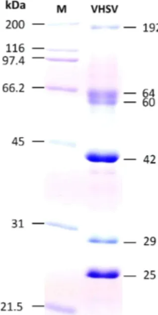
본 연구에서는 현장검사용 VHS 신속 진단키트 개발을 위한 기초연구로서 VHSV (genotype IVa)를
인식하는 MAb를 생산하고자 하였다. 정제한 VHSV 를 사용하여 SDS-PAGE를 실시한 결과, 약 192 kDa, 60-64 kDa, 42 kDa, 29 kDa, 25 kDa의 분자량 이 확인되었다(Fig. 1). 국내에서 분리된 genotype IVa의 속하는 VHSV의 구조 단백질은 약 62 kDa (G), 42 kDa (N), 29 kDa (phosphoprotein, P), 25 kDa (matrix protein, M)으로 보고되어 있다 (Jeong et al., 2017). 본 연구에서 정제한 VHSV의 SDS-PAGE 패 턴은 기존에 보고된 결과와 거의 유사하였다.
정제된 VHSV를 마우스에 면역시킨 후 마우스 의 림프절과 SP2/0Ag14 myeloma cell을 융합시켜 hybridoma를 제작하였다. Hybridoma로부터 생성 되는 항체를 ELISA법으로 선별한 후, 제한 희석법 으로 3회 클로닝하여 최종적으로 5개의 MAb를 선 별하였다 (2C10, 15E10, 18H4, 23H6, 30B7). 선별된 5개의 MAb의 isotyping을 분석한 결과, 2C10과 30B7의 H chain은 IgG2a, 15E10의 H chain은 IgG1, 18H4와 23H6의 H chain은 IgG2b로 나타났으며, L chain은 모두 kappa를 인식하는 것으로 확인되었다 (data not shown).
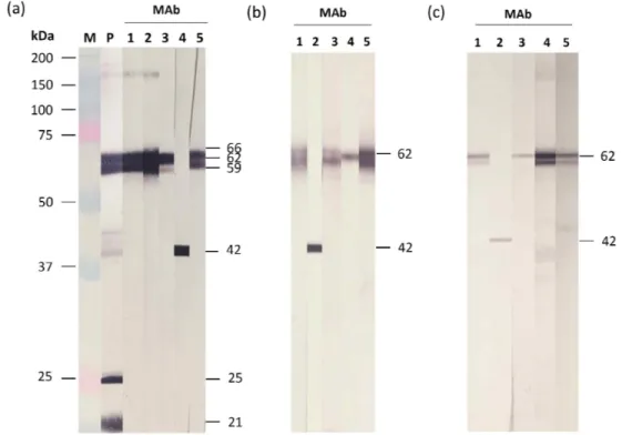
정제된 VHSV와 제작된 MAb를 사용하여 west- ern blot을 실시한 결과, 양성 대조구 (항 VHSV 토
Fig. 1. SDS-PAGE analysis of purified VHSV. M: mo-
lecular marker.
끼 혈청)에서 약 61-66 kDa, 42와 44 kDa, 25 kDa 및 21 kDa에서 밴드가 관찰되었다(Fig. 2a). 제작된 4개의 MAb (2C10, 18H4, 23H6, 30B7)는 59-66 kDa (G로 추정)을 강하게 인식하였고, MAb 15E10은 약 42 kDa (N으로 추정)을 강하게 인식하였다.
VHSV (FYeosu05)에 감염된 FHM 세포와 정상 FHM 세포를 사용하여 western blot를 실시한 결과, VHSV에 감염된 세포에서는 4개의 MAb (2C10, 18H4, 23H6, 30B7)가 약 62 kDa을 인식하였고, 1개 의 MAb (15E10)는 42 kDa을 인식하였다(Fig. 2b).
정상세포에서는 5개의 MAb 모두 반응하지 않았 다(data not shown). VHSV FWando05와 Jeju14 분리 주에 감염된 FHM 세포에서도 Fig. 2b와 동일한 결 과를 보였다(data not shown). VHSV에 감염된 넙치 와 정상 넙치의 조직 마쇄액을 사용하여 western blot을 실시한 결과, VHSV에 감염된 조직 마쇄액 에서는 Fig. 2b보다 약한 반응을 보였으나 인식하 는 부위는 동일하였다(Fig. 2c). 정상 조직 마쇄액
에서는 반응이 관찰되지 않았다(data not shown).
본 연구에서 제작된 항체는 VHSV에 감염된 세포 와 넙치 조직에서만 약 62 kDa과 42 kDa에서 밴드 가 관찰되므로 약 62 kDa을 인식하는 항체는 G를, 42 kDa을 인식하는 항체는 N을 인식하는 것으로 사료된다.
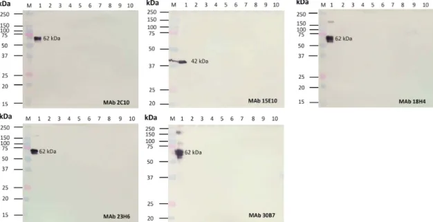
다양한 종류의 어류 바이러스들에 대한 MAb의 특이도를 조사하기 위해 7종의 바이러스(VHSV, IHNV, HIRRV, SVCV, IPNV, MABV, NNV)에 감 염된 세포와 3종의 정상세포(EPC, CHSE-214, SSN- 1)를 사용하여 western blot을 실시하였다(Fig. 3).
제작된 항체 모두 VHSV에 감염된 세포에서만 반 응하였고, 6종의 어류바이러스에 감염된 세포와 3 종의 정상세포에는 반응하지 않았다. 7종의 어류 바이러스를 항원으로 사용하여 ELISA를 실시한 결과, 5개의 MAbs 모두 VHSV에 강하게 반응(OD, 0.38-1.31)하였고, 6종의 다른 어류바이러스(IHNV, HIRRV, SVCV, IPNV, MABV, NNV)에는 반응하
Fig. 2. Western blot analysis using purified VHSV (a), VHSV-infected FHM cells (b), and tissue homogenates of
VHSV-infected olive flounder (c). M: molecular marker, P: Anti-VHSV polyclonal antibody, 1-5: MAbs (2C10,
15E10, 18H4, 23H6, 30B7).
Fig. 4. ELISA using seven fish viruses (VHSV, IHNV, HIRRV, SVCV, IPNV, MABV, and NNV) and five mon- oclonal antibodies (2C10, 15E10, 18H4, 23H6, 30B7).
지 않았다(OD: 0.13 이하)(Fig. 4).
본 연구에서는 정제된 VHSV (genotype IVa)를 사용하여 VHSV에 특이적으로 반응하는 5개의 MAb를 생산하였다. Western blot 상에서 4개의 MAbs (2C10, 18H4, 23H6, 30B7)는 G를 인식하였 고, MAb 15E10은 N을 인식하였다. Jeong et al.
(2017)은 초음파(40 kHz로 1시간)로 처리된 VHSV 를 사용하여 MAb를 제작한 결과, G를 인식하는 항체를 얻을 수 없었으나 N, P, M을 인식하는 항체
를 얻을 수 있었다. 1회 실험한 결과를 단순히 비교 하기에는 한계가 있으나 면역 항원(VHSV)의 처리 하는 방법에 따라 제작된 항체의 인식부위가 달라 질 수 있을 것으로 생각된다.
제작된 5개의 MAb는 in vitro 시료(바이러스에 감염된 세포와 바이러스 배양 상층액)를 사용한 western blot과 ELISA에서 VHSV에 반응하였고, 6 종의 어류바이러스(IHNV, HIRRV, SVCV, IPNV, MABV, NNV)와 3종의 어류주화세포(EPC, CHSE- 214, SSN-1)에는 반응하지 않았다. 또한 in vivo 시 료(VHSV에 감염된 넙치와 정상 넙치 시료)에서도 VHSV에만 반응하였다. 이상의 결과, 제작된 5개 의 MAbs는 VHSV에 특이적으로 반응하는 것이 확 인되었다.
VHSV를 인식하는 MAb는 여러 연구자들에 의 해서 생산된 바 있다(Lorenzen et al., 1988; Ito et al., 2010; 2012). Genotype I에 속하는 VHSV (분리 주: F1)를 사용하여 MAb를 제작한 후 다양한 VHSV 분리주 (genotype I-IV)를 대상으로 특이도 를 평가한 결과, MAb IP5B11은 genotype I-IV에 속 하는 VHSV 분리주 (50개 분리주) 모두에 특이적 으로 반응하였다(Lorenzen et al., 1988; Ito et al.,
Fig. 3. Western blot analysis using infected cells with 7 fish viruses and normal cells. M, molecular marker; 1,
VHSV; 2, IHNV; 3, HIRRV; 4, SVCV; 5, IPNV; 6, MABV; 7, NNV; 8, CHSE-214; 9, EPC; 10, SSN-1.
2010; 2012). Genotype IVa에 속하는 VHSV (JF00 Ehi1)를 사용하여 MAb를 제작한 후 특이도를 평 가한 결과에서는 MAb VHS-10은 genotype IVa에 만 반응하였다(Ito et al., 2010). Ito et al. (2012)은 genotype I-IV에 속하는 VHSV 분리주들을 사용하 여 MAb를 제작한 후 특이도를 평가하였다.
Genotype IVa에 속하는 VHSV를 사용하여 제작한 MAb VHS-1.24와 VHS-9.23은 각각 genotype Ie와 genotype III을 제외한 genotype에 속하는 VHSV에 반응하였다. MAb VHS-3.80 (항원: genotype Ib 분 리주)은 genotype Ib (일부 분리주 제외), Ic, Id (일 부 분리주 제외) 및 genotype II에 속하는 VHSV에 반응하였다. MAb VHS-7.57 (genotype II 분리주)은 genotype II와 IVa에만 반응하였다. MAb VHS-5.18 (genotype Ib 분리주)은 genotype Ib에만 반응하였 다. MAb VHS-3.75 (genotype III 분리주)는 geno- type III (일부 분리주 제외)와 IVb (일부 분리주)에 만 반응하였다. MAb VHS-1.88 (genotype IVb 분리 주)은 genotype IVb (일부 분리주 제외)에만 반응하 였다. 위의 연구결과를 종합해 보면, 특정 genotype 에 속하는 VHSV를 면역할 경우, 동일 genotype에 속하는 VHSV에만 반응하는 항체, 다양한 geno- type에 속해있는 VHSV에 반응하는 항체, 그리고 모든 genotype에 속하는 VHSV에 반응하는 항체가 생산될 수 있음이 확인되었다. 본 연구에서 제작된 MAb는 genotype IVa에 속하는 3개의 VHSV 분리 주들에 대해서만 특이도를 조사했기 때문에 향후 다른 genotype의 VHSV 분리주들에 대한 특이도를 조사할 필요가 있다. 하지만 제작된 MAb는 3개의 국내 VHSV 분리주에 강하게 반응하므로 genotype IVa에 속하는 VHSV를 검출할 수 있는 현장검사용 VHS 신속 진단키트를 개발하는데 사용될 수 있을 것으로 사료된다.
요 약
본 연구에서는 넙치(Paralichthys olivaceus)로부 터 분리한 바이러스성출혈성패혈증바이러스(viral hemorrhagic septicemia virus, VHSV, genotype IVa) 에 대한 단클론 항체(monoclonal antibody, MAb)를 개발하였다. VHSV에 대한 항체를 생산하는 총 5
개의 hybridoma clone을 생산하였다. 4개의 MAbs (2C10, 18H4, 23H6, 30B7)는 glycoprotein을 인식하 였고, MAb 15E10은 nucleocapsid protein을 인식하 였다. 5개의 MAbs는 western blot 상에서 VHSV에 감염된 세포와 넙치시료에 반응하였으나, 정상 세 포와 넙치시료에는 반응하지 않았다. 또한 ELISA 상에서 VHSV에만 반응하였고 6종의 어류바이러 스(IHNV, HIRRV, SVCV, IPNV, MABV, NNV)에 는 반응하지 않았다. 이상의 결과, 본 연구에서 제 작된 MAbs는 VHSV에만 특이적으로 반응하는 것 이 확인되어 VHSV를 검사하는데 사용될 수 있을 것으로 사료된다.
감사의 글
본 연구는 2016년도 정부의 재원으로 한국연구 재단의 지원을 받아 수행된 기초연구사업 (NRF- 2016R1D1A1B03936186)과 2017년 해양수산부 재 원으로 한국해양과학기술진흥원의 지원을 받아 수행되었습니다(수산동물 바이러스 전염병 진단 용 항체 생산).
References