R E S E A R C H Open Access
The influence of bone graft procedures on primary stability and bone change of implants placed in fresh extraction sockets
Sang Ho Jun
1†, Chang-Joo Park
2†, Suk-Hyun Hwang
3, Youn Ki Lee
3, Cong Zhou
3, Hyon-Seok Jang
4and Jae-Jun Ryu
5*Abstract
Background: This study was to evaluate the effect of bone graft procedure on the primary stability of implants installed in fresh sockets and assess the vertical alteration of peri-implant bone radiographically.
Methods: Twenty-three implants were inserted in 18 patients immediately after tooth extraction. The horizontal gap between the implant and bony walls of the extraction socket was grafted with xenografts. The implant stability before and after graft procedure was measured by Osstell Mentor as implant stability quotient before bone graft (ISQ bbg) and implant stability quotient after bone graft (ISQ abg). Peri-apical radiographs were taken to measure peri-implant bone change immediately after implant surgery and 12 months after implant placement. Data were analyzed by independent t test; the relationships between stability parameters (insertion torque value (ITV), ISQ abg, and ISQ bbg) and peri-implant bone changes were analyzed according to Pearson correlation coefficients.
Results: The increase of ISQ in low primary stability group (LPSG) was 6.87 ± 3.62, which was significantly higher than the increase in high primary stability group (HPSG). A significant correlation between ITV and ISQ bbg ( R = 0.606, P = 0.002) was found; however, age and peri-implant bone change were not found significantly related to implant stability parameters. It was presented that there were no significant peri-implant bone changes at 1 year after bone graft surgery.
Conclusions: Bone graft procedure is beneficial for increasing the primary stability of immediately placed implants, especially when the ISQ of implants is below 65 and that bone grafts have some effects on peri-implant bone maintenance.
Keywords: Immediate implant, Bone graft, Primary stability, Peri-implant bone change
Background
Numerous studies over the past 20 years have confirmed the predictability and success of implant placement at the time of extraction [1 – 3], with a number of prospect- ive and retrospective studies confirming high survival rates for more than 95% of immediate post-extraction implants and with observation periods ranging from 1 to 5 years [1, 4 – 7]. Albrektsson et al. [8] reported that pri- mary implant stability and lack of micromovement are
two of the main factors considered necessary for the achievement of predictably high success rates for osseointegrated oral implants. Primary stability of implants placed immediately after extraction strongly influences the long-term success of dental implants [9].
Following tooth extraction, the alveolar bone support- ing tooth undergoes constant atrophy [10]. It is shown that a marked reduction of the height of the alveolar ridge consistently occurred following tooth extraction and that implant installation in the fresh extraction socket did not interfere with the process of bone model- ing [11]. For an improved esthetic and functional pros- thodontic result, it is necessary to preserve the alveolar bone volume after tooth extraction to facilitate the
* Correspondence:
[email protected]Sang Ho Jun and Chang-Joo Park contributed equally to this work.
†
Equal contributors
5
Department of Advanced Prosthodontics, Korea University Anam Hospital, Inchon-ro 73, Seongbuk-gu, Seoul 02841, Republic of Korea
Full list of author information is available at the end of the article
© The Author(s). 2018 Open Access This article is distributed under the terms of the Creative Commons Attribution 4.0 International License (http://creativecommons.org/licenses/by/4.0/), which permits unrestricted use, distribution, and reproduction in any medium, provided you give appropriate credit to the original author(s) and the source, provide a link to the Creative Commons license, and indicate if changes were made.
subsequent placement of dental implants [10]. Chen and Buser [12] reviewed 91 studies and concluded that bone augmentation procedures are effective in promoting bone fill and defect resolution for implants in post- extraction sites.
Although implant primary stability and spontaneous bone graft after immediate implantation are important factors for high success rates and esthetic outcomes, the relationship between them has not been sufficiently eval- uated. The aim of the present study was to evaluate the effect of bone graft after immediate implant placement in fresh extraction socket.
Methods Study sample
This study received approval from the ethics committee of the Korea University Anam Hospital, Seoul, South Korea (AN13025-002). We included a total of 23 implant sites in 18 patients who required immediate implant installation following tooth extraction in this study. All patients were healthy and had no uncontrolled systemic diseases.
After thorough diagnosis and treatment planning, im- mediate implant placement was performed under local anesthesia. A total of 23 implants (Straumann
®Bone Level SLActive
®Implants) with favorable initial stability were used in this study.
Implant stability measurement
Implant placement was performed according to the se- quences recommended by manufacturers (Straumann AG, Basel, Switzerland). Final seating torque value was measured as insertion torque value (ITV) (INTRAsurg 300, KaVo, Germany) during implant placement. In all cases, an intentional bone augmentation procedure was simultaneously performed with xenografts (Bio-Oss
®, Geistlich, Switzerland) of 1–2-mm particle size into the gap between the implant fixture and the residual alveolar bone along the buccal and lingual surface. To standardize the bone graft procedure, a xenograft was packed into the gap by the controlled force minimizing the pulverization of the graft particles up to the remaining crestal level of the extraction socket. Reson- ance frequency analysis (RFA), using Osstell Mentor (Osstell AB, Gothenburg, Sweden), was performed to evaluate the primary stability of implants before and after bone grafting. Each implant was measured three times. The mean value of measurement before bone grafting was designated as the implant stability quotient before bone graft (ISQ bbg), and the mean measurement after bone graft was designated as the implant stability quotient after bone graft (ISQ abg). A healing abut- ment similar in diameter to that of the natural tooth was connected. The reflected flap was carefully su- tured with 5-0 nylon.
Peri-implant bone change analysis
To measure the peri-implant bone changes, peri-apical radiographs were taken both at the time of implant placement and 12 months after placement. Vertical mea- surements were taken from the mesial and the distal shoulder of the implant to the first bone-implant contact in an axis parallel to the implant (Fig. 1). A positive numerical value was recorded when the first bone- implant contact was higher than the implant shoulder, and a negative numerical value was recorded if the con- tact was lower than the implant shoulder. The data col- lected at the time of implant placement were used as the baseline value. Analysis of the peri-implant bone change was computed using image analysis software (ImageJ, 1.33i, National Institutes of Health, Bethesda, MD).
Statistical analysis
Independent t test was used for the comparison of the mean values of ISQ abg and ISQ bbg and peri-implant bone changes according to the implantation sites, and the number of tooth roots was compared by one-way analysis of variance (ANOVA).
The relationships between the initial stability parame- ters (ITV, ISQ abg, and ISQ bbg) and peri-implant bone
Fig. 1 Peri-implant bone measurements, both after implant placement
and at 1-year follow-up
changes were analyzed according to Pearson correlation coefficients. All statistical analyses were performed using SPSS 19.0 statistical software (IBM SPSS, Armonk, NY).
P values less than 0.05 were considered statistically significant.
Results
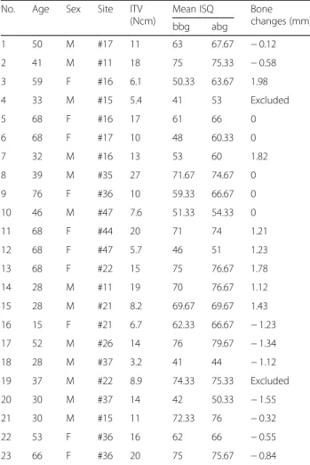
The average age of the 18 patients was 46.1 years, with 11 male and 7 female patients. A total of 23 implants were installed in tooth extraction sockets and healed without complications. Follow-up appointments were scheduled until 12 months after implant placement in order to measure the peri-implant bone changes. Two cases did not meet follow-up visit criteria, and thus, the two implants in these two patients were excluded from measurement of peri-implant bone change. Patient infor- mation and the data of implant stability are listed in Table 1.
Statistical correlations among variables
There was a significant correlation between ITV and ISQ bbg (R = 0.606; P = 0.002) (Fig. 2). Patient age was not found to be significantly related to either implant stability or peri-implant bone change. There was also no significant correlation between peri-implant bone changes and implant stability parameters (ITV, ISQ bbg, and ISQ abg) (Table 2).
The average ISQ bbg was 61.32 ± 12.29 and ranged from 41.00 to 76.00; the average ISQ abg was 66.23 ± 10.16 and ranged from 44.00 to 79.67. A statistically significant in- crease of ISQ was observed 4.91 ± 3.75 (P < 0.001) away from the ISQ bbg. There was a strong correlation between the ISQ bbg and the ISQ abg (R = 0.96; P < 0.01) (Fig. 3).
Influence of bone graft on implant primary stability To investigate how bone graft procedure influenced the various implants’ primary stability, we defined those with a mean ISQ bbg below 65 as the “low primary stability group” (LPSG) and those with a mean ISQ bbg above 65 as the “high primary stability group” (HPSG). Thus, the ISQ bbg of HPSG ranged from 69.97 to 76.00 (mean value 73.00 ± 2.34), while the ISQ bbg of LPSG ranged from 41.00 to 63.00 (mean value 52.33 ± 8.46). The in- crease in implant stability after bone graft was signifi- cantly higher in the LPSG (6.87 ± 3.62) than in the HPSG (2.368 ± 2.05) (P = 0.002) (Figs. 4 and 5).
Peri-implant bone changes
We compared the radiograph images from the day of im- plant installation with those taken at the 1-year follow-up.
The mean change of peri-implant bone was 0.14 ± 1.11 mm in 1 year. The one-sample t test revealed no sig- nificant peri-implant bone changes at 1 year post- implantation (t = 0.57; P = 0.57). In addition, no
Table 1 Patient information and measurements of implant stability and bone changes after 1 year follow-up
No. Age Sex Site ITV
(Ncm)
Mean ISQ Bone
changes (mm)
bbg abg
1 50 M #17 11 63 67.67 − 0.12
2 41 M #11 18 75 75.33 − 0.58
3 59 F #16 6.1 50.33 63.67 1.98
4 33 M #15 5.4 41 53 Excluded
5 68 F #16 17 61 66 0
6 68 F #17 10 48 60.33 0
7 32 M #16 13 53 60 1.82
8 39 M #35 27 71.67 74.67 0
9 76 F #36 10 59.33 66.67 0
10 46 M #47 7.6 51.33 54.33 0
11 68 F #44 20 71 74 1.21
12 68 F #47 5.7 46 51 1.23
13 68 F #22 15 75 76.67 1.78
14 28 M #11 19 70 76.67 1.12
15 28 M #21 8.2 69.67 69.67 1.43
16 15 F #21 6.7 62.33 66.67 − 1.23
17 52 M #26 14 76 79.67 − 1.34
18 28 M #37 3.2 41 44 − 1.12
19 37 M #22 8.9 74.33 75.33 Excluded
20 30 M #37 14 42 50.33 − 1.55
21 30 M #15 11 72.33 76 − 0.32
22 53 F #36 16 62 66 − 0.55
23 66 F #36 20 75 75.67 − 0.84
ITV insertion torque value, ISQ implant stability quotient, bbg before bone graft, abg after bone graft
Fig. 2 The correlation between implant stability quotient (ISQ) and
insertion torque value (ITV)
statistically significant differences in peri-implant bone changes were found according to the implantation sites and the number of tooth roots.
Discussion
Immediate installation of implants in fresh sockets is a challenging and sensitive technique and requires careful case selection [13]. It has been postulated that when the size of horizontal gap surrounding immedi- ately placed implants exceeds the threshold of 1 to 2 mm, bone graft procedures might be recommended to reduce peri-implant bone resorption and improve the esthetic outcome of the soft tissue [10, 12, 14, 15]. The influence of grafting procedure on primary stability of immediately placed implants, nevertheless, has not been given enough attention.
Table 2 Pearson ’s correlations among patient age, implant stability parameters, and peri-implant bone changes
Age ITV ISQ bbg ISQ abg Bone change
Age R 1.000 0.187 0.069 0.147 0.254
P 0.394 0.754 0.503 0.266
ITV R 0.187 1.000 0.606 0.603 − 0.033
P 0.394 0.002 0.002 0.886
ISQ bbg R 0.069 0.606 1.000 0.964 0.036
P 0.754 0.002 < .0001 0.879
ISQ abg R 0.147 0.603 0.964 1.000 − 0.088
P 0.503 0.002 < .0001 0.705 Bone level change R 0.254 − 0.033 0.036 0.088 1.000
P 0.266 0.886 0.879 0.705
ITV insertion torque value, ISQ implant stability quotient, bbg before bone graft, abg after bone graft
Fig. 3 The correlation between implant stability quotient after bone graft (ISQ abg) and implant stability quotient before bone graft (ISQ bbg)
Fig. 4 The mean increase of implant stability quotient (ISQ) after bone graft procedure in both the low and the high primary stability groups
Fig. 5 The changes of mean implant stability quotient (ISQ) for all the implants after bone graft procedures in both the low primary stability group (LPSG) and the high primary stability group (HPSG).
The implants are arranged in the sequence of ISQ before bone graft
(bbg). The mean ISQ bbg below 65 was defined as the LPSG, above
65 was defined as the HPSG
Though RFA suffers from a lack of sensitivity to the quality of surrounding bone, ISQ measurement is a ver- satile technique that can be used repeatedly for quantita- tive stability both intraoperatively and postoperatively [16]. We found a strong correlation between ITV and ISQ in accordance with the previous study that analyzed the ITV and ISQ on a fresh cadaver and concluded that ITV and ISQ were statistically correlated [16]. In another experiment carried out in fresh-frozen pig femoral bones, Zhou et al. [17] obtained a similar result to the present study that correlation between ISQ and ITV was significant. Some studies, however, failed to demonstrate a statistical correlation between ISQ and ITV [18, 19], although the ITV was obtained using some equipment, such as implant engines and various custom-made man- ual torque devices, which may have affected their results.
No significant correlation between peri-implant bone changes and implant stability parameters (ITV, ISQ bbg, and ISQ abg) was detected by the present study. In addition, implantation sites and defect configurations classified by the number of tooth roots showed no sig- nificant effects on peri-implant bone changes after the immediate placement of implant. However, a consensus has been reached that bone graft procedure is beneficial in preventing peri-implant bone loss, which was con- firmed by this study. Araújo et al. [10] evaluated the osseointegration and peri-implant tissue modeling following implant placement in fresh extraction sockets, and he found that in the absence of bone graft, the dimensions of both the buccal and the lingual bone walls around the implant were reduced; even the osseointegra- tion could be in part lost following bone modeling.
Therefore, with the support from experimental evidence, the control group in which bone graft procedure would not be carried out was not included in this study.
In the present study, the xenografts filled the horizontal gap between the bony walls and the implants. It is worth noting that the grafting procedure led to an increase in the primary stability of immediately placed implants, which was more notable for implants in the LPSG, in which the ISQ bbg was below 65 (Fig. 4). In light of the ef- fect of increasing the primary stability of implants, grafting the horizontal gap with bone substitutes could be benefi- cial. The influence of graft procedure on implant primary stability has not been intensively studied, even though Santos et al. [20] evaluated recent animal and human studies of bone substitutes used for peri-implant defects in post-extraction implants. They concluded that although the technique of installing implants in fresh extraction sockets is a reliable alternative to reduce treatment time, the use of a biomaterial is required to increase bone- implant contact and enhance osseointegration.
It has been proven that alveolar bone remodeling ap- peared progressively active following tooth extraction;
the horizontal resorption of the buccal alveolar di- mension amounted to about 56% at 4 months after tooth loss [21]. Based on indications from McGlum- phy and Larsen [22], when the size of the gap is less than 1 mm, the graft procedure is not needed; it is only when the size of gap exceeds 1 mm that bone graft or other guided bone regeneration procedures may be necessary. Although several kinds of bone graft materials, such as autogenous graft, allograft, xenograft, synthetic materials, or any combination of these, have been used or tested to maintain the bone level around immediately placed implants, none have been proven to be superior to the others [20, 23, 24].
In this study, bovine bone was used as the grafting material to augment the horizontal gaps adjacent to immediate implants. We found that the bundle bone was progressively resorbed during the modeling and remodeling stages of the extraction healing process, similar to the previous finding that bundle bone could be observed only until 2 weeks after tooth extraction [25, 26]. It has been found that xenografts play an important part in alveolar bone preservation and can maintain the dimensions of the extraction socket in- stead of the bundle bone, as well as encourage osteo- conduction and space maintenance [27]. In our study, the graft procedure resulted in no marginal bone loss after 1 year of follow-up, which was similar to find- ings in previous studies [15, 23].
Conclusions
The results of this study indicate that the bone graft procedure is beneficial for increasing the primary sta- bility of immediately placed implants, especially when the ISQ of implants is below 65. Both ITV and ISQ are effective and practical methods detecting implant primary stability and are statistically correlated with each other.
Abbreviations
HPSG: High primary stability group; ISQ abg: ISQ after bone graft; ISQ bbg: ISQ before bone graft; ISQ: Implant stability quotient; ITV: Insertion torque value; LPSG: Low primary stability group
Acknowledgements Not applicable
Funding
This work was supported by a Korea University Grant and the Basic Science Research Program through the National Research Foundation funded by the Ministry of Education (NRF-2017R1D1A1B03028418).
Chang-Joo Park was supported by the Basic Science Research Program through the National Research Foundation of Korea (NRF) funded by the Ministry of Education (2017R1D1A1A02019047).
Availability of data and materials
All the data supporting our findings are in the “
Methods” and “
Results”
sections of this manuscript.
Authors ’ contributions
SHJ and SHH wrote this manuscript. CZ and LYK framed and designed the clinical study. HSJ performed and confirmed the statistical analysis of the clinical data. CJP and JJR collected and analyzed most of the clinical data. All authors read and approved the final manuscript.
Ethics approval and consent to participate
The study received approval from the ethics committee of the Korea University Anam Hospital, Seoul, South Korea (AN13025-002). Trial registration:
AN13025-002. Registered on February 28, 2013. The first participant enrolled on May 1, 2013.
Consent for publication
The consent for publication was obtained from all the participants to report their individual data.
Competing interests
The authors declare that they have no competing interests.
Publisher ’s Note
Springer Nature remains neutral with regard to jurisdictional claims in published maps and institutional affiliations.
Author details
1
Department of Dentistry, Korea University Anam Hospital, Seoul, Republic of Korea.
2Division of Oral and Maxillofacial Surgery, Department of Dentistry, College of Medicine, Hanyang University, Seoul, Republic of Korea.
3