http://www.medicinalcrop.org http://dx.doi.org/10.7783/KJMCS.2013.21.4.262
우산고로쇠의 향장효과
손상현·이상원·신유수·김형돈·양승옥·김승유·김영옥† 농촌진흥청 국립원예특작과학원 인삼특작부
The Effect of Cosmetic on Anti-Wrinkle of Acer mono Sap
Sang Hyun Sohn, Sang Won Lee, Yu Su Shin, Hyung Don Kim, Seung Ok Yang, Seung Yu Kim and Young Ock Kim†
Department of Herbal Crop Research, NIHHS, RDA, Eumseong 369-873, Korea.
ABSTRACT : The purpose of this study was to research for anti-oxidation and anti-wrinkle effects of Acar mono Sap (AM).
To cosmetic effect of AM, safety effect (MTT assay), anti-wrinkle effect (elastase, MMP-1 inhibition assay) and anti-oxidant effect (DPPH assay) were measured. When water extract of AM was used for cell viability, it was over 100% at 6% (6 ml/
100 ml in phosphate buffer) concentration. AM showed 45.7% elastase inhibition and 23.7% MMP-1 inhibition at 50% (50 ml/
100 ml in phosphate buffer) concentration so that it had good anti-wrinkle characteristic. And AM showed 68.9% antioxida- tion capacity at 50% concentration by using a DPPH assay. Consequently, AM can be used as natural materials or additives for human skin owing to their beneficial biologic functions, including the anti-wrinkle effect, for cosmetic compositions.
Key Words : Acer mono Sap, Anti-Wrinkle, Elastase
INTRODUCTION
Wrinkles are result of dermal-hypodermal junction and shrinking of the superficial muscles, which have their points of insertion at the dermis. Mechanical and elastic properties of skin associated with skin wrinkling are related to histomorphology of the epidermis (Fujimura T., et al., 2000). And wrinkles are formed and promoted by both internal and external factors. Internal factors include aging, changes in the endocrine system, nerve system and hereditary factors. External factors include exposure to UV rays and the oxidation or drying associated with UV exposure (Sophie et al., 2002, Son et al., 2012). The skin tissue is rich in lipids, which are thought to be vulnerable to oxidative stress from sunlight. Ultraviolet (UV) exposure leads to alterations in the composition of the skin, including the accumulation of elastic fibres (Braverman
and Fonferko, 1982), collagen reduction and degeneration (Oikarnin and Kallionen, 1989). Elastase is the only enzyme that is capable of breaking down elastin, an insoluble elastic fibrous protein that, together with collagen, determines the mechanical properties of connective tissue (Antonicelli et al., 2007). Collagen and elastin provide suppleness and elasticity to the skin and reinforce the fibers of the two fundamental elements which constitute the supporting capacity of the cutaneous layer (Kim et al., 2011).
Of the environmental factors that injure the skin, ultraviolet (UV) irradiation is the most common and pernicious. It leads to alterations in the composition of the skin, including the accumulation of elastic fibers (Braverman and Fonferko, 1982), collagen reduction and degeneration (Oikarnin and Kallionen, 1989). Functional cosmetics are defined in Korean Cosmetics Law as cosmetics including whitening, anti-wrinkle, and UV protection effects
†
Corresponding author: (Phone) +82-43-871-5585 (E-mail) [email protected]
Received 2013 Fabruary 19 / 1st Revised 2013 May 13 / 2nd Revised 2013 June 6 / 3nd Revised 2013 June 25 / Accepted 2013 June 26
This is an open access article distributed under the terms of the Creative Commons Attribution Non-Commercial License (http://creativecom-
mons.org/licenses/by-nc/3.0/) which permits unrestricted non-commercial use, distribution, and reproduction in any medium, provided the original
work is properly cited.
(Cho et al., 2011). As anti-wrinkle agents, retinol, retinyl palmitate, adenosine, and polyethoxylated retinamide are registered in KFDA (
Korea Food and Drug Administration, 2009). The genus Acer comprises 15 species in Korea (Jin et al., 2006). Acar mono Sap (AM) is an endemic Korean maple species, growing in the mountains of Ullung Isaland (Lee, 1996). This plant has some flavonol glycosides and phenolic compounds. AM was reported anti- HIV-1 activity (Kim et al., 1998) and the inhibitory effect of low density lipoprotein (LDL) oxidation (Thuong et al., 2005). In Korea, the sap of Acer mono (painted maple) has been termed ‘bone-benefit-water’ owing to various minerals(calcium, potassium and magnesium ion) and can prevent extreme dehydration and tiredness or fatigue (Kunkel, 1984). AM has been used in East Asian countries for centuries due to its anti-osteoporosis effects (Lee et al., 2008). however, East Asian medical literature claims that the sap has potential anti-oxidative and immunomodulatory efects (Yang et al., 2008). AM has a source of cosmetic agents capable of improving whitening effect and antioxidant activites (Kim et al., 2011).
However, more systematic scientific studies to establish the safety and efficacy of medicinal herb in both rejuvenation and detoxification procedures are needed. Thus, this study may be interesting to modern scientists to understand different approaches and processes that may be useful to progress research on promotive, preventive and therapeutic interventions generally on various degenerating conditions and aging. Therefore, the objectives of the present study was to evaluate its biological activities such as their antioxidant, anti-elastase, and anti-inflammatory activities for application to human skin.
Materials and Methods
1. Materials
The sap of AM was kindly provided by Professor Lee, Hyun Young in Kangwon National University.
AM is the closest relatives of A. okamotoanum (Pfosser et al., 2002), hence suggesting that the latter might have evolved anagenetically from continental populations of the former.
2. Cell Viability Assay
The procedures of cell viability assay were applied by the method of Mosmann (1983). Hs27 was obtained from
the American Type Culture Collection (ATCC, Rockville, MD, USA). Hs27 is one of a series of human foreskin fibroblast lines developed at NBL, California. The material was obtained from a normal new-born Negro, and the cells possess the G6PD type A phenotype. Longevity studies carried out at NBL demonstrated that the cells can be propagated for 42 passages The cell line was cultured according to instructions defined by the ATCC. Hs27 (newborn foreskin, male, human fibroblast) was cultured in Dulbecco’s modified Eagle’s medium (Gibo, Gaithersburg, MD, USA) supplemented with 15% fetal bovine serum (Hyclone, Logan, MA, USA). The culture medium was replaced every 48 h with pre-equilibrated 37 ℃ medium. Cell suspensions were then counted and aliquoted for subsequent passage or lysis. Hs27 cells were used between passage numbers 5 and 20. To examine the effect of AM on cell viability recovery, we performed a Thiazolyl Blue Tetrazolium Bromide (MTT) assay. Following treatment with AM, culture medium was removed and MTT (0.33 g/L) solution was added for 90 min at 37 ℃. The supernatant was discarded and isopropanol was added to dissolve the formazan product.
The intensity was measured colorimetrically at a wavelength of 570 ㎚. The cell viability being calculated as follows:
Cell viability (%) = (OD 570(sample) / OD 570(control) × 100 where OD 570 (sample) is the absorbance of the treated cells at 570 ㎚ and OD 570 (control) is the absorbance of the negative control at 570 ㎚ (non-treated cells).
3. Elastase Inhibition Assay
The activity of porcine pancreatic elastase (E7885, Sigma, USA) was examined using N-Succinyl-(Ala)
3-p-nitroanilide as the substrate, and the release of p–nitroaniline at 410 ㎚ was measured. The reaction was carried out in a 0.05M Tris-HCl buffer (pH 8.2) containing 5 mM N-Succinyl- (Ala)3-p-nitroanilide and elastase (1.0 unit/mL). AM was added to the reaction mixture to reach a final concentration of 500 µg/mL, and elastase inhibition was assessed at 25℃.
The reaction mixture was pre-incubated for 10 min before
adding the substrate (Hong et al., 2008). The change in
absorbance was measured at 410 ㎚ using a 96-well
reader. The percent inhibition of elastase was calculated as
follows: Inhibition (%) = [(A-B)/A] × 100, where A is absorbance
at 410 ㎚ without AM, and B is the change in absorbance
at 410 ㎚ with AM.
4. Assay of Collagen Type I Synthesis by an EIA Kit Fibroblast cells were inoculated into 24-well plates (5
*10
5cells/well) and cultivated for 24 h. After culturing, the culture medium was changed to serum-free IMDM (Iscove’s modified Dulbecco’s medium) and cultivated for 24 h (Van et al., 1981). The control group was cultivated without a compound. After culturing, the supernatant was collected from each well, and the amount of pro-collagen type I was measured with a pro-collagen type I C peptide assay kit (Takara Bio, Japan).
5. ELISA of pro-MMP 1
Human dermal fibroblast cells were exposed to UV-B irradiation at 52 mJ/cm
2followed by incubation in serum free DMEM for 48 h with or without AM. The MMP-1 secreted into the culture medium was determined by a commercially available ELISA assay kit (R & D Systems, Minneapolis, MN, USA).
6. Antioxidant Assay (DPPH assay)
DPPH (Sigma, USA) was dissolved in ethanol (99.5%, Sigma, USA) to a volume of 250 µM followed by sonication for 5 min to obtain the stable free radical DPPH using the method reported by Bors et al. (1992). The test compound was diluted in DPPH solution in a ratio of 1 : 1. Appropriate controls were run in each series and fresh free radical DPPH solution was prepared daily. AM was tested in triplicate at five different concentrations such that a 50% decrease in free radical DPPH absorbance could be calculated. The change in the absorbance at 517 ㎚ was measured in a 96-well reader. The FSC
50(concentration causing 50% radical scavenging activity) value of AM was determined and compared with that of L- ascorbic acid (Sigma, USA) and Trolox (Sigma, USA). The absorbance (A) of the control and samples was measured, and the percentage DPPH scavenging activity was determined as: DPPH scavenging activity (%) = [(A
control- A
sample)/A
control]
× 100 Each assay was performed in triplicate and the concentration required for a 50% reduction (IC
50) in DPPH radical activity was determined graphically.
RESULTS AND DISCUSSION
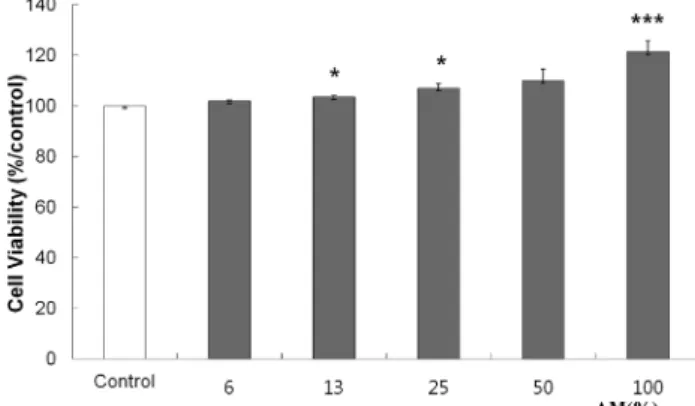
1. AM Protects Dermal Fibroblasts from Cell Death To study whether AM could induce cell viability, we
checked cell viability using a MTT assay. The sample was prepared at various concentrations and used to treat HS27.
The results of this evaluation is shown in Fig. 1. AM showed no cytotoxicity up to the effective concen-tration for anti-wrinkle activity. This finding suggests that AM could be produced as an effective active ingredient with no associated cytotoxicity.
2. Elastase Inhibition Assay
Elastase is known to cause rheumatoid arthritis, pulmonary emphysema, and other chronic inflammatory diseases by the protein degradation of human tissues (Park et al., 1999). It also degrades elastin, which is closely related to the elasticity and restoration of skin, and induces wrinkles
Fig. 1. Cytotoxicity of AM on HS27 human fibroblasts. Control;distilled water treated group with UV-B irradiation 6, 13, 25, 50, and 100; AM treated group. Data represent the mean ± SEM (n = 5). Asterisk indicates a significant difference compared with control at.
*p < 0.5, **p < 0.05, ***p < 0.01.
Fig. 2. Inhibition effect of AM on elastase. All experiments were performed in triplicate and error bars represent mean ± SEM (n = 3).
**p < 0.05, ***p < 0.01.
and a lack of elasticity. The inhibition effect of elastase activity is shown in Fig. 2. The inhibition rate of AM at a 50% concentration was 45.7%. AM was found to have the high elastase inhibition activity compared to adenosine which was used as a positive control. This research offered that AM would have potential as an anti-wrinkle agent for use in cosmetic products.
3. Assay of Collagen Type I Synthesis by an EIA Kit Collagen types I, II, III, IV and V are synthesized as precursor molecules called procollagens. These precursor molecules contain additional peptide sequences, termed
“propeptides”, at both their amino- and carboxy-terminal ends. To evaluate the amount of collagen type I synthesis, collagen type I was quantitatively detected by using a procollagen type I C-peptide assay kit (Takara Bio, Japan).
Collagens are synthesized as precursor molecules, called procollagens. Pro-collagen Type I C-peptide (PIP) has also been extensively referenced in correlation studies between collagen levels and certain health disorders such as bone disease, alcoholic liver disease, liver cirrhosis (Borrmann type IV), and adenocarcinoma of the stomach. AM increased the expression of type I collagen in a dose- dependent manner (up to 33.7% at 100% AM), comparable to that of ascorbic acid (up to 69.2% at 25 µM) as shown in Fig. 3. AM in a concentration range from 6- 100% showed an increase in type I collagen synthesis from 1.6% to 33.7%.
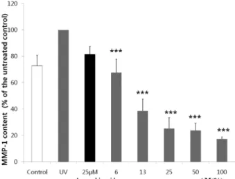
4. AM Reduced the Expression of MMP-1
AM was tested if it has inhibitory effect on the UVB- induced MMP-1 expression. Human dermal fibroblasts were first irradiated with 52 mJ/cm
2UVB and then treated with 6-100% concentrations of AM or 25 µM ascorbic acid for 48 h followed by examination of MMP-1 expression in the culture medium by ELISA assays. AM significantly dose dependently decreased the level of MMP-1 to below the base line level even with irradiation. Ascorbic acid at concentrations 25 µM for 48 h. A dose dependent MMP-1 inhibition capability was observed in AM treated groups.
AM 100% treated groups had the highest inhibition capability, of 17.17 ± 1.6%, while 6, 13, 25 and 50% treated groups had 23.7 ± 5.7%, 25.2 ± 8.0%, 38.5 ± 9.0%, and 67.5 ± 10.3%, respectively (Fig. 4).
5. Antioxidant Assay (DPPH assay)
It has been reported that reactive free radicals was induced by elastase in neutrophil and that they play a role in inflammation. Assays of the free radical scavenging capacity were carried out by the DPPH scavenging effect.
The free radical scavenging capacity of AM was measured at each concentration (6, 13, 25, 50, and 100%). A dose dependent free radical scavenging capability was observed
Fig. 3. Collagen type I synthesis assay in human fibroblastcells using the EIA kit. The control group was cultivated without AM. 25µM L-ascorbic acid was positive control.
All experiments were performed in triplicate and error bars represent mean ± SEM (n = 3).
*p < 0.5, **p < 0.05.
Fig. 4. The effect of AM on the expression of MMP-1 induced by UV-B. HS27 after exposure to UV-B irradiation of 52 mJ/㎠ at 312 ㎚ were treated with AM at concen- trations of 6, 13, 25, 50, and 100% and L-ascorbic acid at concentrations 25µM for 48 h. MMP-1 contents were analyzed by ELISA. All experiments were performed in triplicate and error bars represent mean ± SEM (n = 3). A dose dependent MMP-1 inhibition capability was observed in AM treated groups.
in AM treated groups. AM 100% treated groups had the highest scavenging capability, of 92 ± 2.6%, while 50, 25, 13, and 6% treated groups had 68.9 ± 7.2%, 64.7 ± 8.3%, 56.8 ± 12.1%, and 31.8 ± 8.6%, respectively (Fig. 5).
In order to investigate for wrinkle-care cosmetics, we screened free radical scavenging activities, elastase inhibition activities and MMP-1 assays. Other studies have demonstrated that chronic UV-A and UV-B radiation result in a significant increase in elastase-type enzyme activity (Imayama et al., 1989). Increased elastase activity has been associated with wrinkling, sagging, and laxity of aged skin (Wan et al., 2001). Therefore, inhibition of elastase activity might contribute to an anti-aging effect. AM proved, through in vitro tests, to stimulate collagen production and fibroblast proliferation. In cell cultures, AM showed positive effects on fibroblast proliferation and collagen production. So it might be considered a potential new medicinal plant to use in the future in skin care cosmetics.
ACKNOWLEDGEMENTS
This work was performed with the support of the Cooperative Research Program for Agriculture Science &
Technology Development(PJ0074792012), Rural Development Administration, Republic of Korea.
LITERATURE CITED
Antonicelli F, Bellon G, Debelle L and Hornebeck W. (2007).
Elastin-elastases and inflamm-aging. Current Topics in Developmental Biology. 79:99-155.
Braverman IM and Fonferko E. (1982). Studies in cutaneous aging: I. The elastic fiber network. The Journal of Investigative Dermatology. 78:434-443.
Bors W, Saran M and Elstner EF. (1992). Screening for plant antioxidant. Modern method of plant analysis-plant toxcin.
Analysis-New Series. 13:277-295.
Cho EA, Cho EH, Cho EA, Cho EH, Choi SJ, Park KH, Kim SY, Jeong YJ, Ku CS, Ha BJ, Jang DI and Chae HJ.
(2011). Screening of anti-wrinkle resource from herbal medicinal extracts and stability test of its cosmetic products. Korean Journal of Medicinal Crop Science. 19:126-135.
Fujimura T, Moriwaki S, Takema Y and Imokawa G. (2000).
Epidermal change can alter mechanical properties of hairless mouse skin topically treated with 1alpha, 25-dihydroxyvitamin D(3). Journal of Dermatological Science. 24:105-111.
Hong ES, Ahn GW and Jo BK. (2008). The study on the potential anti-aging properties of Prunella vulgaris extract in vitro and in vivo. Journal of the Society of Cosmetic Scientists of Korea. 34:129-135.
Imayama S and Braverman I M. (1989). A hypothetical explanation for the aging of skin. Chronologic alteration of the three-dimensional arrangement of collagen and elastic fibers in connective tissue. American Association of Pathologists.
134:1019-1025.
Kim YO, Seo YC, Lee HY, Oh SM, Lee SW and Kim HD.
(2011). Anti-wrinkle effect of Ulmus davidiana extracts. Korean Journal of Medicinal Crop Science. 19:508-513.
Jin WY, Min BS, Youn UJ, Hung TM, Song KS, Seong YH and Bae KH. (2006). Chemical constituents from the leaf and twig of Acer okamotoanum Nakai and their cytotoxicity.
Korean Journal of Medicinal Crop Science. 142:77-81.
Kim HJ, Woo ER, Shin CG and Park HK. (1998). A new flavonol glycoside gallate ester from Acer okamotoanum and its inhibitory activity against human immunodeficiency virus- 1(HIV-1) integrase. Journal of Natural Products. 61:145-148.
Kim JS, kim JS, Seo YC, Choi WY, Kim HS, Kim BH, Shin DH, Yoon CS, Lim HW, Ahn JH and Lee HY. (2011).
Enhancement of antioxidant activities and whitening effect of Acer mono sap through nano encapsulation processes. Korean Journal of Medicinal Crop Science. 19:191-197.
Korea Food and Drug Administration(KFDA). (2009). http://
www.kfda.go.kr/index.html.
Kunkel G. (1984). Plants for human consumption. Economic Botany. 39:176-177.
Lee WT. (1996). Coloured standard illustration of Korean Plants.
Academy Publishing House. Seoul, Korea. p.215.
Lee GS, Byun HS, Kim MH, Lee BM, Ko SH, Jung EM, Gwak KS, Choi IG, Kang HY, Jo HJ, Lee HJ and Jeung EB. (2008). The beneficial effect of the sap of Acer mono in an animal with low-calcium diet-induced osteoporosis-like symptoms. Britsh Journal of Nutrition. 100:1011-1018.
Mosmann T. (1983). Rapid colorimetric assay for cellular growth and survival: Application to proliferation and cytotoxicity assays. Journal of Immunological Methods. 65:55-63.
Oikarnin A and Kallionen M. (1989). A biochemical and Fig. 5. DPPH free radical scavenging capability. Control;
control, distilled water treated group. 6, 13, 25, 50 and 100; AM extracts treated group (4, 20, 100, and 500㎎/㎖). All experiments were performed in triplicate and error bars represent mean ± SEM (n = 3).
*p < 0.5, **p < 0.05, ***p < 0.01.
immunohistochemical study of collagen in sunexposed and protected skin. Photo-Dermatology. 6:24-31.
Park SM, Park SI, Huh MD and Hong YK. (1999). Inhibitory effect of green tea extract on collagenase activity and growth of fish pathogenic bacteria. Journal of Fish Pathology. 12:83-88.
Pfosser MF, Guzy-Wrobelska J, Sun BY, Stuessy TF, Sugawara T and Fujii N. (2002). The origin of species of Acer (Sapindaceae) endemic to Ullung island, Korea. Systematic Botany. 27:351-367.
Bosset S, Barre P, Chalon A, Robin Kurfurst R, Bonte F, Andra P, Pierre Perrrie P, Disante F, Varlet B and Nicolas JF. (2002). Skin ageing: Clinical and histopathologic study of permanent and reducible wrinkles. European Journal of Dermatology. 12:247-252.
Son ED, Shim JH, Choi H, Kim H, Lim KM, Chung JH, Byun SY and Lee TR. (2012). Cathepsin G inhibitor prevents ultraviolet B-induced photoaging in hairless mice via inhibition
of fibronectin fragmentation. Dermatology. 224:352-360.
Thuong PT, Na M, Su ND, Seong RS, Lee YM, Sok DE and Bae K. (2005). Inhibitory effect of coumarins from Weigela subsessilis on low density lipoprotein oxidation. Biological and Pharmaceutical Bulletin. 28:1095-1097.
Van Wart HE and Steinbrink DR. (1981). A continuous spectrophotometric assay for Clostridium histolyticum collagenase.
Analytical Biochemistry. 113:356-65.
Wan Y, Belt A, Wang Z, Voorhees J and Fisher G. (2001).
Transmodulation of epidermal growth factor receptor mediates IL-1 beta-induced MMP-1 expression in cultured human keratinocytes. International Journal of Molecular Medicine.
7:329-334.
Yang H, Sung SH and Kim YC. (2005). Two new hepatoprotective stilbene glycosides from Acer mono leaves.
Journal of Natural Products. 68:101-103.