녹용 약침액의 주름 개선 효과에 관한 연구
이주희*․이경민*․김재수*․정태영**․임성철*
*대구한의대학교 한의과대학 침구경혈학교실
**제한동의학술원
목적 : 본 연구는 녹용약침액의 주름 개선 효과에 대하여 여러 가지 in vitro 실험을 통해 확인해 보고자 계획되었다.
방법 : 녹용약침액의 항산화효과를 DPPH(1,1-diphenyl-2-picrylhydrazyl) 자유라디칼 소거법을 이용하여 측정하였고, 또, elastase 억제 효과를 측정하였다. 그리고 녹용약침액이 사람 정상 섬유아세포 HS 68에서 UVB 조사 후 type I procollagen의 생산량 회복에 미치는 효과를 ELISA법을 이용하여 측정하였다.
결과 : 녹용약침액에서 우수한 DPPH 자유라디칼 소거 효과가 관찰되었다. 또한 통계적으로 유의한 elastase 활성 억제 효과를 관찰할 수 있었다. 사람 섬유아세포 HS 68을 이용한 실험에서는 녹용약침액 처 치군에서 UVB 조사로 감소된 type I procollagen이 유의하게 회복되는 것이 관찰되었다.
결론 : 본 연구 결과에 의하면, 녹용약침액에는 유효한 주름개선 효과가 있어, 미용약침 소재로 개발할 수 있을 것으로 사료된다.
핵심 단어 : 녹용약침액, 주름 개선, elastase, DPPH, type I procollagen
1)
Anti-wrinkle Effects of Cervi Pantotrichum Cornu Pharmacopuncture Solution
Lee Ju-hee
*, Lee Kyung-min
*, Kim Jae-su
*, Jung Tae-young
**and Lim Seong-chul
**
Dept. of Acupuncture & Moxibustion, College of Oriental Medicine, Daegu Hanny University
**
Je-Han Oriental Medical Academy
․Acceptance : 2010. 5. 22. ․Adjustment : 2010. 7. 26. ․Adoption : 2010. 7. 26.
․Corresponding author : Lim Seong-chul, Department of Acupuncture and Moxibustion, Pohang Oriental Hospital of Daegu Haany University, 907-8 Daejam-Dong Nam-Gu Pohang-Si Kyungsangbuk-Do Republic of Korea
Tel. 82-54-271-8009 E-mail : [email protected]
국문초록
Original Article
Ⅰ. Introduction
It is the latest trend to be looked younger than the actual age. In these days, aging seems to be treated not as a nature to accept but as a disease or a disorder to overcome.
There are two major theories of aging: the programmatic theorystates that aging is an inherent genetic process, and the stochastic theory states that aging represents random environmental damage.
Processes that are associated with cellular damage and aging are the production of free radicals(a process much enhancedafter ultraviolet(UV) irradiation) and an increasing number of errors during DNA replication. Cellular manifestations of intrinsic aging include decreased life span of cells, decreased res- ponsiveness of cells to growth signals, which may reflect loss of cellular receptors to growth factors, and increased responsiveness to growth inhibitors.
All these findings are more pronounced in cells derived from photodamaged skin1).
UV irradiation is responsible for the cutaneous damage after both acute and chronic exposure, and is believed to be an important etiology in human skin cancer and premature skin aging2). Ultraviolet- B(UVB)(312㎚) has a low level of skin penetration, but it can readily affect macromolecules in the epi- dermal layer, thus altering cellular functions via DNA damage, generation of reactive oxygen species (ROS), decreased in skin content of antioxidant com- pounds3). Recent studies have shown that ROS such as superoxide anion, hydroxyl radical, and hydrogen peroxide are responsible for UV-induced oxidative damage4-6). Nowadays, various natural compounds from both nutritive and non-nutritive sources were reported to protect against UV-induced skin da- mage4,7,8).
In Oriental medicine, skin aging problem is th- ought to be due to the failure of nourishing the essence of the kidneys and insufficient nutrition supply for body organs9).
Cervi Pantotrichum Cornu
is considered asa core tonic ingredient which for- tifies the primal yang and the kidneys, generatesessence and nourishes the qi and blood of the body10). Therefore, it is likely to use
Cervi Pantotrichum Cornu
in skin aging such as premature wrinkle.In the present study, I investigated the anti- wrinkle effects of
Cervi Pantotrichum Cornu
including anti-oxidative activities, elastase inhibitory activities and protective effects against the UVB-induced photodamage in human skin fibroblasts HS 68.Ⅱ. Materials and methods
1. Sample preparation
Cervi Pantotrichum Cornu(Nokyong)
was purchased from Omniherb(Korea).Cervi Pantotrichum Cornu
Pharmacopuncture solution(CPC-HAS) was prepared according to the following steps. 2.0g of dried mix- ture ofCervi Pantotrichum Cornu
was distilled in 100㎖ of saline, and 50㎖ of resulting distillates was filtered three times through micro-filter paper and syringe filter(Whatman # 2, 0.45㎛ to 0.2㎛). Filtered material was placed in the disinfected vial and was sealed for further study.2. Reagents
All reagents were purchased from Sigma-Aldrich (St. Louis, MO, USA).
3. Elastase activity
The elastase activity was evaluated by using a modification of a previously reported method of Kraunsoe et al11). In order to evaluate the inhibition of elastase activity, the amount of released p- nitroaniline, which was hydrolyzed from the sub- strate, N-succinyl-Ala-Ala-Ala-p-nitroanilide, by ela- stase, was read with a maximum absorbance at 410
㎚12). 2mM N-succinyl-Ala-Ala-Ala-p-nitroanilide was prepared in a 0.1M Tris–Cl buffer(pH 8.0), and this solution was added to the stock sample.
Each sample solution was diluted to final concen-
trations of 1000, 100, 10㎍/㎖. The solutions were mixed thoroughly by tapping before an elastase (0.1360unit/㎖) stock solution was added. Solution was incubated for 10min at 37°C and the absor- bance was measured at 410㎚.
4. DPPH free radical scavenging activity
The scavenging effect of sample on 1,1-diphenyl- 2-picrylhydrazyl(DPPH) radicals was assayed according to the procedure described by Shimada et al13). The DPPH radical shows a deep violet color due to its unpaired electron, and radical scavenging capacity can be followed spectrophotometrically by the loss ofabsorbance at 540㎚12). Sample was added to 1㎖
of freshly prepared ethanolic solution containing a final DPPH radical concentration of 0.2mM. After it stood for 30min in the dark, the absorbance of the mixture was measured at 540㎚ against an ethanol control with a spectrophotometer. The percent sca- venging capability was calculated according to the following equation :
DPPH free radical scavenging activity(%)
= [1-{(OD540 of sample) - (OD540 of sample blank)} / {(OD540 of control) - (OD540 of blank)}]
× 100
5. Cell culture
HS 68 human fibroblasts(Health Protection Agency Culture Collections, UK) were cultured in Dulbec- co’s Modified Eagle’s medium(Gibco, USA) containing 10% fetal bovine serum, 1% antibiotics at 37°C in a humidified atmosphere of 5% CO2. When cells reached above confluency, subculture was cond- ucted at a split ration 1:3.
6. UVB irradiation
A UVB lamp(Vilber Lourmat, France) was used as a UVB source. HS 68cells were rinsed twice with phosphate-buffered saline(PBS), and all irradi- ations were performed under a thin layer of PBS
(200㎕/well). Immediately after irradiation, fresh serum-free medium was added to the cells. Res- ponses were measured after an incubation period of 24hours. Mock-irradiated blanks followed the same schedule of medium changes without UVB irradi- ation.
7. Cell viability
General viability of cultured cells was determined by reduction of 3-(4,5-dimethylthiazol-2-yl)-2,5- diphenyltetrazolium bromide(MTT) to formazan14). The human fibroblast cells(HS 68) were seeded in 24-well plates at a density of 2×10⁵/㎖ per well and cultured at 37°C in 5% CO2 Cells were pre- treated with the sample at a concentration of 100, 30, 10㎍/㎖ for 24hours prior to UVB irradiation.
After UVB irradiation, cells were retreated with the sample and incubated for additional 24 hours, before being treated with 0.05㎎/㎖(final concentration) of MTT. The blank and control group was cultivated without sample treatment. The cells were then in- cubated at 37°C or additional 4h. The medium con- taining MTT was discarded, and MTT formazan that had been produced was extracted with 200㎕
of DMSO. The absorbance was read at 595㎚ with a reference wavelength of 690㎚. The cell viability was calculated as follows :
Cell viability(%) = [(OD595 of sample) / (OD595 of control)] × 100
8. Assay of collagen type I synthesis by an ELA kit
HS 68 human fibroblasts were inoculated into 24-well plate(2×10⁵cells/well) and cultured at 37°C in 5% CO2. Cells were pretreated with the sample at a concentration of 100, 30, 10㎍/㎖ for 24hours prior to UVB irradiation. After UVB irradiation, cells were retreated with the sample and incubated for additional 24hours. The blank and control group was cultivated without sample treatment. After culturing, the supernatant was collected from each well, and the amount of pro-collagen type I was measured with
a procollagen type I C-peptide assay kit(Takara Bio, Japan).
9. Statistical analysis
The results were expressed as means ± standard error of the mean(SEM). The data of differences between two groups was analyzed by ANOVA test with a Dunnett’s post-hoc test. All differences(p value) less than 0.05 were considered significant.
Ⅲ. Results
1. Elastase activity
CPC-HAS was found to have the lowest elastase activity at a concentration of 1000㎍/㎖(25.4±1.7%, p<0.001). CPC-HAS 100㎍/㎖ and 10㎍/㎖ treated groups showed 60.1±5.8%(p<0.01) and 89.6±6.1% of elastase activity, respectively(Fig. 1).
Fig. 1. Effect of CPC-HAS on inhibition of ela- stase activity
C : control, distilled water treated.
CPC-HAS :Cervi Pantotrichum Cornu herbal acupuncture solution treated group.
Data are expressed as the mean±SEM of three experi- ments.
** : significantly different from the control, p<0.01.
*** : significantly different from the control, p<0.001.
2. DPPH free radical scavenging capability
CPC-HAS 10㎎/㎖ treated groups had the highest
scavenging capability of 63.9±6.2%, while CPC- HAS 2㎎/㎖ and CPC-HAS 0.4㎎/㎖ treated groups had 33.6±3.9% and 8.3±1.3%, respectively(Fig. 2).
Fig. 2. DPPH free radical scavenging capability of CPC-HAS
CPC-HAS :Cervi Pantotrichum Cornu herbal acupuncture solution treated group.
Data are expressed as the mean±SEM of three experi- ments.
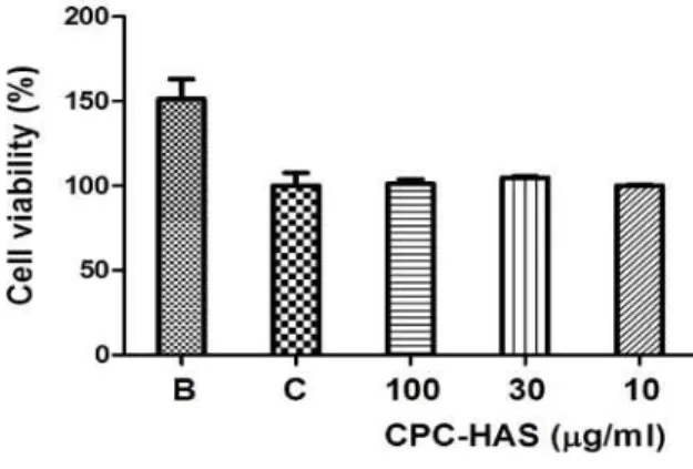
3. Cytotoxicity
The cell viability was recalculated into 100% of control group. The cell viabilities of CPC-HAS 100
㎍/㎖ treated, CPC-HAS 30㎍/㎖ treated and CPC-
Fig. 3. Cell viability of CPC-HAS on HS 68 human fibroblasts
B : blank, distilled water treated group without UVB irradiation.
C : control, distilled water treated group with UVB irradiation.
CPC-HAS :Cervi Pantotrichum Cornu herbal acupuncture solution treated group.
Data are expressed as the mean±SEM of three experi- ments.
HAS 10㎍/㎖ treated are 101.2±2.5%, 104.7± 0.8%, and 100.0±0.8%, respectively. CPC-HAS showed no cytotoxicity up to the effective concentration for anti-wrinkle activity(less than 100㎍/㎖)(Fig. 3).
4. Assay of collagen type I synthesis
The amounts of type I collagen synthesis of CPC-HAS were recalculated into 100% of control group(Fig. 4). CPC-HAS significantly(
p
<0.05) increased the expression of type I collagen at a concentration of 100㎍/㎖(144.4±16.6). CPC-HAS 30㎍/㎖(115.6±7.6) and 10㎍/㎖(126.6±15.1) treated group showed the increase of type I collagen synthesis, but there was no significance.
Fig. 4. Effect of CPC-HAS on collagen type Ⅰ synthesis in human firboblast cells
B : blank, distilled water treated group without UVB irradiation.
C : control, distilled water treated group with UVB irradiation.
CPC-HAS :Cervi Pantotrichum Cornu herbal acupuncture solution treated group.
Data are expressed as the mean±SEM of three experi- ments.
* : significantly different from the control,p<0.05.
Ⅳ. Discussion and Conclusion
The skin aging is one of the most obvious evi- dence of aging. The skin is increasingly exposed to ambient UV-irradiation thus increasing risks for photooxidative damage with long-term detrimental
effects like photoaging, characterized by wrinkles, loss of skin tone and resilience. Photoaged skin displays alterations in the cellular component and extracellular matrix with accumulation of disorga- nized elastin and its microfibrillar component fibrilin in the deep dermis and a severe loss of interstitial collagens, the major structural proteins of the dermal connective tissue. The unifying pathogenic agents for these changes are UV-generated ROS which de- plete and damage non-enzymatic and enzymatic antioxidant defense systems of the skin. As well as causing permanent genetic changes, ROS activate cytoplasmic signal transduction pathways in resident fibroblasts that are related to growth, differenti- ation, senescence and connective tissue degradation15). Elastase is an enzyme from the class of pro- teases or peptidases, that break down proteins. It, specifically, breaks down elastin, an elastic fiber that, together with collagen, determines the mech- anical properties of connective tissue16). Actually, elastase is the only enzyme that is capable of degrading elastin, an insoluble elastic fibrous pro- tein in animal connective tissues. It is capable of hydrolyzing nearly all proteins, including supporting and structural proteins of the connective tissue such as collagen and elastin17). Elastin is the main com- ponent of the elastic fibers of the connective tissue and tendons. The elastic fibers in the skin, together with the collagenous fibers, form a network under the epidermis18). Elastase also plays a critical role in inflammatory processes19). The enzyme has drawn much attention, primarily because of its reactivity and non-specificity. It is able to attack all major connective tissue matrix proteins, including elastin, collagen, proteoglycans, and keratins. Since this el- astic fiber is easily decomposed by elastase secretion and activation caused by exposure to UV light or ROS, an approach that inhibits the elastase activity could also be applied as a useful method to protect against skin aging19).
Cervi Pantotrichum Cornu(Nokyong),
processed young antlers of Cervus nippon Temminck, Cervus elaphus Linne and Cervus canadensis Erxleben10), considered as a nobel ingredient in Korean medicine.It is one of the major restoratives which fortifies the primal yang, generates essence and augments the bonemarrow. Furthermore, it tonifies the kidneys, strengthens the sinews and bones and nourishes the qi and blood. Clinically, it is used to treat fatigues, impotences, cold extremities, sorenesses and lacks of strengths in the lower backs and knees, mental retardations, insufficient growths and so on20).
These days, there are many researches about effects of
Cervi Pantotrichum Cornu
on various kinds of disease. Chen et al reported anti-lipid peroxidation ofCervi Pantotrichum Cornu
21), while Kim et al studied effects ofCervi Pantotrichum Cornu
on Osteoporosis22). Effects ofCervi Panto- trichum Cornu
and fermentedCervi Pantotrichum Cornu
on longitudinal bone growth were reported by Kim et al23), and Lee et al24), and hematopoietic action ofCervi Pantotrichum Cornu
was studied by Kim et al25). ForCervi Pantotrichum Cornu
Pha- rmacopuncture, protective and anti-arthritic effects26) and analgesic effects27) were reported. Also, it was studied in vivo thatCervi Pantotrichum Cornu
Pharmacopuncture improved adrenal cortical insuf- ficiency28), diabetes mellitus29), hypothyroidism30), growth and the intellectual development31). However, there was few study for anti-wrinkle effects so far.In order to investigate the potential of CPC-HAS as an active ingredient for wrinkle-care cosmetics, I measured its DPPH free radical scavenging activity, elastase inhibitory activity, and type I collagen synthesis in normal human fibroblasts HS 68.
The elastase acticvity of CPC-HAS was deter- mined according to the method described previously.
The elastase acticvity was recalculated into 100%
of control group. CPC-HAS showed the elastase inhibitory effect in dose dependent manner. CPC- HAS was found to have the lowest elastase activity at a concentration of 1000㎍/㎖(25.4±1.7%,
p
<0.001).CPC-HAS 100㎍/㎖ and 10㎍/㎖ treated groups showed 60.1±5.8%(
p
<0.01) and 89.6±6.1% of elastase acti- vity, respectively(Fig. 1).It has been reported that free radicals induced by
ultraviolet light or oxidative stress accelerate skin aging32). Assays of the free radical scavenging ca- pacity were carried out by the DPPH method. The free radical scavenging capacity of sample was measured at each concentration(10, 2 and 0.4㎎/㎖).
A dose dependent free radical scavenging capability was observed in sample treated groups. CPC-HAS 10㎎/㎖ treated groups had the highest scavenging capability of 63.9±6.2%, while CPC-HAS 2㎎/㎖ and CPC-HAS 0.4㎎/㎖ treated groups had 33.6±3.9%
and 8.3±1.3%, respectively(Fig. 2).
In order to evaluate the cytotoxicity of CPC- HAS, samples were prepared at various concentrations and used to treat human fibroblasts(HS 68). The results of this evaluation are shown in Fig. 3 at concentrations of 100, 30 and 10㎍/㎖. The cell via- bility was recalculated into 100% of control group.
The cell viabilities of CPC-HAS 100㎍/㎖ treated, CPC-HAS 30㎍/㎖ treated and CPC-HAS 10㎍/㎖
treated are 101.2±2.5%, 104.7±0.8%, and 100.0±0.8%, respectively. CPC-HAS showed no cytotoxicity up to the effective concentration for anti-wrinkle acti- vity(less than 100㎍/㎖).
To evaluate the amount of collagen type I sy- nthesis that occurred upon exposure to the sample, collagen type I was quantitatively detected by using the previously described procollagen type I C-peptide assay kit. Collagens are synthesized as precursor molecules, called procollagens. These molecules contain additional peptide sequences, usually referred to as
‘propeptides’, at both the amino-terminal end and the carboxy-terminal end. These propeptides are cle- aved from the collagen triple-helix molecule during its secretion, after which the triple-helix collagens are polymerized into extracellular collagen fibrils.
Thus, the amount of free propeptide stoichiomet- rically reflects the amount of collagen molecules synthesized12). The amounts of type I collagen synthesis of CPC-HAS were recalculated into 100%
of control group(Fig. 4). CPC-HAS significantly (
p
<0.05) increased the expression of type I collagen at a concentration of 100㎍/㎖(144.4±16.6). CPC-HAS 30㎍/㎖(115.6±7.6) and 10㎍/㎖(126.6±15.1) treated group showed the increase of type I collagen synthesis,but there was no significance.
The significant DPPH free radical scavenging activity was observed in CPC-HAS. Also, elastase activity was significantly inhibited by CPC-HAS.
Furthermore, type I procollagen production reduced by UVB irradiation was recovered by CPC-HAS in HS 68 cells.
In conclusion, CPC-HAS showed the anti-wrinkle effects in vitro. These results suggest that CPC- HAS may have potential as an anti-aging ingredient in cosmetic Pharmacopuncture. I think further studies will be needed to unravel exactly under the molecular mechanisms.
Ⅴ. References
1. Yaar M, Gilchrest BA. Cellular and molecular me- chanisms of cutaneous aging. J Dermatol Surg Oncol. 1990 ; 16(10) : 915-22.
2. Na MK, Min BS, An RB, Song KS, Seong YH, Bae K. Effects of Astilbe koreana on ultraviolet B(UVB)-induced inflammatory response in human keratinocytes. Biol Pharm Bull. 2004 ; 27 : 1301-4.
3. Cimino F, Ambra R, Canali R, Saija A, Virgili F. Effect of cyaniding-3-O-glucoside on UVB- induced response in human ketarinocytes. J Agr Food Chem. 2006 ; 54 : 4041-7.
4. Ho JN, Kim HK, Cho HY, Lim EJ. Effects of aucubin isolated from Eucommia ulmoides on UVB-induced oxidative stress in human kera- tinocytes HaCaT. Food Sci Biotechnol. 2009 ; 18(2) : 475-80.
5. Nishi J, Ogura R, Sugiyama M, Hidaka T, Kohno M. Involvement of active oxygen in lipid peroxide radical reaction of epidermal homo- genate following ultraviolet light exposure. J Invest Dermatol. 1991 ; 97 : 115-9.
6. Erden IM, Kahramant A, Kokent T. Beneficial effects of quercetin on oxidative stress induced by ultraviolet A Clin Exp Dermatol. 2001 ; 26 : 536-9.
7. Katiyar SK, Ahmad N, Mukhtar JH. Green tea
and skin. Arch Dermatol. 2000 ; 136 : 989-94.
8. Rodriguez J, Yanez J, Vicente V, Alcaraz M, Benavente-Garcia O, Castillo J, Lorente J, Loz- ano JA. Effects of several flavonoids on the growth of B16F10 and SK-MEL-1 melanoma cell lines: Relationship between structure and acti- vity. Melanoma Res. 2002 ; 12 : 99-107.
9. Jang MK. Complete compendium of Chinese me- dicine formulas for cosmetic dermatology. Beijing : China Chinese Medicine Publishing Company.
2001 : 34.
10. Kang BS, Kim IR, Kim HC, Kuk YB, Park YK, Seo BI, Seo YB, Song HJ, Shin MK, Lee YJ, Lee YC, Leem KH, Cho SI, Chung JK, Joo YS, and Choi HY. Boncho-Hak. Seoul : Young-Lim Press. 2004 : 588-9.
11. Kraunsoe JA, Claridge TDW, Lowe G. Inhibi- tion of human leukocyte and porcine pancreatic elastase by homologues of bovine pancreatic trypsin inhibitor. Biochemistry. 1996 ; 35 : 9090-6.
12. Kim YH, Chung CB, Kim JG, Ko KI, Park SH, Kim JH, Eom SY, Kim YS, Hwang YI, Kim KH. Anti-Wrinkle Activity of Ziyuglycoside I Isolated from a Sanguisorba officinalis Root Ex- tract and Its Application as a Cosmeceutical Ingredient. Biosci Biotechnol Biochem. 2008 ; 72(2) : 303-11.
13. Shimada K, Fugikawa K, Yahara K, Nakamura T. Antioxidative properties of xanthan on the autoxiation of soy gean oil in cyclodextrin emul- sion. J Agric Food Chem. 1992 ; 40 : 945-8.
14. Mosmann T. Rapid colorimetric assay for cellular growth and survival : application to proliferation and cytotoxicity assays. J Immunol Methods.
1983 ; 65 : 55-63.
15. Wlaschek M, Tantcheva-Poór I, Naderi L, Ma W, Schneider LA, Razi-Wolf Z, Schüller J, Scharffetter-Kochanek K. Solar UV irradiation and dermal photoaging. J Photochem Photobiol B. 2001 ; 63(1-3) : 41-51.
16. Horwitz M, Benson KF, Person RE, Aprikyan AG, Dale DC. Mutations in ELA2, encoding neutrophil elastase, define a 21-day biological clock in cyclic haematopoiesis. Nat Genet. 1999 ;
23(4) : 433-6.
17. Yang ZY, Guon GX, Lin ZF. Elastolytic activity from Flavobacterium odoratum. Microbial screening and cultivation, enzyme production and purification.
Process Biochem. 1994 ; 29 : 427-36.
18. Wiltried M, Klaus N, Lin ZF. Elastic fibre arrange- ment in the skin of the pig. Arch Derm Res.
1994 ; 270 : 390-401.
19. Wiedow O, Schroder JM, Christophers E. An elastase-specific inhibitor of human skin. J Biol Chem. 1990 ; 265 : 14791-5.
20. Dan B and Andrew G. Chinese Herbal Medicine ; Materia Medica. Seattle : Eastland Press Inc.
1992 : 32-4.
21. Chen X, Jin S, Di L, Liu X, Song H. Anti-lipid peroxidaton of the water extract from Cornu Cervi Pantotrichum. Zhong Yao Cai. 2003 ; 26(10) : 733-4.
22. Kim YS. Effects of Cervi Pantotrichum Cornu on diabetes mellitus induced by streptozotocin.
The Journal of East-West Medicines. 1991 ; 16(4) : 88-99.
23. Kim KT, Kim MG, Leem KH. Effects of Cervi Pantotrichum Cornu and Cervi Cornu on Long- itudinal Bone Growth in Adolescent Male Rats.
Korean Journal of Herbology. 2006 ; 21(1) : 63-9.
24. Lee SN, Son JB, Sohn JH, Kim WK, Lee SJ, Lee PJ, Leem KH. Effects of Herbal composi- tion and Fermented Cervi Pantotrichum Cornu on Longitudinal bone growth in adolescent male rats. Korean Journal of Herbology. 2009 ; 24(1) : 121-31.
25. Kim JS and Kim YT. A Study of hematopoietic action of pilose Antler in Senescence Accele- rated Mice. The Korean Society of Pharma- cognosy. 1996 ; 27(4) : 371-7.
26. Kim KS, Choi YH, Kim KH, Lee YC, Kim CH,
Moon SH, Kang SG, Park YG. Protective and anti-arthritic effects of deer antler aqua-acupuncture (DAA), inhibiting dihydroorotate dehydrogenase, on phosphate ions-mediated chondrocyte apoptosis and rat collagen-induced arthritis. Int Immuno- pharmacol. 2004 ; 4(7) : 963-73.
27. Kim YJ, Kang SG, Park DS. Studies on the Analgesic Effect of Aqua-acupuncture with Cervus elaphus Extract Solution in Mice. The Journal of Korean Acupuncture & Moxibustion Society. 1987 ; 4(1) : 63-74.
28. Lee YH and Kim KR. A Study of the Effect of Cervus elaphus Aqua-acupuncture on the Adrenal Cortical Insufficiency in Rats. The Journal of Korean Acupuncture & Moxibustion Society.
1987 ; 4(1) ; 49-62.
29. Park EJ, Shin JC, Na GH, Lee DH, Han SG, Yoon YC, Chae YS, Cho MR. Study on clinical effects of Cervus elaphus Herbal-acupuncture on Osteoarthritis in Knee joint. The Journal of Korean Acupuncture & Moxibustion Society.
2004 ; 21(2) : 275-86.
30. An CB, Song CH, Yang HT, Yoon JH, Kim GS.
Study of effect of cervus nippon temminck aqua- acupuncture on the hypothyroidism induced by thioureain rats. The Journal of Korean Acupun- cture & Moxibustion Society. 1992 ; 9(1) 215- 27.
31. Han SW, Kim YT, Son YS, Jin SH, Sim IS, Lim SBN, Lee HI. The Effects of Cervus elaphus on the Growth and the Intellectual Development of Animals. The Journal of Korean Acupuncture &
Moxibustion Society. 2001 ; 18(5) : 122-34.
32. Jurkiewicz BA and Buettner GR. Ultraviolet light induced free radical formation in skin : an electron paramagnetic resonance study. Photo- chem. Photobiol. 1994 ; 59 : 1-4.