PREVENTIVE EFFECT OF ADHESIVE TAPE SUPPLEMENTED WITH NaF ON ENAMEL EROSION IN VITRO
Sang Ho Lee, Nan Young Lee, In Hwa Lee*
School of dentistry, *Department of environmental engineering, Chosun university
This study examined the effect of adhesive tape supplemented with sodium fluoride on the prevention of den- tal erosion in vitro.
Sound bovine tooth samples were selected and divided randomly into the following 4 groups according to the material treatments: group 1, APF gel; group 2, fluoride varnish; and groups 3 and 4, fluoride tape supple- mented with 5% NaF in either a methyl cellulose or poly vinyl acetate carrier, respectively. All specimens were submitted to alternate cycles of acid exposure in a cola beverage (pH 4.3) and artificial saliva for 6 × 5 min/day over a 5 day period. The micro-hardness was recorded each day and the lesion depth was measured after 5 days. The micro-hardness of the experimental sides of groups 2, 3 and 4 were significantly higher than that of their control sides and the experimental side of group 1 during the experimental period (p<0.05) except on the 5th day. The enamel surfaces of treatment groups 2, 3 and 4 showed significantly higher resistance to mineral loss in terms of the erosion depth (p<0.05) than group 1 and their control sides. There was no statistically sig- nificant difference among group 2, 3 and 4, indicating that the fluoride varnish and tapes produce similar re- sults.
Fluoride adhesive tapes are effective in reducing the progression of erosion and can be recommended for young patients who are more susceptible to dental erosion.
Key words:Fluoride adhesive tape, Fluoride varnish, Dental erosion, Micro-hardness value, Erosion depth Abstract
Ⅰ. Introduction
Dental erosion has attracted more attention from the dental community. Despite the publication of a few pop- ulation-based surveys, erosive defects are not rare. In 1991, Lussi, et al1)reported that 29.9% of young adults in the Swiss population (26-30-year-olds) had at least one severely eroded occlusal surface. In 2000, according to the National Diet and Nutrition Survey in the UK, 58% of 4-6-year-olds and 42% of 11-14-year-olds were affected by dental erosion2). In Saudi Arabia, the erosion problem was reported to be 34% and 26% of children and adolescents, respectively3), and an overall 47% were
reported to be affected in Ireland4). The frequency of ero- sion lesions in the population appears to be increasing and should create more concern.
The etiology of dental erosion involves frequent contact between acids and the tooth surface. These frequent processes result in a loss of dental hard tissue that is etched away chemically from the tooth surface without bacterial involvement5,6). It has been suggested that acidic soft drinks and beverages is a major cause consid- ering the dramatically increased consumption of these products in children and adolescents.
The beneficial effect of fluoride in the prevention of dental caries is well known. Similar to its anti-cariogenic
교신저자 : 이 상 호
광주광역시 동구 서석동 375번지 / 조선대학교 치과대학 소아치과학교실 / 062-220-3865 / [email protected] 원고접수일: 2009년 09월 29일 / 원고최종수정일: 2010년 01월 09일 / 원고채택일: 2010년 01월 18일
properties, the application of fluoride has been suggested as a treatment option in preventing erosion. However, the role of fluoride in the prevention of dental erosion is still controversial7-11). Some in vitro studies12-17)reported a limited erosion-inhibiting effect from topical fluoride treatment. These studies examined agents that have been selected over the years for caries prevention, such as fluoride varnish, fluoride gel and oral toothpaste.
Considerable effort has been made to identify the fluo- ride products that are more effective in preventing den- tal erosion13,17). Bio-adhesive polymer, which is used in drug delivery systems, would also be a suitable choice for fluoride. This material was assumed to be able to de- liver a high concentration of fluoride to the target in con- trolled release18), and promote the formation of a firmer and more poorly permeable remineralized surface.
Furthermore, it is colorless, odorless and has a better taste when applied. Therefore, it is easy for uncoopera- tive children to use. In this study, the fluoride tapes consisting of 5% sodium fluoride and a bio-adhesive polymer, such as a methyl cellulose carrier and poly vinyl alcohol carrier, were used. This study examined whether this experimental fluoride system could prevent the enamel erosion in a similar manner to that of other topical fluoride products.
Ⅱ. Materials and Methods 1. Sample preparation
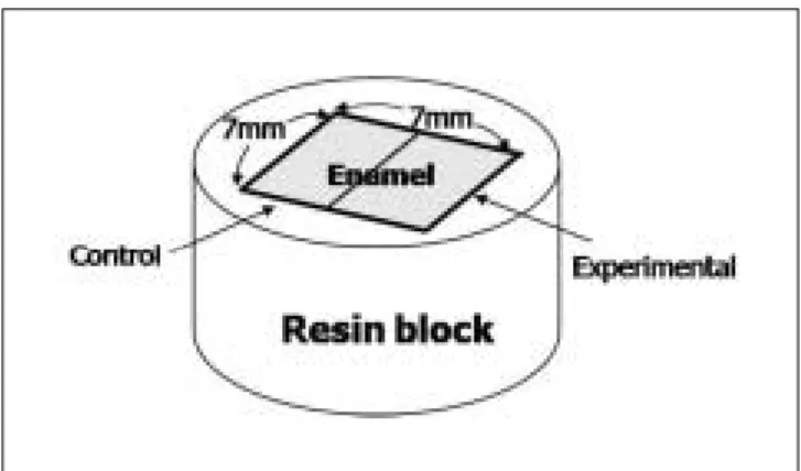
Sound bovine permanent incisors free of caries and hy- po-calcification were selected for this study. After extrac- tion, the teeth were mechanically cleaned and disinfect- ed in 70% alcohol. Crowns were sectioned from the roots and then vertically and horizontally sectioned with a di- amond disc to produce enamel specimens (7×7 mm) from each crown (Fig. 1). Totally, 97 specimens were produced.
All these specimens were embedded in acrylic resin in
moulds with the outer enamel surface exposed. The enamel surfaces of specimens were ground flat with a water-cooled grinding machine (Metpol-1, R&B Inc, Korea) using progressively finer grades of CC-400, 800, 1,200 Cw paper(silicon carbide waterproof abrasive pa- per).
Baseline surface micro-hardness analysis of the enam- el blocks was recorded using a micro-hardness tester(HMV-2, Shimadzu, Japan) with a Vickers dia- mond head under a 100-g load for 5 seconds. Three in- dentations spaced 100 μm for each other were made at the center of the enamel surface. Enamel blocks with baseline micro-hardness value between 230 and 300 VHN (Vickers’s Hardness Number) were selected for this study.
2. Test materials
The specimens were treated with 2 new product fluo- ride tapes in comparison with commercially available an- ti-cariogenic products: fluoride gels - 60 seconds taste� (1.23% APF, Pascal Company Inc., USA) and fluoride varnish - Cavity ShieldTM (5% sodium fluoride, Ominii Pharmaceuticals, USA) (Table 1).
Fig. 1. Schematic diagram of tooth section invested in resin block.
Table 1. Fluoride products used in this study
Group Product Major composition Manufacturer
1 60 seconds taste� 1.23% APF Pascal company Inc., USA
2 Cavity ShieldTM 5% Sodium fluoride Ominii Pharmaceuticals, USA
3 Fluoride tape M* 5% Sodium fluoride Experimental product
4 Fluoride tape P** 5% Sodium fluoride Experimental product
* : Fluoride tape supplemented with 5% NaF in methyl cellulose carrier
** : Fluoride tape supplemented with 5% NaF in poly vinyl alcohol carrier
3. Erosive agent and artificial saliva
The erosive soft drink Coca-Cola�was used as a dem- ineralizing solution. Five bottles of 1.5 L each were tak- en from the same package in supermarket. The pH of each was 2.43 at room temperature measured by pH meter (PMH 210, Radiometer analytical SAS, Cedex, France).
The artificial saliva used was similar to that described by McKnight-Hanes and Whitford19), but it was modified by the exclusion of sorbitol and Methyl-p-hydroxyben- zoate. It contained (in g/L): Sodium carboxymethyl cel- lulose sodium, 10.0; KCl, 0.625; CaCl2∙2H2O, 0.166;
MgCl2∙6H2O, 0.059; K2HPO4, 0.804; KH2PO4, 0.326.
The first component was used to simulate the protein and mucin contents of the natural saliva and increased the viscosity of this artificial saliva, while the other con- stituents provided the inorganic components at levels comparable with that of natural saliva. The pH was ad- justed to 7 using NaOH.
4. Experiment procedure
Every selected block was divided into 2 sides-control and experimental side by a shallow groove in the center of enamel specimens using 1/4 round diamond bur (Fig.
1). With respect to the treatment products, these sam- ples were randomly distributed into 4 groups with the experimental sides treated as following: (1) Group 1 (n
= 18) was applied with fluoride gel by cotton pellet for 4 minutes without rubbing motion and washed with artifi- cial saliva; (2) Group 2 (n = 18) was blasted and paint- ed with a thin layer of fluoride varnish and allowed to dry for 1 minutes; (3) Group 3 (n = 18) was treated
with fluoride tape made by methyl cellulose containing 5% NaF; and (4) Group 4 (n = 18) was treated with fluoride tape made by poly vinyl alcohol containing 5%
NaF. Fluoride tape was attached to enamel by wetting with artificial saliva and let it aside for 1 minute.
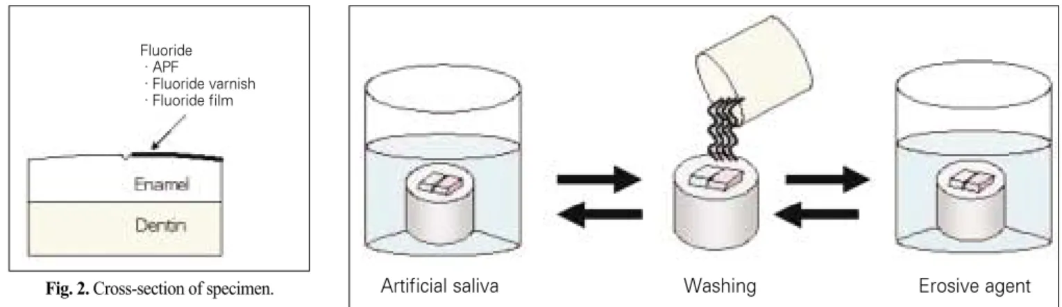
After treating, all specimens were stored for 24 hours in artificial saliva solutions which were changed every day during experiment. The following day, cycling be- tween artificial saliva and erosive agent challenge solu- tions began. The cycle comprised: (1) rinsed specimens in distilled water; (2) soaked in Coca-Cola� for 5 min- utes, pH 2.43, room temperature; (3) washed in dis- tilled water and (4) immersed in artificial saliva for 10 min to stimulate remineralizing (Fig. 3). The beverage was changed every 3 cycles. The containers were put on the lid and were kept under continuous agitation during experiment to simulate the movement of saliva under in- fluence of tongue, lip, buccal. Teeth were cycled between artificial saliva and erosive agent exposure 6 times per day in consecutive 5 days. After the initial application of the fluoride, no further applications were used during experiment.
5. Measurement of micro-hardness
Micro-hardness tester (HMV-2, Shimadzu Co., Japan) with a Vickers diamond head under a load of 100 g force in 5 seconds was used to determine possible changes in surface micro-hardness during experiment. The mea- surements were made initially, after erosion exposures of every experimental day. Three well-formed indentations were measured to calculate the mean Vickers hardness number for each test and control surface. In the cases where remnants of varnish were observed in the optical Artificial saliva Washing Erosive agent Fig. 2. Cross-section of specimen.
Fig. 3. Experiment processing.
Fluoride
∙APF
∙Fluoride varnish
∙Fluoride film
microscope mounted on the hardness tester, care was taken not to make the indentations on such areas.
6. Measurement of erosion depth
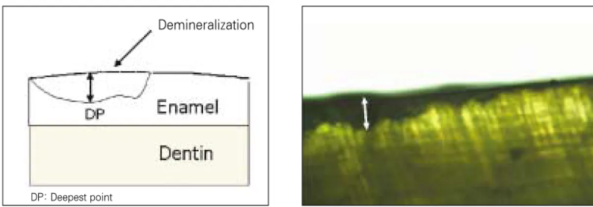
At the end of 5 days test period, specimens were sec- tioned to the thickness of 0.5 mm by low speed diamond blade (Model 650, South Bay Technology, USA). Every section included the experimental and control side. Then further reduction to the thickness of about 100 μm was accomplished by grinding machine (Metpol-1, R&B Inc, Korea) using progressively finer grades of CC- 800, 1,200 silicon carbide abrasive paper. All these proce- dures were cooled properly by water.
Sections were washed with deionized water and ori- ented longitudinally on glass slides. The sections were imbibed with water (refractive index 1.33) for evaluation under polarized light microscopy (Axioskop 40A, Zeiss, Germany). The lesion images were captured and exam- ined using a Axioskop 2 plus program (Express, Media- cybernetics Co., USA). Lesion depth for each section (in μm) was taken as the average of three respective mea- surements from the imagined line connecting two intact points on the surface to the bottom of the lesion.
7. Statistical analysis
The differences in the average erosion depth and Vickers hardness values observed on each specimen in the four groups were analyzed using analysis of variance (ANOVA). The comparisons between individual groups were performed using a Turkey’s post-hoc test. A p val- ue less than 0.05 was considered significant.
Ⅲ. Results 1. micro-hardness value
The mean surface micro-hardness and standard devia- tion values at baseline (before fluoride treatment), and after every acid agent exposure day for each experimen- tal group were summarized in Table 2. Statistical com- parisons between control and its experimental groups and among the experimental groups in connection with the processing duration were displayed in Table 3.
With respect to its control side, group 2 was found having noticeable erosion-inhibiting ability even after the first experiment day (p<0.05). Group 2, 3 and 4 re- vealed their considerable effect from 2ndday and continu- ously until 4thday (p<0.05). On the day 5, there were no significant differences between experimental and control sides in all groups (p>0.05). Group 1 had similar values to its control side in all 5 days (p>0.05).
Observing the change in micro-hardness value among groups(Fig. 5), group 2, 3, 4 remained at the much higher hardness numbers compared with group 1 (p<0.05) through out the first 4 days. Thereafter, on day 5, their hardness values were still higher than group 1 but no significant differences were observed (p>0.05).
Group 2, in comparison with group 3 and 4, showed its stronger effect in first 3 days (p<0.05). Group 3 and 4 had similar hardness value during the experiment (p>0.05). On the day 5, the hardness values were as fol- lowing: group 3 > group 4 > group 2 > group 1, difference without statistical significance (p>0.05).
Demineralization
DP: Deepest point
Fig. 4. Measurement of lesion depth in cross-sectioned specimen.
2. Erosion depth value
The erosion depth and standard deviation values were displayed in table 4.
Treatment group 2, 3 and 4 with fluoride varnish, fluo- ride tape increased the resistance of enamel surfaces from mineral loss significantly (p<0.05) compared to group 1 and their control sides. There were no statistically signifi- cant differences between group 2, 3 and 4 (p>0.05). The lesion depth value in experimental side of group 1 was
not much lesser than its control side (p>0.05)(Fig. 6).
Ⅳ. Discussion
Surface hardness measurements have been used in many studies to obtain information on enamel softening related to acid induced lesion12,20,21). It is known as a use- ful method in assessing the initial stages of erosion when enamel softening starts. This study used hardness tester to observe the softening process during 5 days experi- ment. At the conclusion of day 5, erosion depth values were measured to get knowledge of tooth material loss after immersion periods.
Based on the experiment results, it is clearly revealed that topical fluoride treatment with concentrated sodium fluoride varnish and fluoride tapes inhibited enamel sur- face softening during the acid exposure and, thus, was Fig. 5. Change of micro-hardness number (VHN) according to the time. Fig. 6. Comparison of erosion depth (μm) among groups.
* indicates the significant differences (p<0.05) among experimental sides of groups.
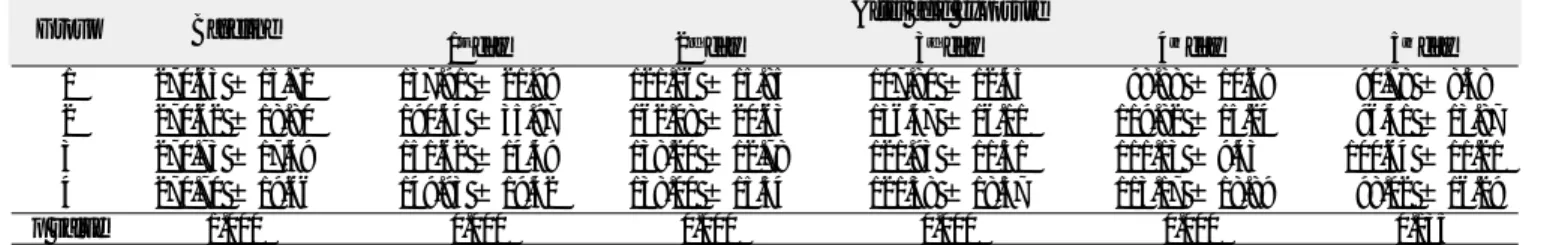
Table 2. Vickers hardness number (VHN) of each group according to the processing duration(mean ± SD)
Group Baseline After acid exposure
1stday 2ndday 3rdday 4thday 5thday
1 270.63 ± 15.71 137.91 ± 21.99 121.16 ± 15.85 107.80 ± 12.45 98.88 ± 10.68 90.78 ± 8.38
2 270.62 ± 18.80 190.44 ± 35.97 162.08 ± 20.63 136.47 ± 16.11 119.82 ± 15.24 96.41 ± 13.87 3 270.73 ± 17.49 151.62 ± 14.49 138.20 ± 12.78 121.93 ± 11.51 111.13 ± 9.43 100.64 ± 11.21 4 270.70 ± 19.66 149.93 ± 19.42 138.00 ± 15.54 121.38 ± 18.57 113.17 ± 18.89 98.02 ± 16.29
p value 1.000 0.000 0.000 0.000 0.000 0.135
Table 3. Comparison of Vickers hardness number (VHN) between groups on day 0/1/2/3/4/5
Control Group 1 Group 2 Group 3 Group 4
Control -/-/-/-/-/- -/+/+/+/+/- -/-/+/+/+/- -/-/+/+/+/-
Group 1 -/+/+/+/+/- -/+/+/+/+/- -/+/+/+/+/-
Group 2 -/+/+/+/-/- -/+/+/+/-/-
Group 3 -/-/-/-/-/-
Group 4
- : not statistically significant (p>0.05) + : statistically significant (p<0.05) Day 0 : before fluoride treatment
1st, 2nd, 3rd, 4th, 5thday : the acid exposure day
Table 4. Comparison of erosion depth (μm) between control and experi- mental side of samples in each group
Group N Control side Experimental side p-value
1 16 2.20 ± 0.63 1.82 ± 0.52 0.068
2 14 2.33 ± 0.99 1.05 ± 0.42 0.000
3 16 1.86 ± 0.74 0.99 ± 0.49 0.000
4 17 1.91 ± 1.10 0.89 ± 0.26 0.001
Micro-hardness
Baseline 1 2 3 4 5 Day
VHN
300 250 200 150 100 50 0
APF gel Fluoride varnish Fluoride tape M Fluoride tape P
Erosion depth
Control Experimental
1 2 3 4 Day 2.5
2 1.5 1 0.5 0 Depth(μm)
able to protect enamel from erosion in initial stages of process. Therefore, these products can be recommended clinically as prophylaxis for erosion patients.
In mean terms for sample side pre-treated with acidu- lated gel at each time point, the Vickers hardness num- ber was always lesser than its control side, but the dif- ferences were small (p>0.05). Besides, its lesion depth results displayed a lesser depth of erosion lesion com- pared with its control side (p>0.05). It can conclude that the acidulated gel can reduce erosion but the actual clin- ical benefit appears low. Huges et al22)also reported the low benefit of acidulated gel.
Our results were in agreement with conclusions drawn by Sorvari et al23) that topical fluoride varnish treatment was able to protect enamel during the initial stages of the erosion process. The study by Kitchens and Owens24), however, did not demonstrate a significant protective ef- fect of fluoride treatment varnish on enamel surfaces. This was because the acid challenge was so severe- up to 350 hours (14 days) which was comparable to 14 years of nor- mal beverage consumption and the sample size was small.
Topical fluoride treatment would maintain its effect for certain duration. Immersion of the fluoride pre-treated enamel surface into acidic beverage in 14 consecutive days could hardly manifest its effect24). Even in caries prevention, generally, fluoride application was recom- mended 2 intervals per year. Therefore, anticipating the effect of fluoride in such long term is unreasonable.
The study of Sorvari et al23)mentioned the time of ef- fect as the enamel surfaces were exposed to cola bever- age totally for 15 minutes. Until the end of the experi- ment (after 15 min), fluoride varnish and fluoride solu- tion still protected the enamel surface from softening ef- fectively. Even after shorter application of fluoride gel or toothpaste, a significant rehardening effect of fluoride has been shown25-30). However, how long the effect does exist still a question. In the present study, fluoride var- nish and fluoride tapes were shown to be able to limit erosion until the forth day of experiment equivalent to 120 min (2 hours) exposure to cola beverage. According to von Fraunhofer and Roger31), average daily consump- tion of soft drink in one person in USA was 24 ounces (two 12 ounce cans) and a residence time in the mouth was about 20 seconds (before salivary clearance). This resulted in an annual exposure of enamel to soft drinks of approximately 25 hours per year. The period time of 120 min was comparable to 1 month of normal soft drink consumption in USA. The fluoride varnish and our prod-
ucts, consequently, were able to inhibit the enamel sur- face from loss of mineral effectively in 1 month if the av- erage daily consumption of soft drink in one person was 24 ounces (or 710 ml).
It must notice that this experiment was done on bovine teeth, which had morphologic difference to that of human such as higher porosity32,33). This would result in higher rates of lesion formation. If this issue was taken into account when interpreting the results, the actual protective time would be lengthened.
In this experiment, acidic challenges were performed 6 times × 5 min per day. This setting was not excessive compared to normal daily routine. Meurman et al34) re- ported that the pH of the oral fluids usually returns to neutral conditions 1-3 min after one single sip of an acidic beverage. In some cases the pH of oral fluid recov- ers only to values around 4.0 within 10 min35). Millward et al36)showed that after dietary acid intake, pH values were slowly recovered to pH 5.5 within 2 min at the in- cisor site and in 4-5 min at the molar site. In addition, if a person could intake 100 ml in his one sip, 6 times of consuming 710 ml beverage would be easily to reach.
Together with the intake time of acidic foods, an experi- mental immersion time of 5 min 6 times daily seems to be close to normal condition.
The role of fluoride in preventing erosions still remains unclear. Under the erosion condition, the acidic chal- lenge is much stronger so that enamel is lost layer by layer from the surface. This irreversible process differs fundamentally from that of a subsurface demineraliza- tion in dental caries37,38). For that reason, fluoride appli- cation is assumed to perform for different purpose.
According to Imfeld38), the effect of fluoride is primarily to harden the enamel surface and thus, reduce its solubility under severe acidic condition. Low fluoride concentra- tions serve better for the purpose of remineralizing the deep subsurface in caries as it does not block enamel pores and facilitates the ion exchange activity to the deeper layer. Contrarily, in case of erosion, low fluoride concentration can not show its effect. It is believed that application of high fluoride concentration promotes the formation of a more poorly permeable remineralized sur- face but only a thin layer is involved39). In the presence of high fluoride concentration, brushite was formed in- stead of calcium fluoride (CaF2)40).
In present experiment, Cavity Shield�varnish and flu- oride tapes contained higher concentration of fluoride than APF gel and displayed better protective effect. On
the first 3 days, Cavity Shield� varnish performed its role best but then, its effect decreased to a similar level of fluoride tapes. Sorvari et al23) suggested that fluoride varnish may have double action. The varnish layer itself was observed very sticky and very hard to remove from enamel40,41), and may act as a barrier against erosion.
This explained the markedly well performance of varnish during first stages of experiment but then rapidly de- creased in the day 4 and 5 due to the resin layer was completely dissolved.
Bio-adhesive polymer has been traditionally used in drug delivery systems. This material was reported to be able to deliver specific release rates of drug to its target18). We suggested applying this special characteris- tic in dentistry. Fluoride tapes containing 5% sodium fluoride in methyl cellulose and poly vinyl acetate carrier were produced for this purpose. High concentration of fluoride was assumed to be delivered to the enamel sur- face in controlled release. We also expected more fluoride products would be permanently bound to dental enamel due to its controlled release rate ability favorable for deeper penetration of fluoride product. The results showed that these products performed rather well if tak- en into account for the whole experimental process. The last result numbers were better for these products even though no significant difference were found. In addition, these products are colorless, odorless and give better taste when applied. These advantages make them easier to indicate for children who are not cooperative.
In conclusion, the present study has shown that fluo- ride varnish and our fluoride adhesive tapes are effective in reducing erosion progression on enamel surface.
Therefore, these products can be recommended clinically as prophylaxis for erosion patients. Further research is needed to evaluate the effective application and appropri- ate time intervals that can gain best benefit to patients.
Ⅴ. Conclusion
The effect of adhesive tape supplemented with sodium fluoride products in preventing dental erosion was inves- tigated in comparison with other fluoride products such as APF gel(60 seconds tasteTM) and fluoride varnish (Cavity ShieldTM). Following results were obtained:
1. Micro-hardness values of experimental sides of group 2, 3 and 4 were significantly higher than those of their control sides and experimental side of group 1 over experimental period (p<0.05) except for the 5th
day. Group 3 and 4 had similar hardness value during the experimental period (p>0.05). Group 1 showed not much difference to its control side (p>0.05).
2. With regard to erosion depth lesion, group 2, 3 and 4 increased the resistance of enamel surfaces from min- eral loss significantly (p<0.05) compared to group 1 and their control sides. There were no statistically sig- nificant difference between group 2, 3 and 4 (p>0.05).
References
1. Lussi A, Schaffner M, Hotz P, et al. : Dental erosion in a population of Swiss adults. Community Dent Oral Epidemiol, 19:286-290, 1991.
2. Dugmore CR, Rock WP : The prevalence of tooth erosion in 12-year-old children. Br Dent J, 196:279- 282, 2004.
3. Al-Majed I, Maquire A, Murray JJ : Risk factor for dental erosion in %-6 year old and 12-14 year old boys in Saudi Arabia. Community Dent Oral Epidemiol, 30:38-46, 2002.
4. Harding MA, Whelton H, O’Mullane DM, et al. : Dental erosion in 5-year-old Irish school children and associated factors: a pilot study. Community Dent Health, 20:165-170, 2003.
5. Ja¨rvinen VK, Ryto¨maa II, Heinonen OP : Risk fac- tors in dental erosion. J Dent Res, 70:942-947, 1991.
6. Magalhães AC, Wiegand A, Rios D, et al.: Insights into preventive measures for dental erosion. J Appl Oral Sci, 17:75-86, 2009.
7. Imfeld T : Dental erosion. Definition, classification and links. Eur J Oral Sci, 104:151-155, 1996.
8. Addy M, Absi EG, Adams D : Dentin hypersensitivi- ty. The effects in vitro of acids and dietary sub- stances on root planed and burred dentine. J Clin Periodontol, 14:274-279, 1987.
9. Grobler SR, Senekal PJ, Laubscher JA : In vitro demineralization of enamel by orange juice, apple juice, Pepsi Cola and Diet Pepsi Cola. Clin Prev Dent, 12:5-9, 1990.
10. Meurman JH, Ha¨rko¨nen M, Na¨veri H, et al. : Experimental sports drinks with minimal dental ero- sion effect. Scand J Dent Res, 98:120-128, 1990.
11. Owens BM, Kitchens M: The erosive potential of soft drinks on enamel surface substrate: an in vitro scanning electron microscopy investigation. J Contemp Dent Pract, 8:11-20, 2007.
12. Wongkhantee S, Patanapiradej V, Maneenut C, et
al. : Effect of acidic food and drinks on surface hard- ness of enamel, dentine, and tooth-coloured filling materials. J Dent, 34:214-220, 2006.
13. Ehlen LA, Marshall TA, Qian F, et al. : Acidic bev- erages increase the risk of in vitro tooth erosion.
Nutr Res, 28:299-303, 2008.
14. Edwards M, Creanor SL, Foye RH, et al: Buffering capacities of soft dinks: the potential influence on dental erosion. J Oral Rehabil, 26:923-927, 1999.
15. 장기택 : 수종 음료수의 법랑질과 상아질침식에 관한 연구.
대한소아치과학회지, 24:719-726, 1997.
16. 김정욱 : 산성 음료수에 의한 법랑질 침식과 구강 내 재경화 에 관한 연구. 대한소아치과학회지, 25:312-322, 1998.
17. Ireland AJ, McGuiness N, Sherriff M : An investiga- tion into the ability of soft drinks to adhere to enam- el. Caries Res, 29:470-476, 1995.
18. Vasir JK, Tambwekar K, Garg S : Bioadhesive microspheres as a controlled drug delivery system.
Int J Pharm, 255:13-32, 2003.
19. McKnight-Hanes C, Whitford GM : Fluoride release from three glass ionomer materials and the effects of varnishing with or without finishing. Caries Res, 26:345-350, 1992.
20. Jensen ME, Donly K, Wefel JS : Assessment of the effect of selected snack foods on the remineraliza- tion/demineralization of enamel and dentin. J Contemp Dent Pract, 1:1-17, 2000.
21. Larsen MJ, Richards A : Fluoride is unable to reduce dental erosion from soft drinks. Caries Res, 36 : 75-80, 2002.
22. Hughes JA, West NX, Addy M : The protective effect of fluoride treatments against enamel erosion in vitro. J Oral Rehabil, 31,357-363, 2004.
23. Sorvari R, Meurman JH, Alakuijada P, et al. : Effect of fluoride varnish and solution on enamel erosion in vitro. Caries Res, 28:227-232, 1994.
24. Kitchens M, Owens BM : Effect of carbonated bever- ages, coffee, sport and high energy drinks, and bottled water on the in vitro erosion characteristics of dental Enamel. J Clin Pediatr Dent, 31:153-159, 2007.
25. Graubart J, Gedalia I, Pisanti S : Effects of Fluoride Pretreatment in vitro on Human Teeth Exposed to Citrus Juice. J Dent Res, 51:1677, 1972.
26. Larsen MJ: Prevention by means of fluoride of enamel erosion as caused by soft drinks and orange juice. Caries Res, 35:229-234, 2001.
27. Van Rijkom H, Ruben J, Vieira A, et al. : Erosion- inhibiting effect of sodium fluoride and titanium
tetrafluoride treatment in vitro. Eur J Oral Sci 111:253-257, 2003.
28. Muñoz CA, Feller R, Haglund A, et al. : Streng- thening of tooth enamel by a remineralizing tooth- paste after exposure to an acidic soft drink. J Clin Dent, 10:17-21, 1999.
29. Vieira A, Ruben JL, Huysmans MC : Effect of titanium tetrafluoride, amine fluoride and fluoride varnish on enamel erosion in vitro. Caries Res, 39:371-379, 2005.
30. Seppa¨L : Effects of a sodium fluoride solution and a varnish with different fluoride concentrations on enamel remineralization in vitro. Scand J Dent Res, 96:304-309, 1988.
31. von Fraunhofer JA, Rogers MM : Dissolution of dental enamel in soft drinks. Gen Dent, 52:308-312, 2004.
32. Featherstone JD, Mellberg JR : Relative rates of progress of artificial carious lesions in bovine, ovine and human enamel. Cares Res, 15:109-114, 1981.
33. Wegehaupt F, Gries D, Wiegand A, et al. : Is bovine dentine an appropriate substitute for human dentine in erosion/abrasion tests? J Oral Rehabil, 35:390- 394, 2008.
34. Meurman JH, Ryto¨maa I, Kari K, et al. : Salivary pH and glucose after consuming various beverages, including sugar-containing drinks. Caries Res, 21:353-359, 1987.
35. Ganss C, Klimek J, Schaffer U, et al. : Effectiveness of two fluoridation measures on erosion progression in human enamel and dentine in vitro. Caries Res, 35:325-330, 2001.
36. Millward A, Shaw L, Harrington E, et al. : Continuous monitoring of salivary flow rate and pH at the surface of the dentition following consumption of acidic beverages. Caries Res, 31:44-49, 1997.
37. ten Cate JM, Imfeld T: Dental erosion, summary.
Eur J Oral Sci, 104:241-244, 1996.
38. Imfeld T : Prevention of progression of dental ero- sion by professional and individual prophylactic measures. Eur J Oral Sci, 104:215-222, 1996.
39. Wei Shy, wefel JS : In vitro interactions between the surfaces of enamel white spots and calcifying solutions. J Dent Res, 55:135-141, 1976.
40. Nelson DG, Jongebloed WL, Arends J : Morphology of enamel surfaces treated with topical fluoride agents: SEM considerations. J Dent Res, 62:1201- 1208, 1983.
41. Petersson LG : Fluoride mouthrinse and fluoride varnishes. Caries Res, 27:35-42, 1993.
불소함유 접착테잎의 법랑질 침식 예방효과
이상호∙이난영∙이인화*
조선대학교 치의학전문대학원, *공과대학 환경공학과
본 연구는 불소함유 접착테잎의 법랑질 침식 예방효과를 평가하기 위해건강한 소의 치아를 절단하여 아크릴 주형에 매몰 하고 정중선에 홈을 형성하여 대조면과 불소 제제를 도포할 실혐면으로 나누었다. 시편을 무작위로 18개씩 4군으로 나누었 다. 1군은 APF gel를 도포하고, 2군은 불소 바니쉬를 도포하였으며, 3군과 4군은 5% NaF를 각각 첨가한 methyl cellu- lose와 poly vinyl alcohol 테잎을 부착하였다. 시편을 콜라에 5분, 증류수에 10분씩 6회 번갈아 처리하며 5일 동안 반복하 였다. 시간 경과에 따라 표면미세경도를 측정하고 5일후 각 시편을 절단하여 침식병소의 깊이를 측정하여 다음과 같은 결과 를 얻었다.
1. 표면미세경도 값은 5일째만 제외하고 2군, 3군, 4군의 실험면이 각각의 대조면에 비해 컸다 (p<0.05). 2군, 3군, 4군 의 실험면 역시 5일째만 제외하고 1군의 실험면에 비해 표면미세경도가 컸다 (p<0.05). 3군과 4군 사이에는 표면미세 경도의 유의한 차이가 없었다(p>0.05). 1군의 실험면과 대조면은 표면미세경도의 유의차가 없었다(p>0.05).
2. 침식병소의 깊이는 2군, 3군, 4군의 실험면이 각각의 대조면에 비해 컸다(p<0.05). 2군, 3군, 4군의 실험면은 1군의 실험면에 비해 침식병소의 깊이가 얕았다 (p<0.05). 2군, 3군, 4군의 실험면 사이의 침식병소의 깊이는 유의차가 없었 다(p>0.05).
상기 결과로 보아 새로 개발된 불소함유 접착테잎은 기존의 불소 바니쉬와 같은 법랑질 침식 예방효과가 분명하게 나타나, 법랑질 침식에 감수성이 높은 어린이 환자에게 임상적인 예방법으로서 활용할 수 있는 가능성이 높다고 사료된다.
Key words: 불소접착테잎, 불소바니쉬, 치아침식, 표면미세경도, 침식깊이 국문초록