443
Copyright © 2020 The Korean Society of Fisheries and Aquatic Science pISSN:0374-8111, eISSN:2287-8815
서 론
우리나라의수산양식업은경제적효율성을위해고밀도양식 을추구해왔으며
,
이로인해질병의발생증가와빠른전파의 문제가발생하였다.
양식현장에서는이러한문제점들을해결 하기위하여항생제를세균성질병의치료에지속적으로이용 해왔으며,
일부항생제는감염성질병의예방이나치료목적 외에도사료와혼합하여어류의성장을촉진하기위해사용되 어왔다(Son et al., 2011).
또한양식장에서는세균성질병을 치료하기위하여현장종사자의임상적경험을토대로항생제 를선택한후무분별하게사용하는경우가빈번하다.
이러한비 전문가적인치료방법으로유발되는항생제오남용은세균의 항생제내성을증가시켜치료가어려워지고,
약물의잔류로인 한양식생물의안전성및수산식품의공중위생학적안전성에도 심각한문제를일으킨다(JSA, 1994; Noga, 1996; Woodward,
1996).
그러므로 양식생물에서질병이발생하게되면원인이되는세균을분리한후
,
동정된세균의항생제감수성검사를 실시하여적절한항생제를선택하고용법과용량에맞도록항 생제를사용하여야한다.
최근까지항생제감수성검사를위하 여disk diffusion susceptibility test
와broth microdilution test
등의방법이주로사용되고있지만,
실험단계가복잡하고시 간이많이소요된다는단점이있다.
지금의항생제감수성검 사방법은질병이발생하고대량폐사로인하여많은경제적피 해가발생한후결과가확인되는경우가많아신속하게적절한 항생제를처리해야하는상황에비효율적인측면을가지고있 다.
따라서양식생물의질병원인을분석함과동시에보다신속 하고효율적인방법을이용하여항생제감수성과내성을구분 할수있는방법이필요한실정이다.
다양한항생제가농도별 로coating
되어있는MIC (minimal inhibitory concentration)
panel
은간편하고신속하게항생제감수성검사를실행할수있도록상품화되어판매되고있지만
,
현재까지는인체및가축에 서유래된세균의조건에맞추어연구되고사용되어왔다.
그러어류 병원성 세균의 MIC 결정을 위한 MIC Panel의 최적화 배양 조건 확립
김예지·전려진·강미래·이다원·우수지
1·김명석
1·정준범*
제주대학교 해양생명과학과, 1국립수산과학원 병리연구과
Establishing of Optimal Culture Conditions for MIC Panels for MIC Determination of Fish Bacterial Pathogens
Ye Ji Kim, Lyu Jin Jun, Mi Rae Kang, Da Won Lee, Soo Ji Woo1, Myoung Sug Kim1 and Joon Bum Jeong*
Department of Marine Life Sciences, Jeju National University, Jeju 63243, Korea
1Pathology Research Division, National Institute of Fisheries Science, Busan 46083, Korea
No established method can be used to select effective antibiotics in antibiotic susceptibility tests for fish bacterial pathogens quickly and accurately. Here, we established the optimal conditions for determining the minimal inhibitory concentration (MIC) of major fish bacterial pathogens ( Streptococcus spp., Edwardsiella tarda, Vibrio spp., Aeromo- nas spp., and Pseudomonas spp.) using the KRAQ1 and CAMPY2 panels. The MIC panel used 18 antibiotics of two types and we conducted experiments to establish the optimal culture medium and temperature for each species. The optimal conditions for incubating Streptococcus spp. were in cation-adjusted Mueller-Hinton broth with TES buf- fer (CAMHBT) at 28°C, using 5% lysed horse blood (LHB) as recommended by the Clinical Laboratory Standards Institute. For Vibrio spp., the optimal culture conditions were 28°C in CAMHBT supplemented with 1% NaCl. The optimal conditions for culturing E. tarda , Aeromonas spp., and Pseudomonas spp. were in CAMHBT at 28°C.
Keywords: Antibiotics susceptibility testing, KRAQ1 panel, CAMPY2 panel, Fish pathogen, MIC
*Corresponding author: Tel: +82. 64. 754. 3426 Fax: +82. 64. 756. 3493 E-mail address: [email protected]
This is an Open Access article distributed under the terms of the Creative Commons Attribution Non-Commercial Licens (http://creativecommons.org/licenses/by-nc/3.0/) which permits unrestricted non-commercial use, distribution, and reproduction in any medium, provided the original work is properly cited.
Received 10 March 2020; Revised 11 April 2020; Accepted 22 June 2020
저자 직위: 김예지(대학원생), 전려진(박사후연구원), 강미래(대학원생), 이다 원(대학원생), 우수지(연구사), 김명석(연구관), 정준범(교수)
https://doi.org/10.5657/KFAS.2020.0443
Korean J Fish Aquat Sci 53(3), 443-450, June 2020
나다양한환경조건을요구하는수산생물에서분리한세균은 배양조건이다르기때문에동일한방법을적용하기에는적합 하지않다
.
또한국외기관인clinical and laboratory standards institute (CLSI, 2020)
와european committee on antimicrobial susceptibility testing (EUCAST, 2020)
에서발간한인체및가 축에서분리한세균의항생제감수성검사표준방법에서권장 되는배양조건은35±2°C
에서16-20
시간동안배양하는것이 지만,
어병세균은이러한배양조건이적합하지않다.
이전의연 구에서어병세균의항생제검사를위한표준방법(CLSI, 2006;
CLSI, 2014a)
으로서22±2°C, 28±2°C
에서24-28
시간배양 을권장한다고명시되어있지만,
다양한어병세균에대한명확 한조건을제시하고있지않으며실험실에서많이사용하고있 는Mueller-Hinton broth (MHB, Difco, Detroit, MI, USA)
배 지의활용성에관한연구도부족한실정이다.
본 연구에서는 수산용항생제들이
96 well plate
에coating
된Sensititre
TMKRAQ1, CAMPY2 panel (TREK Diagnostic system, East Grinstead, UK)
을이용하여,
어류에서분리한세균종에대한
MIC
분석을실시하고효율적인항생제감수성검사를위한최적화조건을확립하고자하였다
.
재료 및 방법
실험 균주
어류에서 분리되는 대표적인 병원성세균인
Streptococcus
spp., Edwardsiella tarda, Vibrio spp., Aeromonas spp.
및Pseu- domonas spp.
를본연구에사용하였다. Streptococcus spp.
와E. tarda, Vibrio spp.
의경우,
넙치병어로부터균을직접분리 하여이용하였으며, Aeromonas spp.
와Pseudomonas spp.
는 국립수산과학원병리연구과로부터 분양을받아서본 실험에 사용하였다.
세균은2%
의NaCl
이 첨가된tryptic soy broth (TSB, Difco, Detroit, MI, USA)
배지상에서증식되었으며,
균 의구분을위해선택배지인thiosulfate citrate bile salts sucrose (TCBS) agar (Difco, Detroit, MI, USA), Salmonella Shigella (SS) agar (MB cell, Seoul, Korea)
와감별배지인blood agar (KOMED, Seongnam, Korea)
에도말하고, 27°C
에서18-24
시 간배양하여균의집락형성및형태를확인하였다. E. tarda
균 주의구분을위해선택배지인SS
배지에서검은색집락을형성 하는것으로규정하였고, Vibrio spp.
균주는구분을위해비브 리오선택배지인TCBS
상에서초록색또는노란색집락을형 성한것으로규정하였다.
실험균은보존을위해20% glycerol (Sigma, St. Louis, MO, USA)
을첨가한후실험에사용하기전 까지-80°C
에서보관하였다.
균주의 동정
세부적인균동정은
polymerase chain reaction (PCR)
방법을 통해확인하였다. PCR
을위한template DNA
는Higene
TMGe-
nomic DNA prep kit (BIOFACT, Daejeon, Korea)
를사용하여 분리하였다.
먼저DNA
의추출을위하여,
균을TSB
에접종하 고27°C
에서18-24
시간배양한후배양액1.5 mL
를microtube
에넣고, 10,000 rpm
에서1
분간원심분리하여pellet
을수집하 였다. Cell re-suspension solution 300 μL
를넣어pellet
을현탁 시킨후, lysozyme 2 μL
를첨가하여37°C
에서1
시간반응시 켰다.
반응후13,000 rpm
에서1
분간원심분리하고, cell lysis solution 300 μL
를넣어pellet
을현탁시킨다음RNase A 1.5 μL
를첨가하여37°C
에서30
분간반응시켰다.
반응후실온에 서식힌후에protein precipitation solution 100 μL
를넣고,
강 하게vortex
하여13,000 rpm
에서5
분간원심분리하였다.
상층 액은100% isopropanol 300 μL
가들어있는새microtube
에넣 고50
회inverting
하여13,000 rpm
에서1
분간원심분리하였다.
상층액을제거한후80% ethanol
로2
번세척하고, 15
분간상온 에서건조시킨후, DNA hydration solution 50 μL
를넣고5
초 간vortex
하여DNA
를추출하였다.
분리된DNA
는실험에사 용하기전까지-20°C
에서보관하였다(Lee et al., 2017).
세균동정을위한
PCR
방법은이전의연구논문을참고하여수행하였다
(Woo et al., 2006; Demircan and Candan, 2006; Sakai et al., 2007; Kim et al., 2014; Kim et al., 2015).
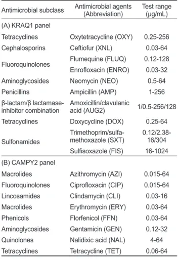
MIC panel을 이용한 항생제 감수성 검사
MIC panel
을 이용한항생제감수성검사방법은국립수산과학원 병리연구과에서 설계하였고
, Thermo
사에서 제작한KRAQ1 panel
의oxytetracycline, ceftiofur, flumequine, en- rofloxacin, neomycin, ampicillin, amoxicillin/clavulanic acid, doxycycline, trimethoprim/sulfamethoxazole
및sulfisoxa- zole 10
종과CAMPY2 panel
의azithromycin, ciprofloxacin, clindamycin, erythromycin, florfenicol, gentamicin, nalidixic acid
및tetracycline 8
종을사용하였다(Table 1).
1% NaCl
이첨가된tryptic soy agar (TSA, Difco, Detroit, MI, USA)
에서순수배양된세균의colony
를멸균loop
를이 용하여3-5
개를취한후, 1% NaCl
이첨가된distilled water
에 희석하여0.5 McFarland (1.5×10
8CFU/mL)
가되도록Sensi- titer
TMNephelometer (TREK Diagnostic system, Cleveland,
OH)
로농도를맞추었다.
농도를맞춘균액은최적화조건확립을위해균종별로 설정한조건에맞춰각각의
broth
배지에100 μL
씩넣었다(Table 2). Streptococcus spp.
의경우, CLSI
에서제시한기준에따라5%
의lysed horse blood (LHB)
가첨 가된cation-adjusted Mueller-Hinton broth with TES buffer
(CAMHBT)
배지에1% NaCl
의첨가유무를다르게하여실험 을진행하였고, MHB
배지에서배양했을때MIC
값의차이점 을확인하고자하였다. E. tarda, Vibrio spp.
그리고Aeromonas
spp.
는CAMHBT
배지, 1% NaCl
이첨가된CAMHBT
배지 그리고1% NaCl
이첨가된MHB
에서배양하였다. Pseudomo-
nas spp.
의경우는CAMHBT
배지에서22°C, 28°C
및35°C
의조건으로만실험을진행하였다
.
준비된균액을96 well panel
에100 μL
씩분주한후kit well
전용필름으로sealing
하고panel
을각조건에맞는배양온도에서24
시간배양하였다.
배양된panel
을Sensititer
TMManual Viewbox
를이용하여육안으로관 찰하였을때,
충분한성장저해가일어나투명한상태로남아있 는well
중가장적은양의항생제를포함하는well
의항생제농 도를최소억제농도(MIC)
로결정하였다.
결 과
균의 동정
본연구에서
PCR
을통해연쇄구균의동정을실시한결과, S.
parauberis 3
균주, S. iniae 2
균주로확인되었다. E. tarda
균주 는선택배지인SS
배지상에서검은색집락이형성되었으며,
정확한동정을위한
PCR
결과에서도동일한결과가나타났다.
이전
Demircan and Candan (2006), Kim et al. (2014), Kim et
al. (2015)
의방법을참고하여 각비브리오균주에대한세부적인동정을실시한결과
, V. alginolyticus, V. anguillarum, V.
parahaemolyticus, Vibrio sp.
및V. harveyi
로동정되었고,
해당 균주를본실험에사용하였다(Table 3).
MIC panel의 최적화 배양 조건
주요어병세균
25
균주를대상으로18
종수산용항생제에대 한다양한환경조건하에서의실험결과를비교한결과,
각균종에대한
MIC panel
의최적화배양조건을결정할수있었다
(Table 4). S. parauberis-3
균주의doxycycline
결과를 확 인하면, 5% LHB
를첨가하고28°C
에서배양한panel
에서는MIC
값이16 μg/mL
로명확하게나타났다.
나머지8
가지의다 른조건에서는탁도가불분명해값을결정하기가어려운경우 가있었고, 8-16 μg/mL
의MIC
값을보였다(data not shown).
S. parauberis
중한균주는1% NaCl
의첨가유무에따라다른 환경조건에비하여neomycin
의MIC
값이3-4
배정도차이를 보이는경우가있었다. Streptococcus spp. 5
균주모두에대한MIC
값을분석한결과, 5% LHB
를첨가하고28°C
에서배양하 는것이가장최적화조건인것으로확인되었으며,
이는CLSI
에서권고하는사항과동일하다. E. tarda
의경우도연쇄구균과유사하게
MIC
값이조건에따라큰차이를보이지는않았으나28°C
에서배양한panel
에서다른조건들에비하여명확한MIC
값을보였다.
Vibrio spp.
는1% NaCl
을첨가한조건의panel
에서bottom
이 더명확하게나타났으며,
염분에민감한Vibrio spp.
는CLSI
에 서권고한1% NaCl
이들어간CAMHBT
배지를이용하는것이
MIC
값분석에가장적절한것으로확인되었다.
온도설정Table 1. Range of concentrations used for minimal inhibitory con- centration (MIC) determination by sensititre panels
Antimicrobial subclass Antimicrobial agents
(Abbreviation) Test range (μg/mL) (A) KRAQ1 panel
Tetracyclines Oxytetracycline (OXY) 0.25-256 Cephalosporins Ceftiofur (XNL) 0.03-64 Fluoroquinolones Flumequine (FLUQ) 0.12-128
Enrofloxacin (ENRO) 0.03-32
Aminoglycosides Neomycin (NEO) 0.5-64
Penicillins Ampicillin (AMP) 1-256
β-lactam/β lactamase-
inhibitor combination Amoxicillin/clavulanic
acid (AUG2) 1/0.5-256/128 Tetracyclines Doxycycline (DOX) 0.25-64
Sulfonamides
Trimethoprim/sulfa-
methoxazole (SXT) 0.12/2.38- 16/304 Sulfisoxazole (FIS) 16-1024 (B) CAMPY2 panel
Macrolides Azithromycin (AZI) 0.015-64 Fluoroquinolones Ciprofloxacin (CIP) 0.015-64 Lincosamides Clindamycin (CLI) 0.03-16
Macrolides Erythromycin (ERY) 0.03-64
Phenicols Florfenicol (FFN) 0.03-64
Aminoglycosides Gentamicin (GEN) 0.12-32
Quinolones Nalidixic acid (NAL) 4-64
Tetracyclines Tetracycline (TET) 0.06-64
Table 2. Culture conditions and medium for representative bacterial species
Species Streptococcus spp. E. tarda, Vibrio spp., Aeromonas spp. Pseudomonas spp.
Panel KRAQ1, CAMPY2 KRAQ1, CAMPY2 KRAQ1, CAMPY2
Medium CAMHBT1
+5% LHB2 CAMHBT CAMHBT
+1% NaCl MHB3
+1% NaCl CAMHBT CAMHBT
+1% NaCl MHB
+1% NaCl CAMHBT
Culture temperature
22°C 22°C 22°C
28°C 28°C 28°C 28°C 28°C 28°C 28°C 28°C
35°C 35°C 35°C 35°C 35°C 35°C 35°C 35°C
1CAMHBT, Cation-adjusted Mueller-Hinton broth with TES buffer. 2LHB, Lysed horse blood. 3MHB, Mueller-Hinton broth. *Incubation time, 24h.
실험에서는
Vibrio sp.
균주가35°C
의조건하에서전혀배양되 지않았으므로, 35°C
는Vibrio spp.
의배양조건에적합하지않 은것으로판명되었다.
그러므로, Vibrio spp.
는1% NaCl
을첨가한
CAMHBT
배지를이용하여실험을실시하고28°C
에서panel
을배양하는것으로최적화조건을설정하였다.
Aeromonas spp.
는6
가지조건을달리하여실험을실시하였 으며,
모든조건에서유사한MIC
값을나타내었지만, A. sobria
는35°C
에서배양한panel
에서는세균이배양되지않았다.
그 러므로, 35°C
의배양조건을제외시켰으며, 1% NaCl
첨가유무 와22°C, 28°C
의조건에서측정된MIC
값을비교한결과, 1%
NaCl
이첨가된결과에서는특정항생제에상이한값이확인되 는경우도있었으며, 22°C
에배양한panel
은세균의탁도가명 확하지않아적절한조건이아닌것으로확인되었다.
Pseudomonas spp.
균주는온도설정조건에대해서만실험 을진행하였으며, 22°C, 28°C
및35°C
모두에서유사한값을확인할수있었으나
,
가장명확한bottom
을나타낸것은28°C
에서배양한panel
이었다.
이와같은결과를바탕으로E. tarda, Aeromonas spp.
그리고Pseudomonas spp.
는CAMHBT
배지를이용하여
28°C
에서배양하는것으로최적화조건을설정하였다
(Table 4).
최적화된조건을적용하여확인한MIC
값은Table 5
와Table 6
에나타내었다.
고 찰
수산업에서는양식현장에서연중발생하는연쇄구균병
,
에드 워드병,
비브리오병등과같은세균성질병에의한피해를막고 생산성향상을위한수단으로서항생제를사용한다.
현재국내 에서승인된수산용항생∙
항균물질은36
종으로다양한항생제 를양식장에서사용하고있으며(NIFS, 2018),
제주지역에서의 수산용항생제사용량은2012
년도에는35
톤이었지만2015
년 도에는48
톤으로약13
톤증가하였다(Kim et al., 2019).
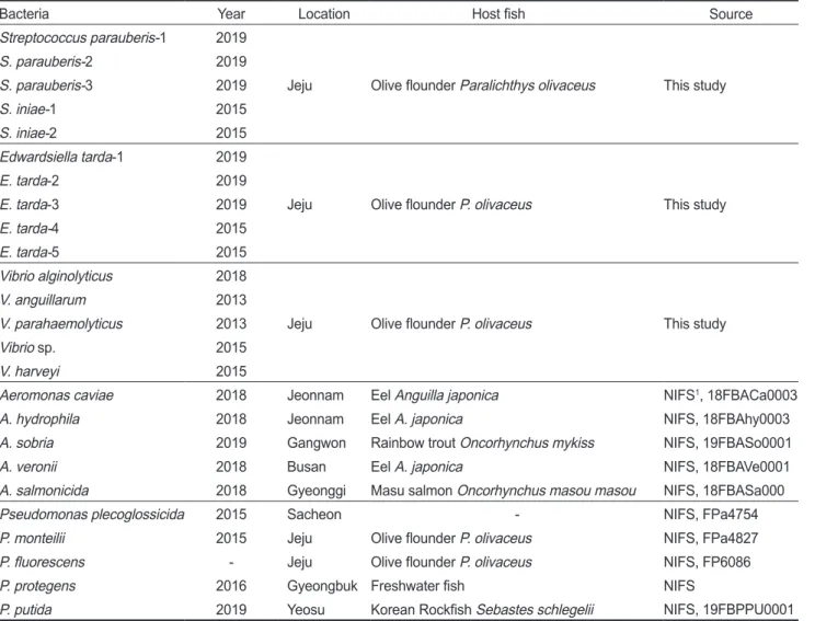
이와Table 3. List of bacterial strains used in this study
Bacteria Year Location Host fish Source
Streptococcus parauberis-1 2019
Jeju Olive flounder Paralichthys olivaceus This study
S. parauberis-2 2019
S. parauberis-3 2019
S. iniae-1 2015
S. iniae-2 2015
Edwardsiella tarda-1 2019
Jeju Olive flounder P. olivaceus This study
E. tarda-2 2019
E. tarda-3 2019
E. tarda-4 2015
E. tarda-5 2015
Vibrio alginolyticus 2018
Jeju Olive flounder P. olivaceus This study
V. anguillarum 2013
V. parahaemolyticus 2013
Vibrio sp. 2015
V. harveyi 2015
Aeromonas caviae 2018 Jeonnam Eel Anguilla japonica NIFS1, 18FBACa0003
A. hydrophila 2018 Jeonnam Eel A. japonica NIFS, 18FBAhy0003
A. sobria 2019 Gangwon Rainbow trout Oncorhynchus mykiss NIFS, 19FBASo0001
A. veronii 2018 Busan Eel A. japonica NIFS, 18FBAVe0001
A. salmonicida 2018 Gyeonggi Masu salmon Oncorhynchus masou masou NIFS, 18FBASa000
Pseudomonas plecoglossicida 2015 Sacheon - NIFS, FPa4754
P. monteilii 2015 Jeju Olive flounder P. olivaceus NIFS, FPa4827
P. fluorescens - Jeju Olive flounder P. olivaceus NIFS, FP6086
P. protegens 2016 Gyeongbuk Freshwater fish NIFS
P. putida 2019 Yeosu Korean Rockfish Sebastes schlegelii NIFS, 19FBPPU0001
1NIFS, National Institute of Fisheries Science, Korea.
으나
,
즉시항생제의사용을줄이는것은불가능하기때문에현 재로서는유효항생제를용법과용량에맞도록사용하는것이 최선의방법이다.
세균의신속한동정과항생제감수성검사는 양식현장에서항생제를선택하는데있어서중요한역할을한 다.
질병의원인세균을동정하고항생제감수성검사시간을단 축하는것은광범위항생제를경험적으로투여하는대신해당 균에대하여효과가검증된항생제를효율적이고신속하게투 여할수있도록한다.
이전의연구에사용했던항생제감수성검사중
broth micro- dilution test
의방법은단계별로희석한항생제와균배양액을96 well plate
에넣고배양한후균의증식여부에따른액체배지 의혼탁도를육안으로확인하여균이자라지않는최소농도를MIC
값으로판독한다. Broth microdilution test
방법에비하여 간편하며이전부터많이사용된disk diffusion test
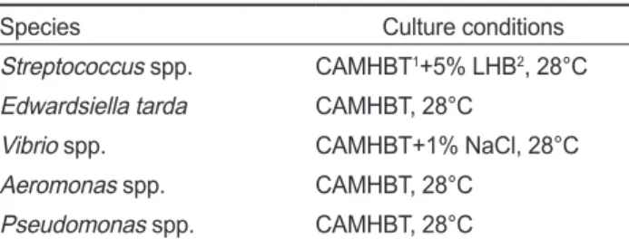
의방법은희 Table 4. Optimized culture conditions of MIC panel for representa-tive bacterial species
Species Culture conditions
Streptococcus spp. CAMHBT1+5% LHB2, 28°C Edwardsiella tarda CAMHBT, 28°C
Vibrio spp. CAMHBT+1% NaCl, 28°C
Aeromonas spp. CAMHBT, 28°C
Pseudomonas spp. CAMHBT, 28°C
1CAMHBT, Cation-adjusted Mueller-Hinton broth with TES buf- fer. 2LHB, Lysed horse blood. *Incubation time, 24h.
Table 5. MIC results for representative fish pathogenic bacteria in optimized culture conditions of the KRAQ1 panel determined in this study Optimizing
Conditions Bacteria Antibiotics (KRAQ1 panel)
OXY XNL FLUQ ENRO NEO AMP AUG2 DOX SXT FIS
CAMHBT1 +5% LHB2 28°C
S. parauberis-1 256 2 128 0.25 8 < 1 < 1/0.5 16 0.5/9.5 > 1024
S. parauberis-2 256 2 64 0.5 16 < 1 < 1/0.5 16 0.5/9.5 > 1024
S. parauberis-3 128 2 64 0.5 8 < 1 < 1/0.5 16 0.5/9.5 > 1024
S. iniae-1 < 0.25 0.06 64 0.5 32 < 1 < 1/0.5 < 0.25 < 0.12/2.38 > 1024 S. iniae-2 0.5 < 0.03 32 0.25 32 < 1 < 1/0.5 < 0.25 < 0.12/2.38 > 1024
CAMHBT 28°C
E. tarda-1 < 0.25 0.06 2 0.25 4 < 1 2/1 1 < 0.12/2.38 > 1024
E. tarda-2 0.5 0.25 2 0.25 2 2 2/1 1 < 0.12/2.38 > 1024
E. tarda-3 < 0.25 0.25 2 0.25 4 4 4/2 1 < 0.12/2.38 < 16
E. tarda-4 256 0.12 2 0.5 2 2 4/2 64 > 16/304 > 1024
E. tarda-5 256 0.12 8 8 4 > 256 8/4 > 64 > 16/304 > 1024
CAMHBT +1% NaCl 28°C
V. alginolyticus 0.5 2 0.5 0.5 8 > 256 8/4 < 0.25 < 0.12/2.38 32
V. anguillarum < 0.25 < 0.03 < 0.12 < 0.03 8 16 16/8 < 0.25 < 0.12/2.38 32
V. parahaemolyticus 128 1 4 0.5 4 4 8/4 8 < 0.12/2.38 1024
Vibrio sp. 0.5 0.25 0.5 0.12 16 > 256 32/16 < 0.25 < 0.12/2.38 512
V. harveyi 0.5 2 128 16 8 > 256 16/8 < 0.25 0.25/4.75 > 1024
CAMHBT 28°C
A. caviae 256 16 > 128 > 32 16 > 256 32/16 32 > 16/304 > 1024 A. hydrophila 256 16 > 128 32 16 > 256 32/16 > 64 > 16/304 > 1024
A. sobria 16 0.25 0.5 0.12 2 128 8/4 1 < 0.12/2.38 < 16
A. veronii 64 1 0.25 0.25 4 > 256 16/8 8 < 0.12/2.38 > 1024
A. salmonicida < 0.25 4 < 0.12 < 0.03 4 > 256 16/8 < 0.25 < 0.12/2.38 < 16
CAMHBT 28°C
P. plecoglossicida 4 32 16 0.5 1 > 256 64/32 2 16/304 > 1024
P. monteilii 4 32 8 1 1 256 64/32 2 16/304 > 1024
P. fluorescens 1 64 8 0.5 1 > 256 > 256/128 1 8/152 1024
P. protegens 4 64 8 1 2 > 256 256/128 4 8/152 1024
P. putida 2 32 8 2 2 256 64/32 1 16/304 > 1024
1CAMHBT, Cation-adjusted Mueller-Hinton broth with TES buffer. 2LHB, Lysed horse blood. *OXY, oxytetracycline; XNL, ceftiofur;
FLUQ, flumequine; ENRO, enrofloxacin; NEO, neomycin; AMP, ampicillin; AUG2, amoxicillin/clavulanic acid; DOX, doxycycline; SXT, trimethoprim/sulfamethoxazole; FIS, sulfisoxazole.
같이수산생물의생산량증가와함께질병발생이증가하면서 항생제의사용량도매년증가하고있는추세이다
.
의약품의사 용량이증가함에따라오남용으로인한위험성이고조되고있석한균주를배지에도말하고항생제
disk
를고착시킨후균의 증식억제대를판독한다(NIFS, 2017). Broth microdilution test
방법은다양한항생제를직접단계희석하여농도별로96 well
plate
에넣어야하는번거로움이 있으며,
실험균의 탁도를맞출때분광광도계를이용하기때문에실험의준비과정이복잡 하다는단점이있다
. MIC panel
을사용한broth microdilution test
방법은항생제가coating
되어나오는제품으로항생제감수 성검사시간을단축시킬수있을뿐만아니라,
연구자에의한 분석절차를줄여서실험적오류를최소화시킬수있다.
또한,
신속하고정확한결과를제공할수있으므로,
양식현장에더욱 효율적인분석방법이될수있을것이다.
현재로서는어병세균에대한정확한배양조건에대한정보가
부족한실정이며
,
본연구에서는최대한많은세균에동일한조 건이적용될수있는범위에서실험을진행하여조건이설정된 후에도실험이간편하게진행될수있도록최적화배양조건을 확립하고자하였다. Streptococcus spp.
는5% LHB
를첨가한CAMHBT
배지, Vibrio spp.
는1% NaCl
을첨가한CAMHBT
배지, E. tarda, Aeromonas spp.
그리고Pseudomonas spp.
는CAMHBT
배지를이용하여KRAQ1 panel
과CAMPY2 panel
에접종하고
28°C
에서24
시간배양하여결과를확인하는것으로최적화조건을설정하였다
.
본연구를통하여확립한배양조건을검증하기위하여
, MIC
panel
의실험에사용한균주중S. parauberis 3
균주와E. tarda 3
균주를disk diffusion test
로실험을실시한후비교분석하였다.
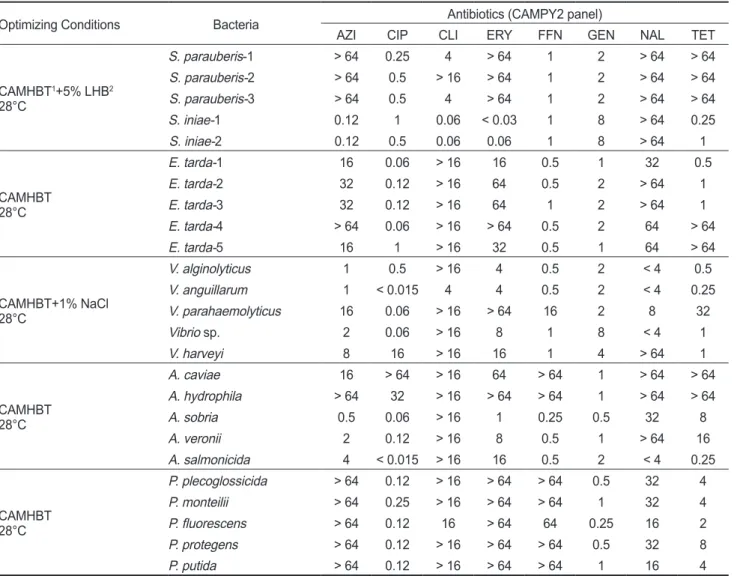
Table 6. MIC results for representative fish pathogenic bacteria in optimized culture conditions of the CAMPY2 panel determined in this study
Optimizing Conditions Bacteria Antibiotics (CAMPY2 panel)
AZI CIP CLI ERY FFN GEN NAL TET
CAMHBT1+5% LHB2 28°C
S. parauberis-1 > 64 0.25 4 > 64 1 2 > 64 > 64
S. parauberis-2 > 64 0.5 > 16 > 64 1 2 > 64 > 64
S. parauberis-3 > 64 0.5 4 > 64 1 2 > 64 > 64
S. iniae-1 0.12 1 0.06 < 0.03 1 8 > 64 0.25
S. iniae-2 0.12 0.5 0.06 0.06 1 8 > 64 1
CAMHBT 28°C
E. tarda-1 16 0.06 > 16 16 0.5 1 32 0.5
E. tarda-2 32 0.12 > 16 64 0.5 2 > 64 1
E. tarda-3 32 0.12 > 16 64 1 2 > 64 1
E. tarda-4 > 64 0.06 > 16 > 64 0.5 2 64 > 64
E. tarda-5 16 1 > 16 32 0.5 1 64 > 64
CAMHBT+1% NaCl 28°C
V. alginolyticus 1 0.5 > 16 4 0.5 2 < 4 0.5
V. anguillarum 1 < 0.015 4 4 0.5 2 < 4 0.25
V. parahaemolyticus 16 0.06 > 16 > 64 16 2 8 32
Vibrio sp. 2 0.06 > 16 8 1 8 < 4 1
V. harveyi 8 16 > 16 16 1 4 > 64 1
CAMHBT 28°C
A. caviae 16 > 64 > 16 64 > 64 1 > 64 > 64
A. hydrophila > 64 32 > 16 > 64 > 64 1 > 64 > 64
A. sobria 0.5 0.06 > 16 1 0.25 0.5 32 8
A. veronii 2 0.12 > 16 8 0.5 1 > 64 16
A. salmonicida 4 < 0.015 > 16 16 0.5 2 < 4 0.25
CAMHBT 28°C
P. plecoglossicida > 64 0.12 > 16 > 64 > 64 0.5 32 4
P. monteilii > 64 0.25 > 16 > 64 > 64 1 32 4
P. fluorescens > 64 0.12 16 > 64 64 0.25 16 2
P. protegens > 64 0.12 > 16 > 64 > 64 0.5 32 8
P. putida > 64 0.12 > 16 > 64 > 64 1 16 4
1CAMHBT, Cation-adjusted Mueller-Hinton broth with TES buffer. 2LHB, Lysed horse blood. *AZI, azithromycin; CIP, ciprofloxacin; CLI, clindamycin; ERY, erythromycin; FFN, florfenicol; GEN, gentamicin; NAL, nalidixic acid; TET, tetracycline.
그결과
,
균의농도를정확하게맞추어실험이진행되는MIC panel
과비교하여disk diffusion test
방법은세균의종류에따 라균이평판배지에서성장하는속도가다르게나타나생성되 는저지대에 차이를보이는경우가있었다(data not shown).
MIC panel
을사용한분석에서는결과를명확하게확인할수있었고다른조건에영향을받지않는것으로나타났다
.
인체나가축에질병을일으키는세균의항생제에대한감수성 과내성을구분할수있는해석기준(interpretive criteria)
에관 한연구들은많이이루어졌으나,
어류의세균에관한내성판단 을위한표준화작업은부족한실정이다.
예를들어현재많은 관련실험실에서참고하는국제프로토콜인CLSI
가이드라인 중VET03/VET04-S2 (CLSI, 2014b)
는어병세균용으로마련 된국제표준방법인데도불구하고A. salmonicida
한종에대 해서만해석기준이마련되어있다.
최근국내에서수산용항생 제에대한내성기준과매뉴얼에대한연구가진행되고있지만(Chun et al., 2019),
다양한어병세균을대상으로실험이이루 어져있지않기때문에세균성질병발생시초기치료로적절한 유효항생제선택에어려움이많다.
또한이러한세균종별항생 제해석기준의부재로인하여내성이증가되고,
내성균에의한 치료실패가중요한임상적문제로써대두됨에따라수산용항 생제에대한내성판정기준을명확히설정할필요가있다.
그리 고,
세균성질병의치료를위한항생제선정과항생제내성연 구를위해동일한방법의검사가필요하며,
다양한검사능력을 갖고있는검사자혹은연구자가쉽게이해하고실행할수있는 검사법의설정이필요하기때문에본연구에서의항생제panel
을이용한액체배지희석법은항생제내성검사를위한표준화 된방법으로제시될수있다.
본연구에서확립한최적화된조 건을사용하여많은균주를대상으로신속하고정확한MIC
값 을분석하고내성균에대한정보를축적함으로써,
향후수산생 물병원성세균에대한항생제내성판정을위한기준설정이가 능할것으로판단된다.
사 사
본연구는국립수산과학원수산과학연구사업
(R2020061)
의 지원에의해수행되었습니다.
References
Chun WK, Lee YH, Kim YJ, Roh HJ, Kim AR, Kim NE, Seo JS, Kwon MG, Lee JH and Kim DH. 2019. Epidemiological cut-off values Generated for disc diffusion data from Strep-
tococcus parauberis. Korean J Fish Aquat Sci 52, 382-388.
https://doi.org/10.5657/KFAS.2019.0382.
CLSI (Clinical and Laboratory Standards Institute). 2006.
Method for antimicrobial disk susceptibility testing of bac- teria isolated from aquatic animals. CLSI document M42-A.
CLSI, Wayne, PA, U.S.A.
CLSI (Clinical and Laboratory Standards Institute). 2014a.
Methods for broth dilution susceptibility testing of bacteria isolated from aquatic animals. CLSI document VET04-A2.
CLSI, Wayne, PA, U.S.A.
CLSI (Clinical and Laboratory Standards Institute). 2014b. Per- formance standards for antimicrobial susceptibility testing of bacteria isolated from aquatic animals; Second informa- tional supplement. CLSI document VET03/ VET04-S2.
CLSI, Wayne, PA, U.S.A.
CLSI (Clinical and Laboratory Standards Institute). 2020. Per- formance standards for antimicrobial susceptibility testing;
thirtieth informational supplement. CLSI document M100- ED30. CLSI, Wayne, PA, U.S.A.
Demircan D and Candan A. 2006. Identification of Vibrio an-
guilliarum by PCR (rpoN Gene) associated with Vibriosis
in marine fish in Turkey. Turk J Vet Anim Sci 30, 305-310.EUCAST (European Committee on Antimicrobial Suscepti- bility Testing) web site. 2020. Antimicrobial susceptibility testing EUCAST disk diffusion method, Version 8.0. Re- trieved from https://eucast.org/fileadmin/src/media/PDFs/
EUCAST_files/Disk_test_documents/2020_manuals/
Manual_v_8.0_EUCAST_Disk_Test_2020.pdf on Jan 10, 2020.
JSA (the Federal Joint Subcommittee on Aquaculture). 1994.
Guide to drug, vaccine, and pesticide use in aquaculture.
Texas A&M University, Texas agricultural extension ser- vice, B-5085.
Kim HJ, Ryu JO, Lee SY, Kim ES and Kim HY. 2015. Multiplex PCR for detection of the Vibrio genus and five pathogenic
Vibrio species with primer sets designed using comparative
genomics. BMC Microbiol 15, 239. https://doi.org/10.1186/s12866-015-0577-3.
Kim MS, Cho JY and Choi HS. 2014. Identification of Vibrio
harveyi, Vibrio ichthyoenteri and Photobacterium damselae
isolated from olive flounder Paralichthys olivaceus in Korea by multiplex PCR developed using the rpoB gene. J Fish Sci 80, 333-339. https://doi.org/10.1007/s12562-014-0702-5.Kim YJ, Seo JS, Park JO, Jeong AH and Lee JH. 2019. Monitor- ing of aquatic medicine managements in South Korea. J Fish Pathol 32, 37-43. https://doi.org/10.7847/jfp.2019.32.1.037.
Lee DW, Jun LJ and Jeong JB. 2017. Distribution of tetracy- cline resistance genes in pathogenic bacteria isolated from cultured olive flounder Paralichthys olivaceus in Jeju in 2016. JFMSE 29, 834-846. https://doi.org/ 10.13000/
JFMSE.2017.29.3.834.
NIFS (National Institute of Fisheries Science). 2017. Antibiotic resistant bacteria inspection manual (11-1192266-000195- 01). GMK communication, Busan, Korea, 34-49.
NIFS (National Institute of Fisheries Science). 2018. Aquatic medicine catalog. Aquatic Disease Control Division, Busan, Korea, 12-101.
Noga EJ. 1996. Diagnosis and treatment. In: Fish disease. Mos-
by-Year Book, Inc., St. Louis, MO, U.S.A., 19-215.
Sakai T, Iida T, Osatomi K and Kanai K. 2007. Detection of type 1 fimbrial genes in fish pathogenic and non-pathogenic
Edwardsiella tarda strains by PCR. Fish Pathol 42, 115-117.
https://doi.org/ 10.3147/jsfp.42.115.
Son KT, Jo MR, Oh EG, Mok JS, Kwon JY, Lee TS, Song KC, Kim PH and Lee HJ. 2011. Residues of Ampicillin and Amoxicillin in Olive Flounder Parlichthys oliveceus Fol- lowing Oral Administration. Korean J Fish Aquat Sci 44, 464-469. http://dx.doi.org/10.5657/KFAS.2011.0464.
Woo SH, Kim HJ, Lee JS, Kim JW and Park SI. 2006. Patho- genicity and classification of streptococci isolated from cul- tured marine fishes. J Fish Pathol 19, 17-33.
Woodward KN. 1996. The regulation of fish medicines - UK and European Union aspects. Aquac Res 27, 725-734.
https://doi.org/10.1046/j.1365-2109.1996.00782..x.