• 검색 결과가 없습니다.

Multicentric T cell lymphoma in a Maltese dog

N/A
N/A
Protected

Academic year: 2021

Share "Multicentric T cell lymphoma in a Maltese dog"

Copied!
5
0
0

로드 중.... (전체 텍스트 보기)

전체 글

(1)

85

Multicentric T cell lymphoma in a Maltese dog

Ji-Youl Jung, Sang-Chul Kang, In-Soon Roh

1

, Hyun-Joo Sohn

1

, Young-Min Yun, Jung-Hun Kim, Kyoung-Kap Lee, Min-Chan An

2

, Jong-Hee Bae, Jae-Hoon Kim*

Department of Veterinary Medicine, Cheju National University, Jeju 690-756, Korea

1

National Veterinary Research & Quarantine Service, Anyang 430-824, Korea

2

Halla Pet Hospital, Jeju 690-832, Korea

(Accepted: January 9, 2007)

Abstract : A case of multicentric high grade T cell lymphoma is reported in a 5-year-old male Maltese dog with generalized lymphadenopathy. The dog showed depression, anorexia, blindness, jaundice, arrhythmia, and hematuria for 8 months. Complete blood count and chemistry profile revealed anemia and increased alanine transferase, alkaline phosphatase, total bilirubin, and total cholesterol. Grossly, most of lymph nodes, spleen, and liver were enlarged and neoplastic masses were occupied in these tissues.

Histologically, massive accumulation of small noncleaved neoplastic lymphocytes with high mitotic figures was observed in all lymph nodes and spleen. Infiltration of neoplastic lymphocytes was also noted in the lung, liver, kidney, eye, skin, muscle, and bone marrow of femur. Immunohistochemistry revealed that tumor cells were CD3-positive and but CD79a-negative, consistent with T-cell lineage. In our best knowledge, this is the first report of multicentric lymphoma clarified the origin of tumor cells in Korea.

Key words : CD3, dog, immunohistochemistry, multicentric lymphoma, T-cell Lymphomas are malignant neoplasms characterized

by the proliferation of cells native to the lymphoid tissues, that is, lymphocytes, histiocytes, and their precursors and derivates [11]. The term “lymphoma”

is one of a misnomer, since this tumor is lethal unless controlled or eradicated through therapy. Based on the human terminology, non-Hodgkin’s lymphomas, also known as canine lymphomas, are very common tumors in dogs, but Hodgkin’s lymphoma had not been discovered in dogs. In the general canine population, the annual incidence of this tumor was estimated from 13 to 24 per 100,000 annually [11]. The mean age of dogs with lymphoma varies from 6.3 to 7.7 years, with no significant sex predilection [13]. Boxers, Saint Bernard, Scottish terriers, basset hounds, Airedale terriers, and bulldogs are reported to be at increase risk, and dachshunds and Pomeranians at reduced risk [13].

Lymphosarcoma, resulting from the malignant transfor- mation of developing lymphocytes, represents one of the most commonly encountered canine neoplasms [11]. These tumors are highly aggressive, and their etiology remains obscure. However, a canine long-term

T-cell line, established from a dog with Sézary syndrome and a canine large granular lymphocyte leukemia, both producing retroviral particles, have been recently reported [6].

According to anatomic and histologic criteria, lymphoma can be classified into 5 different forms such as multicentric, alimentary, subcutaneous, mediastinal, and solitary or extranodal forms [11]. The most common form in the dog is the multicentric type. Extranodal forms involved in the central nervous system or eye are less common type in dogs. Multicentric lymphoma [1, 8], extranodal lymphoma [7], and intraocular lymphoma [15] were previously reported in Korea.

However, the origin cells of their tumors were not clarified in most cases. In this paper, we described a case of multicentric T cell lymphoma in a Maltese dog based on the histopathological characteristics and immunohistochemistry.

A 5-year-old male Maltese dog was presented with an 8-month history of depression, anorexia, blindness, arrhythmia, mild jaundice, and hematuria. Physical examination revealed lymphadenopathy at multiple sites.

*Corresponding author: Jae-Hoon Kim

Department of Veterinary Medicine, Cheju National University, Jeju 690-756, Korea

[Tel: +82-64-754-3387, Fax: +82-64-702-9920, E-mail: kimjhoon@cheju.ac.kr]

(2)

metastatic tumor, lymphoid hyperplasia, and lympha- denitis. A complete blood cell (CBC) count revealed anemia with a packed cell volume of 15%. Abnormal serum biochemical results included elevated alanine transferase (126 U/L), alkaline phosphatase (2,000 U/L), total bilirubin (1.24 mg/dl), and total cholesterol (412.5 mg/dl). The dog was unresponsive to symptomatic, fluid and chemotherapies. Because of poor prognosis the dog was euthanized and submitted to the Veterinary Pathology Laboratory of Cheju National University for postmortem examination.

At necropsy, the dog was in poor physical condition and there was a considerable depletion of adipose tissue at the coronary groove and in the abdominal cavity. The corneal opacity with central perforation was observed in the right eye. Generalized erythemas of well-demarcated red patch were distributed on the skin.

Milky-white gelatinous and pale yellow watery materials were occupied in subcutaneous tissue. The lymph nodes of the mandibular (4

×

3

×

2.5 cm in size), pharyngeal, cervical, inguinal, popliteal (3

×

1.5

×

3 cm in size), iliac, mesenteric, and most of subcutaneous regions were severely enlarged (Fig. 1). The lymph nodes in subcutaneous areas of skin were freely movable, soft or rubbery. These enlarged lymph nodes were occupied with yellowish tan oval masses ranged from 2 mm to 4 cm in diameter. They were bulging and uniformly firm on cut surface. The abdominal cavity contained a large amount of watery blood stained fluid. The spleen was severely enlarged to a size of approximately 19.5

×

4-7

×

2 cm. Two yellowish, fairy firm, and raised nodular masses, 1 to 1.5 cm in diameter were presented in the surface of spleen (Fig. 1). Both masses were surrounded by fibrous capsule. Dull demarcated pale yellow discolorations were scattered in the hepatic parenchyma.

Tissues samples from the neoplastic masses of various lymph nodes, spleen, liver, heart, kidney, femur, pancreas, and other representative parenchymal organs were fixed in 10% neutral phosphate-buffered formalin, routine processed, and stained with hematoxylin and eosin (H&E) for light microscopic examination.

Immunohistochemical staining were performed on paraffin embedded tissue sections with antibodies specific to T- cell marker, polyclonal rabbit anti-human CD3 (Dako,

Denmark), and B-cell and plasma cell marker, monoclonal mouse anti-human CD79a (Dako, Denmark).

For immunohistochemical staining tissue sections were placed on poly L-lysine coated slides and were stained by using the streptavidin-biotin method previously described [3, 5]. Immuno-staining was done using a Benchmark staining platform (Discovery; Ventana Medical System, USA), with the DABMap detection kit (Ventana Medical Systems, USA) using a streptavidin-biotin amplification system. Slides were counterstained with hematoxylin and bluing reagent.

Histopathologically, the tumor masses were charac- terized by massive solid proliferation of lymphoid cells.

All lymph nodes and spleen were composed of dense

population of neoplastic lymphoid cells resulting in

loss of their normal architecture. In lymph nodes,

neoplastic cells were diffusely distributed in cortex and

invaded with cord-like pattern in medullary cords and

sinuses. The tumor cells consisted of relatively uniform

population of small noncleaved lymphocytes, predomi-

nantly 1.5 red blood cells in diameter (Fig. 2). Tumor

cells had large nucleus with marked irregular thickening

of the nuclear membranes, prominent round to branched

hyperchromatic nucleoli, and scant dense eosinophilic

cytoplasm. There was an average of 3 to 5 mitotic

Fig. 1. All mesenteric lymph nodes (black arrows) were

enlarged. Two yellowish masses (white arrows) were found

in the surface of spleen.

(3)

figures per high-power filed. Hemorrhagic infarct areas were sometimes observed in lymph nodes and spleen.

Within most tissues, the distribution of tumor cells generally reflected a vascular tropism. In the lung, large numbers of neoplastic lymphocytes accumulated in the areas of peribronchiolar, perivascular, and alveolar walls (Fig. 3). Tumor cells were markedly infiltrated in portal triads, where they surrounded blood vessels and bile ducts, and forming coalescence to adjacent central veins (Fig. 3). The pancreas and the bone marrow in femur were heavily infiltrated with tumor cells, and they obliterated normal architecture. In the kidney, multifocal small tumor cells foci were scattered in cortex. Tumor cells also found in the perivascular areas in subcutaneous tissues of skin. In the eye, a severe corneal ulceration and diffuse zonal infiltration of lymphocytic tumor cells in subcorneal connective tissue, especially perivascular areas, were observed.

Immunohistochemistry on formalin fixed tissues (lymph nodes, spleen, pancreas, lung, and kidney) showed the tumor cells strongly expressed CD3 (Fig. 4). But they did not express CD79a.

Since the last decade, the National Cancer Institute- Working Formulation has become the standard for classification of human lymphomas in the United States, while the Kiel system is used widely in Europe [9, 11].

Fig. 2. Neoplastic lymphocytes in lymph node.

Note mitotic figures (arrows). H & E,

×

400.

Fig. 3. Infiltration of tumor cells in peribronchiolar area of the lung (up) and portal area of the liver (down).

H & E,

×

100.

Fig. 4. Neoplastic tumor cells express positive reaction for CD3 in the pancreas.

Streptavidin-biotin peroxidase stain,

×

400.

(4)

Wide experience in humans has shown that the biological behavior of lymphoid tumors can be predicted as low, intermediate, and high grade based on the estimation of mitotic index and characterization of tissue architecture and morphologic features such as nuclear size and shape. A high grade lymphoma generally responds best to chemotherapy, yet patients with high grade lymphoma experience shorter survival times than those with low grade one. Based on the anatomical involvement, the characteristic histopatho- logical findings, and the results of immunohistoche- mistry, this case was diagnosed as multicentric high grade small noncleaved T cell lymphoma. According to previously study, the multicentric type accounted for 84% of cases of canine lymphoma [10]. About 80%

of dogs with multicentric type present with bilateral and symmetrical superficial lymphadenopathy. In addition, hepatomegaly, splenomegaly, and subcutaneous edema may be commonly present. Most of the gross findings of this case closely resembled with previous studies [1, 8, 11].

Dogs with lymphoma were classified into 5 stages and 2 sub-stages using modified World Health Organi- zation (WHO) staging system on the basis of clinical examination and additional laboratory tests [2, 11]. The dog in this case presented with stage IV category (involvement of liver and/or spleen, with or without generalized lymph node involvement), which would suggest a poor prognosis.

Determination of the immunophenotype of lympho- sarcomas in humans has become an essential step in their classification because of the relation to biological behavior and response to therapy [9]. All of updated classification of human lymphomas based on immuno- phenotype. After trial and error, veterinary researchers established the standardization of the availability of the different cellular markers for canine lymphoid cells.

Approximately 70% of canine lymphomas are of a B cell phenotype, while T cell lymphomas accounted for 10 to 40% of cases depending on the studies [4, 5, 14].

Dogs with T cell lymphomas, as in humans, have a significantly greater risk of relapse and early death compared with the dogs with B cell lymphomas.

Multicentric lymphoma may be easily diagnosed clinically on the basis of generalized lymphadenopathy.

clinical signs in early stage of lymphomas, the diag- nosis of this tumor in dogs is made usually late. The prognosis for lymphoma is variable and it has been suggested that many factors may influence survival of patients. The most consistent prognostic factors are clinical stage and sub-stage of the tumor progression.

The morphologic type and immunophenotype of tumor cells also greatly influence on the prognosis of the dogs with lymphoma [12]. Therefore cytologic examination for the fine needle aspirates of enlarged lymph nodes is a best way to monitor progress and response to therapy, to assist in staging, and to detect recurrence.

And the application of immunohistochemistry for immunophenotype is very important diagnostic step to determine the prognosis and the therapeutic methods.

References

1. Bak UB, Sung JK, Lee CW, Kim JW. Visceral lymphosarcoma in a dog. Korean J Vet Res 1985, 25 , 175-182.

2. Carter RF, Valli VEO, Lumsden JH. The cytology, histology and prevalence of cell types in canine lymphoma classified according to the National Cancer Institute Working Formulation. Can J Vet Res 1986, 50 , 154-164.

3. Fernandez NJ, West KH, Jackson ML, Kidney BA.

Immunohistochemical and histochemical stains for differentiating canine cutaneous round cell tumors. Vet Pathol 2005, 42 , 437-445.

4. Fournel-Fleury C, Magnol JP, Bricaire P, Marchal T, Chabanne L, Delverdier A, Bryon PA, Felman P.

Cytohistological and immunological classification of canine malignant lymphomas: comparison with human non-Hodgkin’s lymphomas. J Comp Pathol 1997, 117 , 35-39.

5. Fournel-Fleury C, Ponce F, Felman P, Blavier A, Bonnefont C, Chabanne L, Marchal T, Cadore JL, Goy-Thollot I, Ledieu D, Ghernati I, Magnol JP.

Canine T-cell lymphomas: a morphological, immuno- logical, and clinical study of 46 new cases. Vet Pathol 2002, 39 , 92-109.

6. Ghernati I, Corbin A, Chabanne L, Auger C,

Magnol JP, Fournel C, Monier JC, Darlix JL, Rigal

(5)

D. Canine large granular lymphocyte leukemia and its derived cell line produce infectious retroviral particles.

Vet Pathol 2000, 37 , 310-317.

7. Kim JE, Eom KD, Bae JS, Jin HK, Jang KH. An extranodal form of malignant lymphoma in the left retrobulbar area in a dog. J Vet Clin 2004, 21 , 214-218.

8. Kim JE, Jang KH, Chae HG, Lim JH, Kwon YS, Lee KW, Jang IH. Multicentric form of lympho- sarcoma in a dog. Korean J Vet Clin Med 1999, 16 , 492-496.

9. Lennert K, Feller CA. Histologie des Lymphomes Malins Non Hodgkiniens Selon la Classification de Kiel Actualisée. pp. 1-50, Doin, Paris, 1991.

10. MacEwen EG, Young KM. Canine lymphosarcoma and lymphoid leukemias. In: Oncology, 2nd ed. pp.

451-478, Saunders, Philadelphia, 1996.

11. Meuten DJ. Tumors of the hemolymphatic system. In:

Tumors in Domestic Animals. 4th ed. pp. 119-144, Iowa State Press, Ames, 2002.

12. Ponce F, Magnol JP, Ledieu D, Marchal T, Turinelli V, Chalvet-Monfray K, Fournel-Fleury C. Prognostic significance of morphological subtypes in canine malignant lymphomas during chemotherapy. Vet J 2004, 167 , 158-166.

13. Teske E. Canine malignant lymphoma: a review and comparison with human non-Hodgkin’s lymphoma. Vet Q 1994, 16 , 209-219.

14. Teske E, van Heerde P, Rutteman GR, Kurzman ID, Moore PF, MacEwen EG. Prognostic factors for treatment of malignant lymphoma in dog. J Am Vet Med Assoc 1994, 205 , 1722-1728.

15. Woo HM. Intraocular lymphoma in a dog. J Vet Clin

2001, 18 , 429-433.

수치

Fig. 4.  Neoplastic tumor cells express positive reaction for CD3 in the pancreas.

참조

관련 문서

this study analysed cast speed and solidification in thermal and flow perspectives and based on the results, conducted a confidence test on the high-speed general purpose

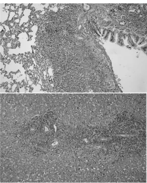
We investigated upper gastrointestinal tract involvement and characteristic endoscopic findings in scrub typhus, and we also determined the correlation between

This paper is a case study on how Venture and Small-Business Executives managers can take advantage of their intuitions in situations where the

This chapter aims to discuss the organic relations between Shakespeare’s literature and Star Trek series based on The Sonnets that is characteristic of the

Second, the seventh educational course applied to the practical curriculum in 2001, and it applied to the first- grade students of high school , so as

Just as we achieved good control with a joint-based controller that was based on a linearizing and decoupling model of the arm, we can do the same for the Cartesian case.

출처 : IAEA 발표 자료(Comprehensive inspection exercise at bulk handling facilities, “U-235 Enrichment measurements by gamma-ray spectroscopy”) 13.  Uranium

Based on the findings of this study, it was considered that the senior welfare centers should manage and operate various health promotion programs to