Tuberc Respir Dis 2010;68:140-145
CopyrightⒸ2010. The Korean Academy of Tuberculosis and Respiratory Diseases. All rights reserved.
한 대학병원에서 급성 폐색전증으로 진단된 환자들의 임상적 특성 및 예후
계명대학교 의과대학
1내과학교실,
2영상의학교실,
3흉부외과학교실
채진녕1, 최원일1, 박지혜1, 노병학2, 김재범3Acute Pulmonary Embolism: Clinical Characteristics and Outcomes in a University Teaching Hospital
Jin Nyeong Chae, M.D.
1, Won-Il Choi, M.D.
1, Jie Hae Park, M.D.
1, Byung Hak Rho, M.D.
2, Jae Bum Kim, M.D.
3Departments of
1Internal Medicine,
2Diagnostic Radiology,
3Thoracic Surgery, Keimyung University School of Medicine, Daegu, Korea
Background: Pulmonary embolism (PE) is a common clinical problem in the West that is associated with substantial morbidity and mortality. The diagnostic modality has been changed since 2001. This study retrospectively reviewed the PE mortality with the aim of identifying the risk factors associated with mortality since the multidetector computed tomography (MDCT) was introduced.
Methods: We analyzed 105 patients with acute PE proven by multidetector CT or ventilation perfusion scan. The primary outcome measure was the all-cause mortality at 3 months. The prognostic effect of the baseline factors on survival was assessed by multivariate analysis.
Results: The main risk factors were prolonged immobilization, stroke, cancer and obesity. Forty nine percent of patients had 3 or more risk factors. The overall mortality at 3 months was 18.1%. Multivariate analysis revealed low diastolic blood pressure and the existence of cancer to be independent factors significantly associated with mortality. Forty two PE patients were examined for the coagulation inhibitors. Four of these patients had a protein C deficiency (9.5%), and 11 had a protein S deficiency (26%).
Conclusion: PE is an important clinical problem with a high mortality rate. Close monitoring may be necessary in patients with the risk factors.
Key Words: Pulmonary Embolism; Venous Thrombosis; Prognosis; Risk Factors; Thrombophilia
Address for correspondence: Won-Il Choi, M.D.
Department of Internal Medicine, Keimyung University School of Medicine, 216, Dalseong-ro, Jung-gu, Daegu 700-712, Korea
Phone: 82-53-250-7572, Fax: 82-53-250-7434 E-mail: [email protected]
Received: Nov. 24, 2009 Accepted: Dec. 29, 2009
서 론
폐색전증은 질병 자체로 인한 치명적인 결과를 초래할 수 있고, 치료 중의 출혈에 의한 합병증, 그리고 재발로 인한 위험성을 고려할 때 임상적으로 매우 중요한 질환이 다. 국내에서는 폐색전증의 임상 양상에 대한 연구가 있
으나 대부분 다채널 검출기 전산화 단층 폐혈관 조영술을 시행하기 이전의 연구가 대부분이었다1-4.
대한결핵 및 호흡기학회에서 1998년부터 2000년까지 3 년간 35개 병원이 참여하여 808명의 폐색전증 환자의 임 상양상을 분석한 바 있다4. 이 연구에서는 43%의 환자를 흉부 전산화단층촬영(computed tomography, CT)으로 진단하였는데 다채널(multichannel) 검출기를 사용하기 전이라 진단의 민감도와 특이도가 다채널 전산화 단층촬 영기 보다 낮을 수 있다5. 다기관 무작위 임상시험결과에 서도 multidetector computed tomography (MDCT)를 이 용한 군이 폐 환기 관류 스캔을 이용한 군에 비해 폐색전 증 진단율이 5% 가량 높음이 보고된 바 있고6, MDCT 사 용이 폐색전증의 발생률 증가와 사망률 감소에 기여함이
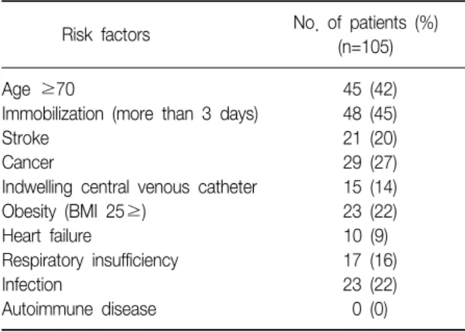
Table 1. Risk factors
No. of patients (%) Risk factors
(n=105)
Age ≥70 45 (42)
Immobilization (more than 3 days) 48 (45)
Stroke 21 (20)
Cancer 29 (27)
Indwelling central venous catheter 15 (14)
Obesity (BMI 25≥) 23 (22)
Heart failure 10 (9)
Respiratory insufficiency 17 (16)
Infection 23 (22)
Autoimmune disease 0 (0)
BMI: body mass index.
밝혀진 바 있다7. 국내의 많은 병원에서 MDCT를 이용하 여 폐색전증을 진단하고 있으며, 현재 computed tomo- graphic pulmonary angiography (CTPA)는 폐색전증의 진단에 표준 진단법으로 자리매김 되었다8.
이에 저자는 진단방법이 변화된 시점에서 폐색전증의 예후 및 임상양상을 기술하고자 본 연구를 계획하였다.
연구는 2년 반 동안 단일 병원에서 16채널(channel) CT가 도입된 이후 진단 된 폐색전증 환자의 자료를 수집하였고, 폐색전증의 예후 및 위험인자 등을 분석하고자 하였다.
대상 및 방법
1. 연구 대상
본 연구는 계명대학교 동산병원 의학연구윤리심의위원 회(Institutional Review Board)의 승인을 받아 시행되었 다. 2005년 1월부터 2007년 3월까지 계명대학교 동산병원 의 입원 및 외래 환자 혹은 응급실 방문 환자 중에서 CTPA를 시행한 후 폐색전증으로 확진 받은 환자 102예와 폐 환기 관류 스캔을 통해서 확진된 3예를 포함하여 총 105예를 대상으로 하였다. 모든 환자는 폐색전증이 처음 진단된 환자를 대상으로 하였으며, 만성 폐색전증 환자는 제외하였다.
2. 연구 방법
본원에서 폐색전증의 진단은 임상적으로 폐색전증이 의심되는 환자에서는9 Siemens사의 16 또는 64개의 검출 기(detector)를 가진 스캐너를 이용하여 CTPA를 시행하 고, CTPA에서 폐색전증의 소견이 관찰되지 않은 경우 폐 색전증이 없는 것으로 해석하였다. 환자가 심부정맥 혈전 을 가지고 있으면서 호흡기 증상이 동반된 경우 다시 CTPA로 폐색전증 유무를 평가하였다. 신부전이 있거나 조영제에 부작용의 병력이 있는 경우 폐환기관류 스캔을 시행하였다. CTPA 판독은 폐동맥 중심부 혹은 주변부의 충만결손(filling defect)이 있을 때 폐색전증으로 진단하 였다.
3. 응고인자억제제 측정
C단백(protein C)의 활성도와 S단백(protein S)의 활성 도는 응고인자 V를 기질(substrate)로 사용하여 응고 기능 을 평가하여 측정하였다(Diagnostica Stago, Asnieres, France). 혈장의 총 S단백과 자유 S단백은 radial im- munodiffusion 방법으로 측정하였다.
4. 응고인자억제제 결핍증의 분류
결핍증의 유형은 다음과 같이 분류하였다. C단백 결핍 의 경우 1형(type 1)은 기능성 활성도(functional activity) 와 항원이 같이 감소된 경우이고, 2형(type 2)은 기능성 활성도는 감소되어 있으나 항원의 농도는 정상인 경우로 하였다. S단백 결핍의 경우 기능성 활성도는 감소되어 있 으면서 S항원의 유리형과 총 항원 치도 감소된 경우를 1 형(type 1), 기능성 활성도는 감소되어 있으면서 S항원의 유리형 항원과 총 항원치는 정상인 경우를 2형(type 2), 그리고 기능성 활성도는 감소되어 있으면서 S항원의 유리 형 항원은 감소되었지만 총 항원치는 정상인 경우를 3형 (type 3)으로 하였다.
5. 통계 처리
연속변수는 평균과 표준편차로 표시하였다. 위험인자는 단변량분석법과 다변량분석법으로 분석하였고, 유의 수준 0.05 미만일 때 통계적으로 유의한 것으로 판정하였다. 통 계프로그램은 SPSS version 13.0 (SPSS Inc., Chicago, IL, USA)을 사용하였다.
결 과
1. 임상양상의 분석
연구기간 동안 총 105예의 폐색전증 환자가 진단되었 다. 여자가 58명, 남자가 47명이었다. 이 기간 동안 본 병 원에서는 정맥혈전의 예방은 하지 않았다. 환자들의 임상 양상은 Table 1, Table 2와 같다. 환자의 남녀 비는 1:
Table 2. Baseline clinical characteristics
No. of patients Clinical finding
(Mean±SD) Respiratory frequency, breath/min 22.2±4.6
Heart rate, breath/min 90.2±17.6
Systolic blood pressure, mm Hg 115.4±23.6 Diastolic blood pressure, mm Hg 72.0±13.6
Temperature,
oC 36.6±0.58
PaO
2, mm Hg 75.3±24.3
D-dimer, μg/mL 8.2±10.0
Pro-BNP, pg/mL 4,010.5±5,319.0
Troponin-I, ng/mL 0.4±1.2
RV dysfunction, n (%) 43 (41%)
Table 3. The number of risk factors in patients with multi- ple risk factors
No. of risk factors No. of patients (%) 5 or more risk factors 13 (12)
4 risk factors 15 (14)
3 risk factors 24 (23)
2 or less risk factors 53 (50)
Figure 1. Distribution of disease by age and sex in 105 patients with acute pulmonary thromboembolism.
Table 4. Thrombus site in CTPA
No. of involved artery (%) Site
(n=102) Lt
Main artery 33 (32.4)
Lobar artery 46 (45.1)
Segmental artery 50 (49.0)
Subsegmental artery 20 (19.6) Rt
Main artery 55 (53.9)
Lobar artery 62 (60.8)
Segmental artery 61 (59.8)
Subsegmental artery 25 (24.5) CTPA: computed tomographic pulmonary angiography.
Figure 2. All-cause cumulative mortality.
1.23으로 여자에서 많았고, 평균 발생 연령은 63.7±14.9 세였다. 총 45명(42%)의 환자가 70세 이상으로 고령에서 발생하였고 남녀 모두 70대에서 가장 많은 환자가 발생하 였다. 70대까지 연령이 증가하면서 환자 수도 증가하는 경향을 보였다(Figure 1). 위험인자로 3일 이상의 거동 제 한(immobilization)이 있는 경우가 48명(45%)으로 가장 많았으며, 암(27%), 비만(22%), 감염(23%) 등의 순으로 관찰되었다. 위험인자를 가지고 있지 않은 환자는 없었으 며, 수술 후 발생한 환자가 15명(14%), 입원기간 중 발생 한 환자는 26명(25%)이었다. 52명(50%)의 환자에서 3가 지 이상의 위험인자를 가지고 있었다(Table 3). 암의 종류 는 폐암이 10예(34.5%)로 가장 많았고 간암(10.3%), 위암 (10.3%), 췌장암, 대장암 등의 순이었다. 심장초음파를 시 행하여 우심실 기능부전이 있는 경우가 43명(41%)이었고, CTPA로 진단받은 환자 102예에서 침범된 혈관을 분석한
결과 우엽폐동맥이 62예(60.8%)이고 우분절폐동맥 61예 (59.8%) 순이었다(Table 4). 90일째 사망률은 18.1%로 관 찰되었다(Figure 2). 사망과 관련된 위험인자를 단일변량 분석 및 다변량분석법으로 조사하였다. 단일변량분석에 서 낮은 이완기 혈압과 낮은 혈액 내 중탄산염이 유의한 위험인자로 관찰되었고, 혈액 내 높은 빌리루빈과 요소질 소 그리고 높은 맥박수 또한 유의한 위험인자로 관찰되었
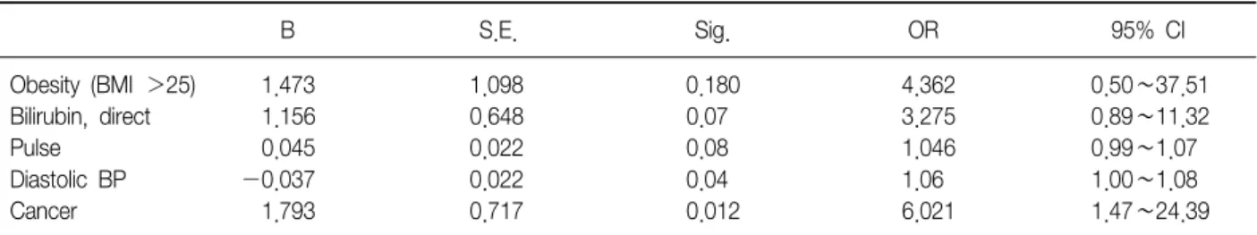
Table 5. Factors associated with death in patients with pulmonary embolism by multiple logistic regression analysis
B S.E. Sig. OR 95% CI
Obesity (BMI >25) 1.473 1.098 0.180 4.362 0.50∼37.51
Bilirubin, direct 1.156 0.648 0.07 3.275 0.89∼11.32
Pulse 0.045 0.022 0.08 1.046 0.99∼1.07
Diastolic BP -0.037 0.022 0.04 1.06 1.00∼1.08
Cancer 1.793 0.717 0.012 6.021 1.47∼24.39
B: regression coefficient; S.E.: standard error; Sig.: significance; OR: odds ratio; CI: confidence interval; BMI: body mass index.
Table 6. Summary of protein C and protein S deficiency
Deficiency
Protein C Protein S
Type I Type II Type I Type II Type III
PE Patients 2 2 2 7 2
(n=42)
Mean age, 52.5 67 51.5 66.9 66.5
yr
Age range, 48∼57 58∼76 48∼55 42∼80 57∼76 yr
다. 암이 동반된 경우에도 유의한 위험인자로 관찰되었 다. 다변량분석에서는 낮은 이완기혈압(p<0.04; odds ratio [OR], 1.06)과 암이 동반된 경우(p<0.012; OR, 6.02)만이 유의한 사망 예측인자로 관찰되었다(Table 5).
2. 응고인자억제제(coagulation factor inhibitor) 결핍
총 42명의 환자에서 응고인자억제제를 검사하였다. 응 고인자억제제 이상은 정상 하한치의 80% 미만인 경우로 정하였고, 기준은 다음과 같다. C단백 활성도 44% 이하, C단백 총 항원치 0.14 mg/dL 이하, S단백 활성도 44%이하, S단백 총 항원치 여자 0.752 mg/dL, 남자 1.08 mg/
dL 이하로 하였다. 이 기준을 적용할 때 C단백 결핍은 4명 (9.5%), S단백 결핍은 11명(26%)이었다. C단백과 S단백 이 같이 결핍된 예는 4명으로 관찰되었다. C단백의 활성 도가 감소한 4예의 활성도는 정상범위의 25∼44%였고, S단백의 활성도가 감소한 11예의 활성도의 범위는 2.8∼
42%였다. C단백과 S단백이 동시에 결핍된 경우가 4예에 서 관찰되었다(Table 6).
고 찰
1999년에 발표한 International Cooperative Pulmonary
Embolism Registry (ICOPER) 연구에서 90일째 사망률이 17.4%였고10, 대한결핵 및 호흡기학회 연구에서는 90일째 사망률이 11%였다4. 본 연구에서 폐색전증 환자의 90일째 사망률이 18.1%로 관찰되었다. 단일기관의 연구이지만, 예상과는 달리 사망률이 여전히 높았다. 대한결핵 및 호 흡기학회에서 진단 당시의 환자의 평균 연령은 58세였고, ICOPER 연구의 진단 당시 평균 연령은 62세였으나, 본 연 구에서는 평균 64세였고, 70세 이상의 환자 비율을 보면 ICOPER 연구의 경우 28.4%이나 본 연구에서는 70세 이상 환자 비율이 42%로 높아 대상 환자의 연령이 예후에 영향 을 주었을 것으로 추측할 수 있다. 또한 본 연구에는 대상 군의 27%에서 암이 동반되었고, 이는 ICOPER 22.5%, 대 한결핵 및 호흡기학회 연구 15.8%에 비해 높은 것으로 3개월째 사망률이 줄지 않은 또 다른 인자로 생각할 수 있다.
대한결핵 및 호흡기학회 조사에서는 고혈압이 27.9%로 위험인자 중에서 가장 빈도가 높았으나, 본 연구에서는 3일 이상의 거동 제한(immobilization)이 45%로 가장 빈 도가 높은 위험인자로 관찰되었다. ICOPER 연구의 경우 5일 이상 거동 제한의 빈도는 28%였고, 대한결핵 및 호흡 기학회 보고에서는 거동 제한의 빈도가 23%로 본 연구에 비해 낮은 경향을 보여, 본 연구에서는 진단 연령의 고령 화와 더불어 거동 제한이 주요한 위험인자로 대두되고 있 음을 추측할 수 있다. 2000년 이후에 폐색전증에 대한 인 지도가 높아지고 다채널검출기를 장착한 전산화단층촬영 을 시행하여 진단율이 높아지면서 이에 대한 역학도 변화 가 있을 것으로 추측할 수 있다. 대한결핵 및 호흡기학회 조사에서는 전산화단층촬영으로 진단한 비율이 42.9%였 지만, 본 연구에서는 105명 중에서 102명(97%)의 환자가 전산화단층촬영으로 폐색전증을 진단하여 이러한 의견을 뒷받침 한다고 볼 수 있다.
국내에서 다기관 공동 연구를 통해 대사성 증후군이 심 부 정맥 혈전증과 관련이 있음을 밝힌 바 있으며11, 본 연
구의 대상군에서도 신체질량지수(body mass index, BMI) 25 이상의 예가 22%였고, ICOPER 연구에서는 BMI 29 이 상인 예가 29%로 관찰되었으나, 대한결핵 및 호흡기학회 조사에서는 대상환자의 7.4%만이 BMI 25 이상으로 관찰 되었다. 식생활 형태가 점차 서구화 되는 경향을 보이는 점을 고려할 때, 대사성증후군과 연관된 질병이 점차로 두드러질 것으로 생각한다.
본 연구의 다변량분석에서 암의 동반 유무가 사망과 유 의한 연관성을 보인 예후인자로 관찰되었다. 폐암과 동반 된 폐색전증의 경우 환자 대조군 연구에서는 폐색전증이 전체적인 예후에 영향을 미치지 못한다는 보고도 있으나12, 암 환자에서 혈전증이 동반될 경우 예후가 좋지 않은 것은 이미 잘 밝혀져 있다13. ICOPER 연구나 대한결핵 및 호흡 기학회 조사 등에서도 암이 유의한 예후인자임을 밝힌 바 있다4,10.
C단백과 S단백 결핍의 역학은 대상 환자군에 따라 차이 가 있는데, 건강인에서는 결핍의 빈도가 매우 낮지만, 50 세 미만의 재발성 심부정맥혈전증 혹은 가족력이 있는 환 자군에서는 C단백 결핍의 빈도가 4.8%, S단백 결핍의 빈 도가 4.3%로 높게 관찰된다14. 국내에서는 정맥혈전증에 서 C단백 결핍증은 10.3%, S단백 결핍증은 6.9%로 관찰 된 바 있고15, 만성폐색전증 환자에서는 C단백이 결핍된 환자는 20명 중에서 5명(25%)이었고 S단백이 결핍된 환 자는 20명 중 1명(5%)이었다16. 본 연구에서는 C단백 결 핍은 4예(9.5%), S단백 결핍은 11예(26%)로 높게 관찰되 었다. 일본에서의 한 연구는, 심부정맥혈전증 환자에서 C 단백 결핍은 113예 중 10예(9%), S단백 결핍은 113예 중 21예(19%)로 높게 관찰되어, 본 연구와 유사한 빈도로 관 찰된 바 있다17. 향후 다 기관 연구를 통해 본 연구 대상 환자군의 선택에 문제가 있는지, 국내의 폐색전증 환자에 서 일반적으로 높은 빈도로 존재하는지는 추가 연구가 필 요하겠다. S단백과 C단백 결핍이 심부정맥혈전의 위험도 를 높이는 것은 잘 알려져 있어서18,19, 응고인자억제제 결 핍이 발견될 경우 치료 및 추적 관찰에서 각별한 주의가 요망된다.
결론적으로, 폐색전증은 사망률이 높은 질환으로, 혈전 증 발생과 관련 있는 위험인자를 가진 경우 폐색전증 발생 유무를 적극적으로 확인하려는 노력이 필요하겠고, 응고 인자억제제 결핍에 대한 관심과 주의가 필요할 것으로 생 각한다.
참 고 문 헌
1. Park YS, Ha JW, Kwon KH, Jang YS, Chung NS, Shim WH, et al. Clinical characteristics and predictors of in-hospital mortality for patients with acute major pul- monary embolism. Korean J Med 2000;58:293-300.
2. Lee GL, Kim JY, Park JS, Yoo CG, Kim YW, Shim YS, et al. Clinical study of the patients, in whom pulmo- nary embolism was suspected by lung perfusion scan.
Tuberc Respir Dis 1997;44:889-98.
3. Lee SS, Lim CM, Song KS, Sung KB, Koh YS, Lee SD, et al. The value of spiral computed tomography in the diagnosis of pulmonary embolism. Korean J Med 1997;
53:787-94.
4. Lee S, Jeong H, In K, Yoo S, Kim S, Kim J, et al.
Clinical characteristics of acute pulmonary throm- boembolism in Korea. Int J Cardiol 2006;108:84-8.
5. Stein PD, Fowler SE, Goodman LR, Gottschalk A, Hales CA, Hull RD, et al. Multidetector computed tomog- raphy for acute pulmonary embolism. N Engl J Med 2006;354:2317-27.
6. Anderson DR, Kahn SR, Rodger MA, Kovacs MJ, Morris T, Hirsch A, et al. Computed tomographic pulmonary angiography vs ventilation-perfusion lung scanning in patients with suspected pulmonary embolism: a randomized controlled trial. JAMA 2007;298:2743-53.
7. DeMonaco NA, Dang Q, Kapoor WN, Ragni MV.
Pulmonary embolism incidence is increasing with use of spiral computed tomography. Am J Med 2008;121:
611-7.
8. British Thoracic Society Standards of Care Committee Pulmonary Embolism Guideline Development Group.
British Thoracic Society guidelines for the management of suspected acute pulmonary embolism. Thorax 2003;
58:470-83.
9. Wells PS, Anderson DR, Rodger M, Stiell I, Dreyer JF, Barnes D, et al. Excluding pulmonary embolism at the bedside without diagnostic imaging: management of patients with suspected pulmonary embolism present- ing to the emergency department by using a simple clinical model and d-dimer. Ann Intern Med 2001;135:
98-107.
10. Goldhaber SZ, Visani L, De Rosa M. Acute pulmonary embolism: clinical outcomes in the International Coop- erative Pulmonary Embolism Registry (ICOPER). Lancet 1999;353:1386-9.
11. Jang MJ, Choi WI, Bang SM, Lee T, Kim YK, Ageno W, et al. Metabolic syndrome is associated with venous thromboembolism in the Korean population. Arteriosc-
ler Thromb Vasc Biol 2009;29:311-5.
12. Lee JW, Cha SI, Jung CY, Choi WI, Jeon KN, Yoo SS, et al. Clinical course of pulmonary embolism in lung cancer patients. Respiration 2009;78:42-8.
13. Sorensen HT, Mellemkjaer L, Olsen JH, Baron JA.
Prognosis of cancers associated with venous throm- boembolism. N Engl J Med 2000;343:1846-50.
14. Seligsohn U, Lubetsky A. Genetic susceptibility to ve- nous thrombosis. N Engl J Med 2001;344:1222-31.
15. Lee A, Song KS. Screening results of AT III, protein C and S, and APC resistance for diagnosis of inheritied thrombophilia. Korean J Thromb Hemost 1995;2:147- 53.
16. Kim HK, Na JO, Ahn JJ, Park YB, Lim JM, Hong SB, et al. Comparison of clinical features between idio-
pathic pulmonary arterial hypertension and chronic th- romboembolic pulmonary hypertension. Tuberc Respir Dis 2005;59:170-8.
17. Suehisa E, Nomura T, Kawasaki T, Kanakura Y.
Frequency of natural coagulation inhibitor (antithrom- bin III, protein C and protein S) deficiencies in Japane- se patients with spontaneous deep vein thrombosis.
Blood Coagul Fibrinolysis 2001;12:95-9.
18. Tabernero MD, Tomas JF, Alberca I, Orfao A, Lopez Borrasca A, Vicente V. Incidence and clinical character- istics of hereditary disorders associated with venous thrombosis. Am J Hematol 1991;36:249-54.
19. De Stefano V, Finazzi G, Mannucci PM. Inherited th- rombophilia: pathogenesis, clinical syndromes, and ma- nagement. Blood 1996;87:3531-44.