Tuberc Respir Dis 2011;70:498-503
CopyrightⒸ2011. The Korean Academy of Tuberculosis and Respiratory Diseases. All rights reserved.
만성 폐쇄성 폐질환(COPD) 환자에서 운동 시 발생하는 산소 불포화 반응과 관련된 인자
계명대학교 의과대학 1내과학교실, 2예방의학교실, 3영상의학교실
심상우1, 조준연1, 권용식1, 채진녕1, 박지혜1, 이미영2, 노병학3, 최원일1
Factors Related to Exertional Oxygen Desaturation in Patients with COPD
Sang Woo Shim, M.D.1, Jun Yeon Jo, M.D.1, Yong Sik Kwon, M.D.1, Jin Nyeong Chae, M.D.1, Jie Hae Park, M.D.1, Mi-Young Lee, M.D.2, Byung Hak Rho, M.D.3, Won-Il Choi, M.D., Ph.D.1
Departments of 1Internal Medicine, 2Preventive Medicine, and 3Diagnostic Radiology, Keimyung University School of Medicine, Daegu, Korea
Background: The causes of exertional desaturation in patients with COPD can be multifactorial. We aimed to investigate factors predict exertional desaturation in patients with moderate to severe COPD.
Methods: We tested 51 consecutive patients with stable COPD (FEV1/FVC, 40±13% predicted). Patients performed a six minute walk test (6MWT). Pulse oxymetric saturation (SpO2) and pulse rate were recorded.
Results: Oxygen desaturation was found in 15 subjects after 6MWT, while 36 subjects were not desaturated. Lung diffusing capacity was significantly lower in desaturation (DS) group (62±18% predicted) compared with not desaturated (ND) group (84±20, p<0.01). However there was no statistical difference of FEV1/FVC ratio or residual volume between two groups. The pulse rate change was significantly higher in the desaturated compared with the not desaturated group. Six minute walking distance, subjective dyspnea scale, airflow obstruction, and residual volume did not predict exertional oxygen desaturation. Independent factors assessed by multiple logistic regression revealed that a pulse rate increment (odd ratio [OR], 1.19; 95% confidence interval [CI], 1.01∼1.40; p=0.02), a decrease in baseline PaO2 (OR, 1.105; 95% CI, 1.003∼1.218; p=0.04) and a decrease in lung diffusing capacity (OR, 1.10; 95% CI, 1.01∼1.19; p=0.01) were significantly associated with oxygen desaturation. Receiver operator characteristic (ROC) analysis showed that an absolute increment in pulse rate of 16/min gave optimal discrimination between desaturated and not desaturated patients after 6MWT.
Conclusion: Pulse rate increment and diffusion capacity can predict exertional oxygen desaturation in stable COPD patients with moderate to severe airflow obstruction.
Key Words: Pulmonary Disease, Chronic Obstructive; Heart Rate; Anoxia
Address for correspondence: Won-Il Choi, M.D., Ph.D.
Department of Internal Medicine, Keimyung University School of Medicine, 216, Dalseong-ro, Jung-gu, Daegu 700-712, Korea
Phone: 82-53-250-7572, Fax: 82-53-250-7434 E-mail: [email protected]
Received: Jan. 22, 2011 Accepted: May 21, 2011
서 론
만성 폐쇄성 폐질환(chronic pulmonary obstructive disease, COPD) 환자에서 발생하는 호흡곤란은 환자의 건강상태에 부정적인 영향을 주고, 사회적 활동범위를 결 정하는 중요한 요소이다1,2. COPD 환자에서 활동 시 산소 불포화(oxygen desaturation) 반응이 발생하면, 이로 인해 활동이 제약된다3,4. 저산소혈증이 동반된 COPD 환자에 산소 치료를 할 경우 사망률이 감소되며5,6, 국내에서도 안
정 시 저산소혈증이 있는 환자에서 산소가 널리 처방되고 있다.
COPD 환자에서 저산소혈증은 기류제한과 연관된 환기 관류 장애에 의하여 주로 발생한다고 알려져 있지만7,8, COPD 환자에서 쌕쌕거림이나 가래가 없는 상황에서도 산소포화도의 차이가 있는 경우를 고려하면, 기류제한에 의한 환기관류 장애만으로는 설명되지 않는 부분이 있다.
COPD 환자에서 운동 시 발생하는 산소불포화 반응은 환 기관류 장애7, 폐확산능의 제한9,10, 혼합정맥혈의 단락, 폐 포 저환기, 폐로 회귀하는 혼합정맥혈의 감소된 산소 등 다양한 원인에 의해 발생할 수 있으며, 운동 전 산소포화 도는 운동 후 산소불포화 반응과 유의한 상관관계가 있다 는 보고도 있다9-12.
COPD 환자에서 6분 보행 검사(6-minute walk test, 6MWT)는 활동 시 산소불포화를 발견하는데 운동부하 검 사에 비해 더 민감한 것으로 알려져 있으며13, 6분 보행검 사의 운동거리와 산소소모량은 COPD 환자의 생존율과 연관되어 있다4,14. 이에 본 연구는 COPD 환자에서 6분 보행 검사를 시행하여, 산소불포화 반응과 관련된 요인들 을 조사하고자 한다.
대상 및 방법 1. 연구대상
본 연구는 계명대학교 동산병원 의학연구윤리심의위원 회(Institutional Review Board)의 승인을 받아 시행되었 다. 연구대상은 COPD로 진단받고 최근 2개월간 기침, 가 래, 호흡곤란 등 호흡기 증상의 변화 없이 안정적으로 외 래에서 추적관찰 중인 환자를 대상으로 하였다. 2006년 6월부터 2008년 12월 사이에 계명대학교 동산의료원 호 흡기내과를 방문한 환자 중 COPD 환자 51명이 연구에 참여하였다. 호흡기 질환 이외의 장애로 보행을 하지 못 하는 경우는 대상에서 제외하였다. 모든 환자들은 폐기능 검사와 6분 보행 검사를 하였다.
2. 폐기능 검사
폐활량은 미국 SensorMedics사(Sensormedics Corp., Anaheim, CA, USA)의 6200 Autobox DL Pulmonary Function Laboratory를 이용하였다. Plethysmographic 방 법으로 기능적 잔기량(functional residual capacity, FRC) 을 측정하였다. 폐활량의 추정 정상치는 한국인을 대상으 로 제시한 식으로 계산하였다15.
3. 6분 보행 검사(6-Minute Walk Test)
6분 보행 검사는 미국흉부학회(American Thoracic So- ciety, ATS)에서 제시하는 방법을 바탕으로 45 미터의 거 리, 직선으로 된 복도를 반복하여 시행하였다16. 6MWT 전 혈압 측정과 동맥혈 가스 분석을 하였다. 6MWT 시행 전과 후에 맥박수, 산소포화도, medical research council (MRC) 호흡곤란 척도로 호흡곤란 정도를 측정하였다. 산 소포화도는 손가락형 맥박 산소 측정기(Onyx 9,500, Nonin Finger Pulse Oximeter)를 이용하여 측정하였다.
본 연구에서는 6MWT 직후에 측정한 산소포화도가 기저 치에 비해 2% 이상 감소한 경우를 산소불포화군(desatur- ated, DS)으로, 기저치에 비해 감소하지 않거나 증가한 경 우를 산소불포화가 없는 군(not desaturated, ND)으로 정 의하였다17.
4. 통계분석
결과값은 평균값±표준편차로 나타내었다. DS와 ND, 두 군 사이의 평균치 비교는 독립표본 t-검정, 그리고 6분 보행 검사 시행 전후의 산소포화도 감소에 미치는 인자는 다중 로지스틱 회귀분석법을 이용하였고, 이때 단변량 분 석에서 통계적인 유의성이 있거나, 중요한 영향을 미칠 것으로 여겨지는 변수들(Hct, FEV1, residual volume, baseline PCO2, baseline PaO2, baseline O2 saturation)을 함께 투입하였다. 6분 보행 검사 전 후 맥박수의 변화 및 산소포화도는 피어슨 상관계수를 구하여 상관관계를 분 석하였다. 통계 프로그램은 SPSS version 18.0 (SPSS Inc., Chicago, IL, USA)을 이용하였고, 0.05 미만의 p값을 통계 적으로 유의한 것으로 판단하였다.
결 과 1. 환자 특성
연구 대상자는 모두 51명으로 남자가 45명(88%), 여자 가 6명(12%)이었다. 전체 환자의 연령범위는 43세부터 82 세였으며 평균연령은 66±9세였다(Table 1). 흡연량은 평 균 39갑년이었다.
2. 폐기능 검사
대상자에서 1초간 노력성 호기량의 노력성 폐활량에 대 한 비(forced expiratory volume in 1 second/forced vital capacity, FEV1/FVC)의 평균은 49%였으며, FEV1 예측치
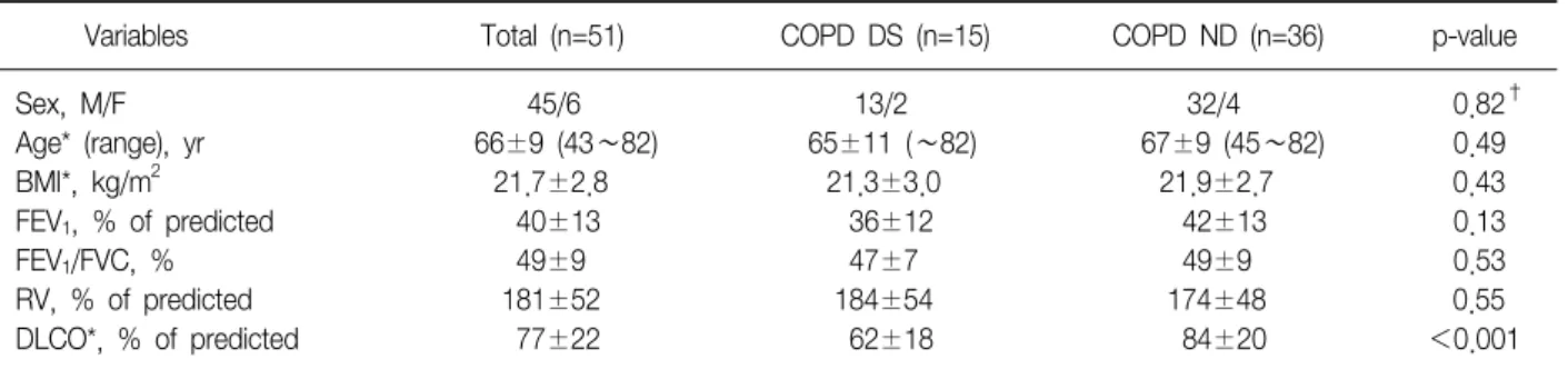
Table 1. Patient characteristics and pulmonary function data
Variables Total (n=51) COPD DS (n=15) COPD ND (n=36) p-value
Sex, M/F 45/6 13/2 32/4 0.82†
Age* (range), yr 66±9 (43∼82) 65±11 (∼82) 67±9 (45∼82) 0.49
BMI*, kg/m2 21.7±2.8 21.3±3.0 21.9±2.7 0.43
FEV1, % of predicted 40±13 36±12 42±13 0.13
FEV1/FVC, % 49±9 47±7 49±9 0.53
RV, % of predicted 181±52 184±54 174±48 0.55
DLCO*, % of predicted 77±22 62±18 84±20 <0.001
*Mean±SD, †Chi-square test for comparison.
COPD: chronic pulmonary obstructive disease; DS: desaturated; ND: not desaturated; BMI: body mass index; FEV1: forced expiratory volume in one second; FEV1/FVC: forced expiratory volume to forced vital capacity ratio; RV: residual volume; DLCO: diffusing capacity of the lung for carbon monoxide; SD: standard deviation.
Table 2. Oxygen saturation and pulse rate of pre and post 6MWT
Variables
COPD (n=51)
p-value Desaturation No desaturation
Number 15 36
O2 saturation, %
Pre 94.0±3.9 95.2±1.9 0.13
Post 87.5±7.6 95.8±2.3 <0.001
Pulse, /min
Pre 83±18 82±13 0.83
Post 104±22 98±14 0.12
Pulse difference 21±12 13±8 0.02
6MWD, m 326±80 339±93 0.65
Hct 41.3±4.5 40.5±4.2 0.51
Baseline blood gas
pH 7.40±0.03 7.42±0.03 0.14
PCO2, mm Hg 42.4±5.3 39.1±6.3 0.09 HCO3−
, mEq/L 26.2±2.4 25.0±3.6 0.28
PaO2, mm Hg 70.5±13.6 85.7±17.5 0.03 Baseline MRC grade 3.6±0.8 3.1±0.7 0.08 Values are presented as Mean±SD unless otherwise indicated.
COPD: chronic pulmonary obstructive disease; 6MWT: 6-mi- nute walk test; 6MWD: 6-minute walk distance; MRC: medical research council; SD: standard deviation.
의 평균은 40%였다. Global Initiative for Chronic Ob- structive Lung Disease (GOLD) 진료지침에 따라 COPD 의 중증도를 분류하면 중등증 20명(37%), 중증 25명 (46%), 고도 중증 9명(17%)이었다. 폐확산능(diffusing capacity of carbon monoxide, DLCO)의 예측치는 산소불 포화군에서 산소불포화가 발생하지 않은 군에 비해 22%
정도 낮았고, 통계적으로 유의하였다(Table 1). 그러나 FEV1, FEV1/FVC과 잔기량(residual volume) 등은 두 군 사이에 유의한 차이가 관찰되지 않았다.
3. 6분 보행 검사
검사 전후 손가락형 맥박 산소 측정기를 사용하여 산소 포화도와 맥박수를 측정하였다. 15명(29%)에서 산소포화 도가 감소하였고, 평균 감소치는 −6.4±5.7%였다. 산소 포화도가 감소하지 않은 환자 36명은 검사 전보다 평균 0.6±.6% 증가하였다. 산소포화도가 감소되지 않은 군 7 명에서 2% 이상 산소포화도가 증가하였다. 51명의 환자 중 48명이 검사 후에 맥박수가 1회/분 이상 증가하였고, 1명은 변화가 없었으며, 2명은 검사 전보다 1회/분 이상 감소하였다. 맥박수가 10회/분 미만으로 증가한 환자는 10명(19%), 10∼19회/분 증가한 환자는 20명(43%), 20∼
29회/분 증가한 환자는 12명(23%), 30회/분 이상 증가한 환자는 4명(7%)이었다. 산소불포화 반응이 발생한 환자 군(DS)에서 검사 후 맥박수는 평균 21회가 증가하여, 평 균맥박이 14회 증가한 ND군에 비해 통계적으로 유의한 차이가 관찰되었다(p=0.019). 6분 보행 검사 보행거리, MRC 호흡곤란 척도, 적혈구 용적률(hematocrit), 운동 전 산소포화도, pH, HCO3−
, PaCO2 등은 양 군 사이에 유의 한 차이가 관찰되지 않았다. 기저 동맥혈 산소분압(PaO2)
은 산소포화도가 감소한 군에서 감소하지 않은 군에 비해 유의하게 낮았다(p=0.03) (Table 2).
4. 산소포화도 감소와 관련된 인자
산소포화도 감소에 영향을 미칠 수 있는 변수들을 다중 로지스틱 회귀방법으로 분석한 결과에서 맥박수 증가와, 기저 동맥혈 산소분압, 그리고 폐확산능 감소만이 유의한
Figure 1. Correlation between the change in oxygen satu- ration and pulse rate before and after 6-minute walk test.
Table 3. Multiple logistic regression analysis for factors as- sociated with oxygen desaturation in patients with COPD after 6-minute walk test
Variable OR 95% CI
Hct 0.908 0.702∼1.175
FEV1, % predicted 0.979 0.905∼1.058 A decrease in DLCO, % predicted 1.101 1.011∼1.198
RV, % predicted 0.988 0.966∼1.010
Pulse rate difference 1.190 1.010∼1.402
Baseline PaCO2 1.028 0.844∼1.253
A decrease in baseline PaO2 1.105 1.003∼1.218
Baseline O2 sat 1.098 0.755∼1.599
COPD: chronic pulmonary obstructive disease; OR: odd ratio;
CI: confidence interval; FEV1: forced expiratory volume in 1 sec- ond; DLCO: diffusing capacity of the lung for carbon monoxide;
RV: residual volume.
Table 4. Sensitivity, specificity of pulse rate increment after 6-minute walk test
Pulse rate change, per min Sensitivity Specificity
>5 93 0
>11 87 47.2
>16 67 61.1
>20 53 80.5
>24 33 83.3
Total number of patients is 51 and rates are represented as percent.
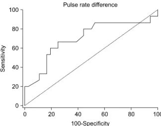
Figure 2. ROC curve of pulse rate change. Area under the ROC curve=0.704 (95% CI, 0.56 to 0.82). ROC: re- ceiver operating characteristic; CI: confidence interval.
인자로 관찰되었다(Table 3). 6분 보행 검사 전 후의 맥박 수 변화와 산소포화도 변화는 상관계수는 0.53 (p<
0.001)으로 서로 유의한 상관관계가 있었다(Figure 1).
5. 산소포화도 감소 진단의 민감도와 특이도
맥박 수가 16회 이상 증가한 경우를 기준으로 했을 때 15명의 산소포화도 감소 환자 중에서 10명을 양성으로 판 정하여 민감도 67%, 36명의 산소포화도가 변동이 없거나 증가한 환자 중에서는 22명에서 음성으로 판정하여 특이 도는 61%로 관찰되었고(Table 4), 이때 receiver operat- ing characteristic (ROC) 곡선의 하부 영역(area under the ROC curve)은 0.70으로 중등도의 정확한 검사수준으로
관찰되었다(Figure 2).
고 찰
COPD 환자에게 발생하는 만성 저산소혈증과 고이산화 탄소혈증은 전신질환의 발생위험도를 증가시킨다18. 안정 시 산소포화도는 정상범위이나 운동 시 발생하는 저산소 혈증이 있는 COPD 환자에게 이동용 산소치료를 하면 운 동능력이 개선된다19.
본 연구는 COPD 환자에서 운동 시에 발생하는 산소포 화도 감소와 연관된 인자를 분석하고자 했다. 산소불포화 반응이 일어난 환자 군은 산소포화도의 변화가 없는 군에 비하여 폐확산능이 통계적으로 유의하게 낮았으며 이는 이전의 연구와 잘 일치하였다9-12.
본 연구에서 6MWT 후 맥박수의 변화 정도는 산소불포
화 반응이 일어난 군이 분당 평균 21회가 증가되었고, 산 소포화도의 변화가 없었던 군은 분당 평균 14회 증가되었 으며 유의한 차이가 있었다(p=0.02). 맥박수의 증가 폭이 클수록 산소불포화 정도가 심하여 이들은 음의 상관관계 (R2=0.288)로 관찰되었다(Figure 1). 운동 후 발생하는 맥 박수의 변화는 비교적 간결하게 측정할 수 있으며, 환자의 생리적인 상황을 대변할 수 있는 장점이 있어서 임상적인 유용성이 있으리라 생각한다. 본 연구에서는 6분 보행 검 사 후에 맥박수가 분당 16회 이상 증가할 경우 산소포화 도 감소를 진단할 수 있는 민감도가 67%로, 맥박수 증가 의 기준을 12회로 할 경우 산소포화도 감소를 진단할 수 있는 민감도가 87%로 증가하였다(Table 4). 이에 비해서 6분 보행 검사 보행거리 또는 운동 전 호흡곤란의 정도는 산소포화도 감소를 예측하는데 도움을 주지 않았다.
이전 연구에서는 운동 후 발생한 산소불포화를 기저치 에 비해 2∼4% 감소한 것으로 정의하였다9,20. 본 연구에 서 사용한 산소포화도 측정기(Onyx 9,500; Nonin Finger Pulse Oximeter, Nonin Medical, Inc., Minneapolis, MN, USA)는 평균오차가 0.3% 내외로 낮은 점을 고려하여17, 6분 보행 검사 전후를 비교하여 산소포화도가 2% 이상 감소된 환자를 산소불포화가 있는 군으로 정의하였다. 산 소불포화의 기준을 2%로 했을 때 폐확산능과 운동 후 산 소포화도 감소의 정도 및 상관관계는 이전의 보고들과 잘 일치하였다9-12.
FEV1, FEV1/FVC, 그리고 잔기량 등은 6분 보행 검사 후 산소포화도가 감소한 군과 산소포화도가 감소하지 않 은 군 사이에 차이가 없었다. FEV1/FVC과 FEV1이 운동 후 산소포화도 감소를 예측할 수 있다는 보고가 있었지 만9,21, 본 연구에서는 두 변수 모두 다중 회귀분석에서 유 의한 결과를 보이지 않았다. DS군과 ND군의 잔기량(RV) 과 FEV1, FEV1/FVC 등의 유의한 차이가 없고(Table 1), 다중 로지스틱 회귀분석에서 유의한 결과를 보이지 않은 점을 고려한다면, 본 연구에서 기류제한으로 인한 산소포 화도 감소의 정도는 매우 적었을 것으로 보인다.
운동 전 산소포화도가 운동 후 산소포화도 감소를 예측 할 수 있다는 보고가 있고12, 본 연구에서도 운동 전 산소 포화는 운동 후 산소포화도 감소와는 통계적 유의성이 없 었으나 산소포화도 감소 군에서 산소포화도 감소가 없는 군에 비해 기저 PaO2가 유의하게 낮았다. 이는 산소포화 도 보다는 동맥혈 산소분압이 혈중 산소농도를 더 민감하 게 반영해서 생긴 것으로 보인다. 따라서 기저 동맥혈 산 소농도를 통해서 운동 후 산소포화도 감소를 예측할 수
있다.
COPD 환자에서 안정 시 동맥혈 산소분압이 55 mm Hg 이하인 환자에게 하루 18시간 이상의 산소치료는 사망률 을 감소시킨다6. 그러나, 안정 시에는 저산소혈증이 없으 나, 운동 시에 저산소혈증이 발생하는 COPD 환자에서는 산소치료가 증상호전에 명백한 이득이 없다는 연구도 있 다3,22. 따라서 향후 연구를 통해 산소포화도 감소의 원인 이 저산소혈증으로 설명되는 군과 다른 원인, 예를 들면 이산화탄소 분압이 증가해서 생기는 경우 등으로 나누어 서, 평가할 필요가 있다.
7명의 COPD 환자에서는 운동 후 산소포화도가 2% 이 상 증가하였다. 이는 운동 시에 폐 혈류관류 불균형(venti- lation perfusion mismatch)이 호전되어 발생하는 현상으 로 보인다.
결론적으로, 폐확산능은 COPD 환자에서 운동 시 산소 포화도 감소를 예측할 수 있으며, 운동 전후 맥박수의 증 가 폭은 산소포화도 감소를 예측할 수 있다. 중등도 이상 의 COPD 환자에서 6분 보행 검사를 기준으로 검사 전후 에 16회 이상 맥박수가 증가할 경우 산소포화도 감소를 진단하는 민감도는 67%이다.
참 고 문 헌
1. Reardon JZ, Lareau SC, ZuWallack R. Functional status and quality of life in chronic obstructive pulmonary disease. Am J Med 2006;119(10 Suppl 1):32-7.
2. Weaver TE, Richmond TS, Narsavage GL. An ex- planatory model of functional status in chronic ob- structive pulmonary disease. Nurs Res 1997;46:26-31.
3. Nonoyama ML, Brooks D, Guyatt GH, Goldstein RS.
Effect of oxygen on health quality of life in patients with chronic obstructive pulmonary disease with tran- sient exertional hypoxemia. Am J Respir Crit Care Med 2007;176:343-9.
4. Cote CG, Pinto-Plata V, Kasprzyk K, Dordelly LJ, Celli BR. The 6-min walk distance, peak oxygen uptake, and mortality in COPD. Chest 2007;132:1778-85.
5. Continuous or nocturnal oxygen therapy in hypoxemic chronic obstructive lung disease: a clinical trial.
Nocturnal Oxygen Therapy Trial Group. Ann Intern Med 1980;93:391-8.
6. Long term domiciliary oxygen therapy in chronic hy- poxic cor pulmonale complicating chronic bronchitis and emphysema. Report of the Medical Research Council Working Party. Lancet 1981;1:681-6.
7. Wagner PD, Dantzker DR, Dueck R, Clausen JL, West JB. Ventilation-perfusion inequality in chronic ob- structive pulmonary disease. J Clin Invest 1977;59:
203-16.
8. West JB. Causes of carbon dioxide retention in lung disease. N Engl J Med 1971;284:1232-6.
9. Owens GR, Rogers RM, Pennock BE, Levin D. The dif- fusing capacity as a predictor of arterial oxygen desatu- ration during exercise in patients with chronic ob- structive pulmonary disease. N Engl J Med 1984;310:
1218-21.
10. Sue DY, Oren A, Hansen JE, Wasserman K. Diffusing capacity for carbon monoxide as a predictor of gas ex- change during exercise. N Engl J Med 1987;316:1301-6.
11. Hadeli KO, Siegel EM, Sherrill DL, Beck KC, Enright PL.
Predictors of oxygen desaturation during submaximal exercise in 8,000 patients. Chest 2001;120:88-92.
12. Knower MT, Dunagan DP, Adair NE, Chin R Jr.
Baseline oxygen saturation predicts exercise desatura- tion below prescription threshold in patients with chronic obstructive pulmonary disease. Arch Intern Med 2001;161:732-6.
13. Poulain M, Durand F, Palomba B, Ceugniet F, Desplan J, Varray A, et al. 6-minute walk testing is more sensi- tive than maximal incremental cycle testing for detect- ing oxygen desaturation in patients with COPD. Chest 2003;123:1401-7.
14. Casanova C, Cote C, Marin JM, Pinto-Plata V, de Torres JP, Aguirre-Jaíme A, et al. Distance and oxygen desatu- ration during the 6-min walk test as predictors of
long-term mortality in patients with COPD. Chest 2008;134:746-52.
15. Choi JK, Paek D, Lee JO. Normal predictive values of spirometry in Korean population. Tuberc Respir Dis 2005;58:230-42.
16. ATS Committee on Proficiency Standards for Clinical Pulmonary Function Laboratories. ATS statement:
guidelines for the six-minute walk test. Am J Respir Crit Care Med 2002;166:111-7.
17. Torre-Bouscoulet L, Chávez-Plascencia E, Vázquez- García JC, Pérez-Padilla R. Precision and accuracy of
"a pocket" pulse oximeter in Mexico City. Rev Invest Clin 2006;58:28-33.
18. Agustí AG, Noguera A, Sauleda J, Sala E, Pons J, Busquets X. Systemic effects of chronic obstructive pul- monary disease. Eur Respir J 2003;21:347-60.
19. Bradley JM, O'Neill B. Short-term ambulatory oxygen for chronic obstructive pulmonary disease. Cochrane Database Syst Rev 2005;(4):CD004356.
20. Kelley MA, Panettieri RA Jr, Krupinski AV. Resting sin- gle-breath diffusing capacity as a screening test for ex- ercise-induced hypoxemia. Am J Med 1986;80:807-12.
21. Ries AL, Farrow JT, Clausen JL. Pulmonary function tests cannot predict exercise-induced hypoxemia in chronic obstructive pulmonary disease. Chest 1988;93:
454-9.
22. Ram FS, Wedzicha JA. Ambulatory oxygen for chronic obstructive pulmonary disease. Cochrane Database Syst Rev 2002;(2):CD000238.