돌연변이 p53 단백질의 Silencing에 의한 사람유방암세포의 in vivo 항 종양 효과
박원익1, 박세라1, 박현주1, 배윤희1, 유현수1, 장혜옥1, 배문경2, 배수경1*
Silencing of Mutant p53 Leads to Suppression of Human Breast Xenograft Tumor Growth in vivo
Won Ick Park1, Se-Ra Park1, Hyun-Joo Park1, Yun-Hee Bae1, Hyun Su Ryu1, Hye-Ock Jang1, Moon-Kyoung Bae2, and Soo-Kyung Bae1*
Received: 18 January 2016 / Revised: 19 February 2016 / Accepted: 28 February 2016
© 2016 The Korean Society for Biotechnology and Bioengineering
Abstract: Mutant p53 (R280K) is highly expressed in MDA- MB-231 triple-negative human breast cancer cells. Currently, we reported the role of mutant p53-R280K in mediating the survival of MDA-MB-231 cells in vitro. The present study was undertaken to determine whether mutant p53-R280K affects breast cancer cell growth in vivo. To this end, we used small interfering RNA to knockdown the level of mutant p53- R280K in MDA-MB-231 cells. Silencing of mutant p53- R280K in MDA-MB-231 cells causes substantial tumor reg- ression of established xenografts in vivo. In xenograft model for breast cancer, silencing of mutant p53-R280K in MDA- MB-231 cells significantly inhibited the tumor growth. More- over, TUNEL assay showed more occurrence of apoptotic cells in mutant p53-R280K silenced tumors compared to con- trol. Our data indicate that mutant p53-R280K has an impor- tant role in mediating tumor growth of MDA-MB-231 cells in vivo. Taken together, this study suggests that endogenous mu-
tant p53-R280K could be used as a therapeutic target for breast cancer cells harboring this TP53 missense mutation.
Keywords: Mutant p53, Breast cancer, Apoptosis, Xenograft tumor model
1. INTRODUCTION
TP53 은 p53 단백질의 유전정보를 암호화하고 있는 암억제 유전자이다. 전사인자로 잘 알려져 있는 p53 단백질은 DNA 손상, 저산소, 그리고 암유전자 발현 등과 같은 세포 손상 유 발원에 의해 활성화되며 타깃 유전자들의 전사를 증가시킴 으로써 DNA 수선, 세포성장 지연, 손상 세포의 세포자멸사 (apoptosis) 유발 등의 과정을 통해 종양 발생을 억제하는데 기여한다 [1-3].
TP53 유전자는 사람 암종의 50 % 이상에서 돌연변이가 관 찰되며 이러한 돌연변이로 인해 야생형 (wild type) p53 단백 질은 암을 억제하는 고유 기능을 잃거나 우성음성 (dominant negative) 활성을 나타내는 것으로 알려져 있다 [1-5]. 또한 발 암 기능-획득 (oncogenic gain-of-function) 활성을 새롭게 가지 게 된 p53 돌연변이 단백질들은 DNA 손상 후 나타나는 세포 주기 억류 (cell cycle arrest)와 세포자멸사를 방해함으로써 결 국 암세포 생존율의 증가와 종양 발생을 초래하게 된다 [6,7].
p53 돌연변이 단백질 중에 p53-R280T는 5637 방광암세포 주의 세포 생존을 증가시키며 siRNA를 이용한 실험에서 암
1부산대학교 치의학전문대학원 치과약리학교실
1Department of Dental Pharmacology, School of Dentistry, Pusan Na- tional University, Yangsan 50612, Korea
Tel: +82-51-510-8253, Fax: +82-51-510-8233;
e-mail: [email protected]
2부산대학교 치의학전문대학원 구강생리학교실
2Department of Oral Physiology, School of Dentistry, Pusan National University, Yangsan 50612, Korea
연구논문
세포 내 p53-R280T의 발현 양을 감소시켰을 때 항암제인 cisplatin에 대한 민감도가 증가한다고 알려져 있다 [8]. 또 다 른 p53 돌연변이 단백질인 p53-L194F는 유방암세포주인 T47D의 생존 조절에 관여하며 p53-R273H는 유방암세포주 MDA-MB-468 의 생존 조절을 매개한다고 발표되었다 [9]. 이 러한 연구결과들은 p53 돌연변이 단백질에 의한 암세포의 세 포자멸사 억제에 관한 실험적 증거가 되며 향후 이들 p53 돌 연변이 단백질을 타깃으로 한 항암 치료제 개발에 응용 가능 할 것으로 여겨진다.
트리플-음성 유방암(triple-negative breast cancers, TNBC:
negative for estrogen receptor, progesterone receptor, and HER2 expression) 은 초기 진단이 어렵고 높은 전이능을 가진 악성 종양으로 알려져 있다 [10]. 따라서 이러한 특징을 가진 유방 암세포의 증식과 생존에 영향을 미치는 인자나 유전자에 대 한 연구는 매우 중요하며 현재 활발히 진행 중이다. 정상 p53 단백질의 코돈 280번 자리가 아르기닌에서 리신으로 치환된 p53-R280K은 트리플-음성 유방암 세포인 MDA-MB-231에 서 발견되는 p53 돌연변이 단백질이다 [10-12].
본 연구진은 최근 p53-R280K의 MDA-MB-231 생존 조절 기능에 대한 in vitro 연구결과를 보고하였다 [11,12]. 하지만 지금까지 MDA-MB-231의 in vivo 종양 성장에 미치는 p53- R280K의 효과에 관한 연구는 발표된 바 없다. 따라서 본 연 구에서는 p53-R280K의 in vivo 효과를 확인하기 위해 siRNA 실험기법과 MDA-MB-231을 누드생쥐에 이식하여 종양의 성장을 연구하는 이종이식 모델 (human tumor xenograft mo- dels)을 이용하여 실험을 진행하였으며, 향후 트리플-음성 유 방암에 대한 유전자 치료 타깃으로서 p53-R280K의 활용 가 능성을 탐색하고자 한다.
2. MATERIALS AND METHOD
2.1. 실험재료
Mouse monoclonal anti-p53과 anti-β-actin 항체는 Calbiochem (San Diego, CA)과 Abcam (Cambridge, MA)에서 각각 구입하 였다. Rabbit polyclonal anti-cleaved Notch1 (c-Notch1) 항체는 Milipore (Billerica, MA)에서 구입하였다. Rabbit polyclonal anti-PARP, Bcl-2, p21, Cyclin D1 그리고 mouse monoclonal anti-Caspase-3 항체들은 Santa Cruz Biotechnology (Santa Cruz, CA)에서 구입하였다. Alexa Fluor 488-conjugated anti-rabbit IgG 와 Alexa Fluor 594-conjugated anti-mouse IgG는 Molecu- lar Probes (Eugene, OR)에서 구입하였다. Horseradish peroxi- dase-conjugated goat anti-rabbit IgG와 goat anti-mouse IgG는 Santa Cruz Biotechnology (Santa Cruz, CA)에서 구입하였다.
2.2. 세포배양
인간유방암세포인 MDA-MB-231 세포는 American Type Cul- ture Collection (Manassas, VA)에서 분양 받았다. 열처리로 불 활성화시킨 10% 우태아혈청 (fetal bovine serum, FBS, GIBCO
BRL, Grand Island, NY)과 1% 항생제가 포함된 Dulbecco's modified eagle's medium (DMEM: GIBCO BRL, Grand Island, NY) 을 사용하였고, 37
oC 로 유지되는 5%의 CO
2배양기를 사 용하여 배양하였다.
2.3. Transient transfection of small interfering (si)RNA 실험에 사용된 p53-R280K의 double-strand siRNA oligonu- cleotides (5'-GACUCCAGUGGUAAUCUACTT-3' and 5'-GU AGAUUACCACUGGAGUCTT-3')와 negative-control siRNA 는 Bioneer (Daejeon, Korea)에서 구입하였다. Oligofectamine (Invitrogen, Carlsbad, CA)을 사용하여 200 nM의 siRNA를 MDA-MB-231 세포에 형질주입시켰다.
2.4. RNA 분리 및 역전사 중합효소 연쇄반응 (RT-PCR) MDA-MB-231 세포로부터 total RNA의 분리는 TRIzol reagent kit (Invitrogen, Carlsbad, CA)를 사용하여 진행하였고, cDNA 는 2 μg의 total RNA를 사용하여 역전사 반응 키트(reverse transcription kit- Promega, Madison, WI) 로 합성하였다. PCR 에 사용된 oligonucleotide primers는 다음과 같다.
β-actin; Forward-5'-GACTACCTCATGAAGATC-3, Rever- se-5-GATCCACATCTGCTG GAA-3' p53; Forward- 5'-GGC CCACTTCACCGTACTAA-3, Reverse-5'-GTGGTTTCA AGG CCAGATGT-3'
2.5. Western blot analysis
MDA-MB-231 세포와 동결보관된 종양조직으로부터의 단백 질 분리는 lysis buffer (40 mM Tris-Cl, 10 mM EDTA, 120 mM NaCl and 0.1% NP-40 with protease inhibitor cocktail (Sigma, St Louis, MO))를 사용하여 진행하였다. 분리된 단백 질 (30 μg/lane)은 SDS/PAGE에서 전기영동 후 nitrocellulose membrane (Amersham Pharmacia Biotech, Piscataway, NJ)으 로 이동시켰다. 그리고 5% skim milk에 blocking을 하였고 0.1% tween-20 이 들어있는 PBS에 1차 항체와 2차 항체를 순 차적으로 처리하여 적당한 시간 동안 반응시킨 후 enhanced chemiluminescence (ECL, Amersham Pharmacia Biotech, Pis- cataway, NJ) 용액을 사용하여 암실에서 X-ray 필름에 감광시 켜 특정 단백질 양을 분석하였다.
2.6. 이종이식 종양형성 (Tumor xenograft growth)
모든 동물 실험은 동물 보호를 위한 제도적 지침과 미국 국립
보건연구소 (NIH publication No. 85-23 revised 1996)에서 출
판된 실험 동물의 사용 설명서에 따라 실시하였다. 그리고 동
물보호기관과 국립부산대학교 위원회 승인을 받았다. 6주된
수컷 BALB/c 누드 생쥐는 Orient Bio Inc. (Sungnam, Korea)
에서 구입하였다. 실험에는 p53 siRNA 또는 control siRNA를
형질주입한 5×10
6개의 MDA-MB-231 세포를 사용하였고 생
쥐의 왼쪽 측면에 이들 세포를 피하주사하였다. 모든 생쥐들
은 병원체가 없는 환경에서 유지하였고 종양이 자라는 것을
주기적으로 관찰하였다. 이종이식으로 형성된 MDA-MB-231
세포의 종양 부피와 생쥐의 무게는 30일동안 5일 간격으로 측정하였다. 종양의 부피는 (length×width
2)/2로 계산하였다.
2.7. 조직학적 분석
파라핀 처리된 조직의 절편은 4 μm 두께로 잘라주고 hemat- oxylin과 eosin (H&E)으로 염색한 뒤 Nikon Eclipse 55i 현미 경으로 분석하였다. 면역조직화학법으로 염색하기 위해 조 직절편을 -20
oC 에서 20분동안 아세톤에 고정시켰다. 그리고 5% goat serum과 0.1% Triton X-100이 들어있는 PBS에 60분 간 blocking하였다. 그런 다음 1% goat serum과 0.1% Triton X-100이 들어있는 PBS에 mouse monoclonal anti-p53 항체 (1:300)와 rabbit polyclonal anti-c-Notch1 항체 (1:200)를 첨가 하여 반응시킨다. 이 항체들을 확인하기 위해 Alexa Fluor 594- conjugated anti-mouse IgG (1:400)과 Alexa Fluor 488-conju- gated anti-rabbit IgG (1:400)을 사용하였고, 면역형광염색은 Carl Zeiss Axio imager M2 형광 현미경으로 확인하였다.
2.8. Terminal deoxynucleotidyl transferase dUTP nick end labeling (TUNEL) assay
세포자멸사 측정을 위한 실험은 제조사의 지침에 따라서 DeadEndTM Fluorometric TUNEL System (Promega, Madison,
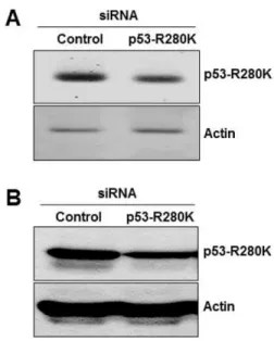
Fig. 1. Effects of siRNA on p53 expression. MDA-MB-231 cells were transfected with control siRNA or with p53 siRNA for 48 h.
(A) The mRNA levels of human p53 and b-actin were detected by RT-PCR analysis; representative RT-PCR. (B) The protein levels of human p53 and β-actin were detected by Western blot analysis;
representative Western blot.
Fig. 2. Effects of p53 silencing on tumor growth in vivo. MDA-MB-231 cells were transfected with control siRNA or p53 siRNA and then subcutaneously injected in the flank of 6-week-old male athymic nude mice (n=5 in each treatment group). After 30 days, the tumor masses were removed, photographed, and used for preparation of paraffin-embedded sections. (A) Images of representative animals with solid tumor and tumor masses. The tumor volume (B) and body weight (C) of mice were measured. *p<0.05 vs. control siRNA.
WI)으로 확인하였다. OCT 처리된 조직의 절편을 4 μm 두께 로 잘라주고 4
oC에서 25분동안 4% 파라포름알데하이드 (pa- raformaldehyde) 에서 고정시켰다. 그런 다음 실온에서 5분간 0.2% Triton X-100으로 반응시킨다. DNA 3'- 말단효소에 TdT- mediated dUTP nick end labeling (TUNEL) 혼합물을 37
oC humidified chamber에서 60분동안 반응시킨다. 라벨링된 DNA 조각 (Labeled DNA fragment)은 형광 현미경 (Nikon, Instech Co., Ltd., Kanagawa, Japan) 으로 확인하였다.
2.9. 통계처리
본 실험에 대한 실험결과는 세 번 실험하여 얻어진 평균치 및 표준편차를 나타내었고 그룹간의 통계적 차이는 Student’s t- test 를 이용하여 분석하였다.
3. RESULTS AND DISCUSSION
3.1. siRNA 에 의한 세포 내 p53-R280K 발현 감소 본 연구진은 최근 p53-R280K의 유방암세포 증식에 미치는 in vitro 실험결과를 발표 [11,12]한 바 있으며 이를 토대로 본 연구에서는 in vivo 종양 성장에 미치는 p53-R280K의 영향에 대해 조사하고자 한다. 먼저 siRNA를 사용하여 MDA-MB- 231 세포 내 p53-R280K의 발현 양을 조절하였으며 이에 대한 확인은 RT-PCR과 western blot 분석을 통해 진행되었다. 그 결과, control siRNA로 형질주입된 대조군에 비해 p53 siRNA 로 형질주입된 MDA-MB-231 세포 내 p53-R280K의 mRNA (Fig. 1A)와 단백질 (Fig. 1B)의 발현 양이 현저히 감소되어 있 음을 확인할 수 있었다. 본 연구를 통해 확립한 p53-silencing MDA-MB-231 세포는 in vivo 종양 성장에 미치는 p53-R280K 의 작용에 관한 다음의 연구에 사용되었다.
3.2. p53 silencing 이 in vivo 종양 형성에 미치는 효과 p53-R280K 단백질 발현 양의 감소가 in vivo에서 MDA-MB- 231 유방암 세포의 성장에 어떠한 영향을 미치는지를 조사 하기 위하여 이종이식 생쥐 모델을 사용하였다. 가슴샘이 제 거된 누드 생쥐 측면의 표피와 진피 사이에 p53-silencing MDA-MB-231 세포를 주입한 뒤 30일 동안 5일 간격으로 종 양의 부피와 생쥐의 몸무게를 측정하였고 주입 후 30일째 되 는 날에 종양을 분리해 내었다. 종양의 크기 (Fig. 2A)와 날짜 별 부피 (Fig. 2B)는 대조군에 비해 p53-silencing MDA-MB- 231 세포를 주입한 종양의 경우에서 현저한 감소를 보였다.
반면, 생쥐의 무게 (Fig. 2C)는 두 그룹에서 차이를 나타내지 않았다. 이러한 연구결과를 통해 in vivo에서 p53-silencing MDA-MB-231 세포의 종양 형성능이 대조군에 비해 저하된 것을 알 수 있다.
3.3. p53-silencing 종양 내 p53과 Notch1의 발현
MDA-MB-231 세포를 생쥐에 이종이식하여 자라난 종양을 분리해내어 조직학적 분석을 진행하였다. 조직 내 세포의 분
포와 밀도를 확인하기 위하여 헤마톡실린과 에오진 염색을 실시하였고 그 결과, p53-silencing MDA-MB-231 세포를 이 식하여 생겨난 종양 (p53-silencing 종양)에서는 대조군에 비 해 세포밀도가 낮게 나타났다 (Fig. 3A). 종양 내 p53-R280K 단백질의 발현 정도를 조사하기 위하여 면역조직화학법으 로 형광염색을 한 결과, p53-silencing 종양 조직에서 p53 단 백질의 발현이 대조군에 비해 낮음을 확인할 수 있었다 (Fig.
3B). 최근 본 연구진은 전사인자로 작용하는 p53-R280K 단 백질의 하류타깃 유전자 중 하나로 Notch1을 새롭게 동정하 여 발표한 바 있다 [11]. 따라서 본 연구에서는 p53-R280K와 Notch1 간의 상관성이 in vivo에서도 나타나는지를 조사하기 위하여 Notch1 단백질의 발현 정도를 관찰하였다. 그 결과, p53-R280K 의 발현이 낮게 나온 종양조직에서는 Notch1의 발현도 함께 급격히 감소함을 확인하였다 (Fig. 3C). DAPI로 염색하였을 때 조직 내 세포의 전체 분포도는 대조군과 p53 silencing 종양 간에 큰 차이가 없었고 DAPI-양성 세포에서 p53-R280K 단백질과 Notch1 단백질이 발현되고 있었다 (Fig. 3D). 이러한 연구 결과를 바탕으로 볼 때, p53 silencing 은 in vitro에서 뿐만 아니라 in vivo에서도 유방암세포의 증
Fig. 3. Effects of p53 silencing on Notch1 expression in human breast xenograft tumor. MDA-MB-231 cells transfected with cont- rol siRNA or p53 siRNA were subcutaneously injected in the flank of athymic nude mice (n=5 in each treatment group). The tumor tis- sue was removed from mice at 30 days and embedded in paraffin.
Tissue sections from xenograft tumors were stained with hematoxylin and eosin (H&E) or immunostained with antibodies against p53 or Notch1. DAPI (blue) stains nuclear DNA. Scale bar: 100 mm.
식에 영향을 미치며 이는 일부 Notch1의 발현을 억제함으로 써 항종양 효과를 나타내는 것으로 사료된다.
3.4. p53 silencing 이 in vivo 종양 내 세포자멸사 유발에 미 치는 효과
p53 siRNA-silencing MDA-MB-231 세포의 in vitro 증식 억제 는 세포자멸사와 관련이 있다는 이전의 연구결과 [11,12]를 바탕으로 하여 p53-silencing 종양 조직에서 세포자멸사 발생 정도를 확인하고자 TUNEL 분석을 실시하였다. 그 결과, 대 조군에 비해 p53 silencing 종양 조직에서 더 많은 수의 TUNEL-양성 세포가 관찰되었다 (Fig. 4A, 4B). 세포자멸사 의 분자생물학적 검증을 위하여 종양조직 내 세포자멸사 관 련 단백질들의 발현 양을 western blot으로 분석한 결과, 대조 군에 비해 p53 silencing 종양 조직에서는 세포자멸사 억제와 세포증식에 관여하는 Bcl2와 Cyclin D1의 발현 양이 감소하
였고 p21의 경우엔 서로 간에 큰 차이가 없음을 관찰하였다.
반면, 세포자멸사 발생 여부를 확인할 수 있는 마커로 사용 되는 PARP와 Caspase-3의 활성화 정도가 크게 증가한 것을 확인할 수 있었다 (Fig. 4C). 이러한 연구결과들은 p53 silen- cing 에 의해 세포자멸사가 유도되고 이로 인해 in vivo에서 종양 형성이 감소되었음을 시사해준다.
4. CONCLUSION
본 연구를 통해 트리플-음성 유방암 세포주인 MDA-MB-231 에서 다량 발현되고 있는 p53 돌연변이 단백질인 p53-R280K 은 MDA-MB-231 세포의 in vivo 종양형성에 중요함을 확인 하였다. siRNA를 이용한 유전자 silencing 방법을 통해 MDA- MB-231 세포 내 R280K 발현 양을 인위적으로 감소시킨 p53-
Fig. 4. Effects of p53 silencing on cellular apoptosis in human breast xenograft tumor. MDA-MB-231 cells transfected with control siRNA or p53 siRNA were subcutaneously injected in the flank of athymic nude mice (n=5 in each treatment group). After 30 days, the tumor masses were removed, and embedded in OCT compound or used for preparation of total protein extraction. (A and B) Apoptotic cells of tissue sections from xenograft tumors were detected by the TUNEL assay. The stained cells (green) were counted, and the percentage of positive cells was calculated. DAPI (blue) stains nuclear DNA. Scale bar: 50 mm. **p<0.001 vs. control siRNA, n=3. (C) Western blot analysis for detecting Bcl-2, Caspase 3, PARP, Cyclin D1, p21, and b-actin protein levels in the xenograft tumor.
silencing MDA-MB-231 세포를 확립하여 누드생쥐에 이종이 식한 뒤 종양형성을 관찰한 결과, 대조군과 비교 시 종양의 크기와 부피 면에서 큰 감소를 나타냈다. 이종이식으로 형성 된 종양조직에서 p53-R280K과 p53-R280K의 타깃유전자로 작용하는 Notch1의 발현을 조사하였고 그 결과, p53-silencing 종양 조직에서 p53-R280K 단백질 발현 양이 감소하였으며 동일 조직에서 Notch1 단백질의 발현 양도 현저히 감소해 있 음을 확인할 수 있었다. 또한 TUNEL 에세이를 통해 대조군 에 비해 p53-silencing 종양조직 내에서 세포자멸사가 더 많이 발생하였음을 관찰하였고 western blot을 통해 PARP와 casp- ase-3 등 세포자멸사 관련 단백질들의 발현 양이 증가하였음 을 확인하였다. 따라서 본 연구결과들을 종합해 볼 때 향후 트리플-음성 유방암 치료를 위한 새로운 유전자치료법이나 항암제 개발에 p53 돌연변이 단백질인 p53-R280K과 Notch1 을 타깃으로 하는 접근이 유효할 것으로 판단된다.
Acknowledgements
이 논문은 부산대학교 기본연구지원사업 (2년)에 의하여 연 구되었음.
REFERENCES
1. Selivanova, G. (2004) P53: Fighting Cancer. Curr. Cancer. Drug Targets 4: 385-402.
2. Horn, H. F. and K. H. Vousden (2007) Coping with stress: multi- ple ways to activate p53. Oncogene. 26: 1306-1316.
3. Molchadsky, A., N. Rivlin, R. Brosh, V. Rotter, and R. Sarig (2010) P53 is balancing development, differentiation and de-differentia- tion to assure cancer prevention. Carcinogenesis. 31: 1501-1508.
4. Brosh, R. and V. Rotter (2009) When mutants gain new powers:
news from the mutant p53 field. Nat. Rev. Cancer. 9: 701-713.
5. Walerych, D., M. Napoli, L. Collavin, and G. Del-Sal (2012) The rebel angel: mutant p53 as the driving oncogene in breast cancer.
Carcinogenesis. 33: 2007-2017.
6. Wijnhoven, S. W., E. N. Speksnijder, X. Liu, E. Zwart, C. T. van Oostrom, R. B. Beems, E. M. Hoogervorst, M. M. Schaap, L. D.
Attardi, T. Jacks, H. van Steeg, J. Jonkers, and A. deVries (2007) Dominant-negative but not gain-of-function effects of a p53.R270H mutation in mouse epithelium tissue after DNA damage. Cancer Res. 67: 4648-4656.
7. Mehta, S. A., K. W. Christopherson, P. Bhat-Nakshatri, J. R. Gou- let-RJ, H. E. Broxmeyer, L. Kopelovich, and H. Nakshatri (2007) Negative regulation of chemokine receptor CXCR4 by tumor sup- pressor p53 in breast cancer cells: implications of p53 mutation or isoform expression on breast cancer cell invasion. Oncogene. 26:
3329-3337.
8. Zhu, H. B., K. Yang, Y. Q. Xie, Y. W. Lin, Q. Q. Mao, and L. P.
Xie (2013) Silencing of mutant p53 by siRNA induces cell cycle arrest and apoptosis in human bladder cancer cells. World J. Surg.
Oncol. 28: 11-22.
9. Lim, L. Y., N. Vidnovic, L. W. Ellisen, and C. O. Leong (2009) Mutant p53 mediates survival of breast cancer cells. Br. J. Cancer.
101: 1606-1612.
10. Bayraktar, S. and S. Glück (2013) Molecularly targeted therapies for metastatic triple-negative breast cancer. Breast Cancer Res.
Treat. 138: 21-35.
11. Bae, Y. H., J. H. Ryu, H. J. Park, K. R. Kim, H. J. Wee, O. H. Lee, H. O. Jang, M. K. Bae, K. W. Kim, and S. K. Bae (2013) Mutant p53-notch1 signaling axis is involved in curcumin-induced apopto- sis of breast cancer cells. Korean J. Physiol. Pharmacol. 17: 291- 297.
12. Bae, Y. H., J. M. Shin, H. J. Park, H. O. Jang, M. K. Bae, and S. K.
Bae (2014) Gain-of-function mutant p53-R280K mediates survival of breast cancer cells. Genes & Genomics 36: 171-178.