pISSN 1738-1843
© 2013 The Korean Society of Pathologists/The Korean Society for Cytopathology
Simian virus 40 (SV40) is a polyomavirus that originates from the rhesus macaque and was discovered in 1960 as a con- taminant of human polio vaccines produced in monkey cells.
1Recently, there has been considerable interest in the identifica- tion of SV40 in human tumors, including malignant mesothe- lioma (MM) and other rare tumors such as osteosarcoma, epen-
No Detection of Simian Virus 40 in Malignant Mesothelioma in Korea
Minseob Eom · Jamshid Abdul-Ghafar Sun-Mi Park · Joung Ho Han
1Soon Won Hong
2· Kun Young Kwon
3Eun Suk Ko
4· Lucia Kim
5Wan Seop Kim
6· Seung Yeon Ha
7Kyo Young Lee
8· Chang Hun Lee
9Hye Kyoung Yoon
10· Yoo Duk Choi
11Myoung Ja Chung
12· Soon-Hee Jung
Department of Pathology, Yonsei University Wonju College of Medicine, Wonju; 1Department of Pathology, Samsung Medical Center, Sungkyunkwan University School of Medicine, Seoul; 2Department of Pathology, Yonsei University College of Medicine, Seoul; 3Department of Pathology, Keimyung University School of Medicine, Daegu; 4Department of Pathology, Soonchunhyang University College of Medicine, Bucheon;
5Department of Pathology, Inha University School of Medicine, Incheon; 6Department of Pathology, Konkuk University School of Medicine, Seoul;
7Department of Pathology, Gil Medical Center, Gachon University of Medicine and Science, Incheon; 8Department of Pathology, The Catholic University College of Medicine, Seoul; 9Department of Pathology, Busan National University School of Medicine, Busan; 10Department of Pathology, Inje University College of Medicine, Busan;
11Department of Pathology, Chonnam National University Medical School, Gwangju;
12Department of Pathology, Chonbuk National University Medical School, Jeonju, Korea
*Minseob Eom and Jamshid Abdul-Ghafar contributed equally to this work.
Background: Simian virus 40 (SV40), a polyomavirus, was discovered as a contaminant of a hu- man polio vaccine in the 1960s. It is known that malignant mesothelioma (MM) is associated with SV40, and that the virus works as a cofactor to the carcinogenetic effects of asbestos. However, the reports about the correlation between SV40 and MM have not been consistent. The purpose of this study is to identify SV40 in MM tissue in Korea through detection of SV40 protein and DNA. Methods: We analyzed 62 cases of available paraffin-blocks enrolled through the Korean Malignant Mesothelioma Surveillance System and performed immunohistochemistry for SV40 protein and real-time polymerase chain reaction (PCR) for SV40 DNA. Results: Of 62 total cases, 40 had disease involving the pleura (64.5%), and 29 (46.8%) were found to be of the epithelioid subtype. Immunostaining demonstrated that all examined tissues were negative for SV40 protein.
Sufficient DNA was extracted for real-time PCR analysis from 36 cases. Quantitative PCR of these samples showed no increase in SV40 transcript compared to the negative controls. Con- clusions: SV40 is not associated with the development of MM in Korea.
Key Words: Immunohistochemistry; Mesothelioma; Polymerase chain reaction; Simian virus 40 Received: January 10, 2013
Revised: February 1, 2013 Accepted: February 6, 2013 Corresponding Author Soon-Hee Jung, M.D.
Department of Pathology, Yonsei University Wonju College of Medicine, 20 Ilsan-ro, Wonju 220-701, Korea
Tel: +82-33-741-1551 Fax: +82-33-731-6590 E-mail: [email protected]
dymoma, and choroid plexus tumors.
2-6Subsequently, it has been found that SV40 may play an important role in the etiolo- gy of these tumors.
2-6However, SV40 was not detected in ma- lignant lymphomas in Korean patients.
7Oncogenesis driven by SV40 is mediated by the large tumor antigen (T-Ag) oncopro- tein, which is capable of transforming different types of cells in the absence of other viral genes.
8,9T-Ag induces DNA synthesis in host cells and prolongs the onset of the S-phase through the inhibition of tumor suppressor proteins p53 and Rb.
8MM is a rare and extremely aggressive neoplasm that arises from the serosal surfaces of pleural, peritoneal, and pericardial cavities and is very likely associated with amphibole asbestos exposure.
10,11In recent years, the incidence of MM has been gradually increasing in concert with industrial development and increased exposure to asbestos.
12However, only a small number of people who have been exposed to asbestos during their lifetime will develop MM.
11This suggests that SV40 may act as an independent carcinogenic agent or as a cofactor of as- bestos to augment the risk of MM.
13Studies investigating the relation of SV40 to MM have been inconsistent and differ by geographical location.
6The incidence of MM varies according to geographic loca- tion, and the highest incidence rates are reported from Austra- lia, Belgium, and Great Britain.
14In Korea, the incidence of MM is very low compared with other developed countries, with only about one case per million in the last decade.
14,15However, the incidence is recently increasing. Korea has a higher propor- tion of females (33.8%) among MM cases compared with other nations such as Canada (15.4%) or Italy (27.6%), and most of the patients are diagnosed when they are in their 50s, which is younger compared to cases in other countries.
15Although the majority of MM patients have had occupational (36.8%) and/or environmental (20.4%) asbestos exposure, the remaining pa- tients (42.8%) had no known asbestos exposure.
15The role of SV40 in the oncogenesis of MM following asbes- tos exposure has not yet been studied in a Korean population.
The purpose of this study is to identify SV40 DNA and protein in tissues extracted from MM from a Korean population using quantitative polymerase chain reaction (PCR) and immunohis- tochemical staining.
MATERIALS AND METHODS
Study populationUsing the Korean Malignant Mesothelioma Surveillance (KMMS) tissue bank, 356 cases of MM that occurred between
the years 2001 and 2009 were available. Of these, 62 formalin- fixed and paraffin-embedded tissue tumor blocks were selected.
The selection was based on the availability of adequate neoplas- tic tissue within the paraffin blocks. Original diagnoses were based on morphological evaluation in the primary centers and confirmed by three expert pathologists. All cases were analyzed using an immunohistochemical panel (positive marker: cal- retinin and/or D2-40, WT-1; negative marker: carcinoembry- onic antigen). Each tumor slide was re-evaluated to verify the original diagnosis.
Immunohistochemistry
Detection of SV40 protein was performed by an automatic staining procedure using the Ventana Benchmark XT (Roche Diagnostics, Basel, Switzerland). The sections were deparaf- finized, then pretreated with CC1 (Roche Diagnostics) for 60 minutes at 42˚C. The sections were then washed with reaction buffer and incubated with anti-SV40 antibody (Cell Marque Corp., Rocklin, CA, USA) at a 1:100 dilution for 60 minutes at 42˚C. Antibody detection was performed using the UltraView Universal DAB kit (Roche Diagnostics) according to the manu- facturer’s recommendations. Finally, the slides were counter- stained with hematoxylin (Roche Diagnostics) and mounted.
As a positive control for SV40 sections from paraffin-embedded tissue, kidney tissue confirmed to contain polyomavirus was used (kindly provided by Professor Young-Mee Cho from the Department of Pathology, Asan Medical Center, Seoul, Korea).
The negative controls were performed without the primary an- tibody.
DNA extraction from paraffin-embedded tissues
The paraffin-embedded tissue blocks were de-paraffinized us- ing xylene. Briefly, the paraffin was removed by two extractions with 1 mL of xylene for 30 minutes and two with 0.5 mL of ethanol and subsequently dried at 37˚C. DNA was extracted using the DNeasy Blood and Tissue Kit (Qiagen, Valencia, CA, USA) according to the manufacturer’s instructions. Of the par- affin-embedded tissues analyzed, sufficient DNA was only ex- tracted from 36 samples.
β-actin PCR
To ensure the quality of the extracted DNA, all samples were
analyzed for the presence of the
β-actin gene. DNA (200 ng)
was amplified using a HotStar Taq Plus master mix kit (Qia-
gen). After a denaturing step of 15 minutes at 95˚C, 30 ampli-
fication cycles were performed. Each cycle consisted of 1 minute
at 94˚C, 30 seconds at 56°C, and 1 minute at 72˚C, followed by a final extension step of 5 minutes at 72˚C using
β-actin (for- ward 5´-CCT TCC TGG GAC TGG AGT CCT-3´ and reverse 5´-GGA GCA ATG ATC TTG ATC TTC-3´) primers.
SV40 quantitative PCR
A real-time PCR method (TaqMan) for SV40 was previously established which uses a primer pair and an oligonucleotide probe with the fluorescein reporter dye FAM attached to the 5´
end and a rhodamine dye (TAMRA) quencher linked to the 3´
end. A threshold cycle value (Ct) was calculated for each sample by determining the point at which the fluorescence exceeded the threshold limit chosen for the specific plate.
The real-time PCR assay used the forward primer 5´-CAC AGC ATG ACT CAA AAA ACT TAG CA-3´ and reverse pri- mer 5´-GAC TCT CAA CAT TCT ACT CCT CCA AAA-3´
and the fluorogenic TaqMan probe 5´-ACC CCA AGG ACT TTC-3´.
Negative controls (water) and positive controls (DNA from the HEK293T cell line) were included on each plate, and a stan- dard curve (SV40 standard solutions) was produced from which the number of genomes in the samples could be calculated. The SV40 quantities used in the standards were 100,000, 10,000, 1,000, 100, and 10 copies per 1 μL (i.e., per well). Thermal cy- cling, fluorescence detection, and data analysis were performed on an ABI PRISM 7900 Sequence Detector (Applied Biosys- tems, Foster City, CA, USA) using the software provided by the manufacturer.
The standard stock solution, plasmid pDRIVE, containing the SV40 genome was cultivated in medium, and the culture was purified according to the manufacturer’s instructions (Qia-
gen). The concentration of the plasmid solution was calculated using the absorbance at 260 nm.
Ethics statement
This study was approved by the Institutional Ethics Com- mittee of Yonsei University, Wonju College of Medicine, Won- ju, South Korea (IEC No. YWMR-12-4-033) and was in com- pliance with the Helsinki Declaration.
RESULTS
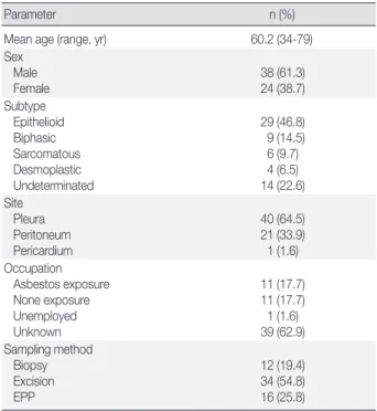
Clinicopathological characteristicsThe study included 38 (61.3%) males and 24 (38.7%) fe- males between the ages of 34 and 79 years, with a mean age of 60.2 years. The majority of patients developed MM when they were in their 50s, followed by patients over 70, patients in their 60s, 40s and finally in their 30s (Fig. 1). Most cases of MM oc- curred in the pleura (40 cases, 64.5%), followed by the perito- neum (21 cases, 33.9%) and the pericardium (one case, 1.6%).
With regard to histologic classification, the epithelioid type was the most common histologic type (29 cases, 46.8%), fol- lowed by the biphasic (nine cases, 14.5%), sarcomatoid (six cas- es, 9.7%), desmoplastic types (four cases, 6.5%), and other un- determined variants (14 cases, 22.6%). The clinicopathological data are summarized in Table 1.
20 18 16 14 12 10 8 6 4 2 0
No. of cases
Age of patients (yr)
30-39 40-49 50-59 60-69 70-79 Men Women Total
Fig. 1. According to the age distribution, the majority of cases de- veloped malignant mesothelioma when patients are in their 50s, followed by those over 70, those in their 60s, 40s and finally in their 30s.
Table 1. Clinicopathologic characteristics of malignant mesothelio- ma cases
Parameter n (%)
Mean age (range, yr) 60.2 (34-79)
Sex Male
Female 38 (61.3)
24 (38.7) Subtype
Epithelioid Biphasic Sarcomatous Desmoplastic Undeterminated
29 (46.8) 9 (14.5) 6 (9.7) 4 (6.5) 14 (22.6) Site
Pleura Peritoneum Pericardium
40 (64.5) 21 (33.9) 1 (1.6) Occupation
Asbestos exposure None exposure Unemployed Unknown
11 (17.7) 11 (17.7) 1 (1.6) 39 (62.9) Sampling method
Biopsy Excision EPP
12 (19.4) 34 (54.8) 16 (25.8) EPP, extended pleuropneumonectomy.
Table 2. Occupational history of malignant mesothelioma patients
Occupation No. of
patients Comments
Construction 3 O ne patient also worked in copper pipe fabrication
Automobile repair 1 Shipbuilding repair 2 Environmental asbestos 1
Office worker 6 A dministrative, education office worker, Red Cross staff, teacher
Farmer 3
Trading 1
Housewife 4
Unemployed 1
Others 1 Production company
Unknown 39 Occupation history is not available
Total 62
A B
C D
Fig. 2. Immunohistochemical findings for simian virus 40 (SV40) antibody; (A) positive control tissue from kidney infected with SV40 shows a positive reaction, while tissue sections from malignant mesothelioma show a negative result for SV40 antibody (B, epithelioid type; C, bipha- sic type; D, sarcomatoid type).
The occupational history was available for only 23 (37.1%) patients out of 62 patients, and among these, only 11 (17.7%)
had a job that placed them at risk for asbestos exposure such as construction, automobile repair and production, shipbuilding and repair, or farming (Table 2).
Immunohistochemistry and real-time PCR findings
None of the 62 MM tissue samples studied showed a positive immunohistochemical staining for SV40, while the positive con- trol kidney tissue for each slide was strongly positive (Fig. 2).
Of the 36 samples that yielded sufficient DNA following ex-
traction and purification, real-time PCR analysis failed to show
SV40 DNA amplification in any case. The mean cycle thresh-
old (Ct) value for the 36 available blocks was 34.133
±1.440
and was not significantly different from the negative control
(positive control, 28.413
±0.476; negative control, 34.437
±0.160).
DISCUSSION
The possible association between SV40 and MM has inspired much interest, especially after Carbone et al.
16reported the pres- ence of SV40 in approximately 60% of mesotheliomas. Several subsequent studies have investigated the relation of mesothelio- ma with SV40, but reported results have varied considerably.
One study which investigated the presence of SV40 T-Ag in 18 autopsied MM patients in Japan involved eight positive sam- ples; however, none of these cases were likely to have received a contaminated polio vaccine due to their age.
12A recent review showed that the association of SV40 and MM was positive in only three cases out of 12.
6Another three studies showed that an average of 22% of tumor tissues were positive for SV40.
6,10A strong association between exposure to asbestos and the development of MM has long been established, but there exist cases with unclear etiologies that appear unrelated to asbestos exposure. MM is known to occur in only 10% to 20% of indi- viduals heavily exposed to asbestos, and about 20% of patients lack a history of any exposure.
17In a case-control study, PCR analysis revealed the presence of SV40 DNA in eight of 19 cas- es of MM among individuals exposed to asbestos, suggesting that SV40 may increase the risk of MM among patients exposed to asbestos.
13In hamsters, intrapleural injection of SV40 virus resulted in the development of MM in 100% of cases within two to six months.
18Additionally, a strong association between asbestos and SV40 was found among hamsters exposed to low levels of asbestos, as only the SV40-infected animals developed MM.
19To understand the roles of both T-Ag and t-Ag in the develop- ment of MM in hamsters, t-Ag mutant SV40 virus was injected intracardially into newborn hamsters, after which only one ani- mal developed MM, while the majority developed histiocytic lymphomas.
20While T-Ag binds and deactivates tumor sup- pressor proteins, t-Ag may play a key role in mesothelial cell transformation and may be necessary to complete inactivation and allow mesothelial cells to enter S phase.
11,20In humans, the role of SV40 in the development of MM is controversial. In total, more than 26 studies have identified SV40 in human mesotheliomas or other human tumors;
11how- ever, many recent investigators have been unable to detect SV40 in human mesothelioma tissues.
10,11In a study by Manfredi et al.,
21of 69 human MM tissues screened, none contained detect- able SV40 T-Ag sequences, arguing against the role of SV40 in the development of human MMs.
In this study, no evidence of SV40 was found in any of the
MM tissues examined, either by immunohistochemistry or real- time PCR analysis. These results are consistent with those of most recent investigations. The discrepancy found among these different studies may suggest a possible role for geographical differences in the association between SV40 and MM. Alterna- tively, contamination of laboratory plasmids may have led to false positive results in some cases.
22Occupational and environmental exposure to asbestos is the main causes of MM in Korea.
15Our results do not support an association between MM and SV40 or the proposed function of SV40 as a cofactor to asbestos carcinogenicity. If SV40 is indeed a cofactor for asbestos carcinogenicity, viral transcripts and pro- teins would have been detectable in the cases examined in our study.
We acknowledge some advantages and limitations of our study. We performed immunohistochemistry and real-time PCR separately to detect SV40 protein and DNA, respectively, in MM tissues. Frozen or fresh tissues are preferred for PCR de- tection of viral DNA. However, acquiring frozen or fresh MM tissues is impractical due to the infrequency of MM. In addi- tion, our sample size may not have been large enough to detect an association between SV40 and MM in the Korea.
In conclusion, no detection of SV40 in MM tissues suggests that the virus does not have a role in development of MM in Korea; therefore, a more active epidemiologic study for asbes- tos-related occupational and environmental causes in MM pa- tients is needed.
Conflicts of Interest
No potential conflict of interest relevant to this article was reported.
Acknowledgments
This study was supported by the Korean Ministry of Envi- ronment as “The Environmental Health Action Program.” The authors gratefully acknowledge financial support by the Korean Ministry of Environment. The authors also would like to thank the members of the Cardiopulmonary Pathologists Society, Ko- rean Society of Pathologists for reporting malignant mesotheli- oma cases.
REFERENCES
1. Sweet BH, Hilleman MR. The vacuolating virus, S.V. 40. Proc Soc Exp Biol Med 1960; 105: 420-7.
2. Ziadi S, Boughamoura H, Ben Maitig M, et al. Immunodetection of
SV40 T/t-antigens in human osteosrcoma in a series of Tunisian pa- tients. Pathol Oncol Res 2012; 18: 691-6.
3. Gazdar AF, Carbone M. Molecular pathogenesis of malignant me- sothelioma and its relationship to simian virus 40. Clin Lung Can- cer 2003; 5: 177-81.
4. Shivapurkar N, Wiethege T, Wistuba II, et al. Presence of simian vi- rus 40 sequences in malignant mesotheliomas and mesothelial cell proliferations. J Cell Biochem 1999; 76: 181-8.
5. Pershouse MA, Heivly S, Girtsman T. The role of SV40 in malignant mesothelioma and other human malignancies. Inhal Toxicol 2006;
18: 995-1000.
6. Shah KV. SV40 and human cancer: a review of recent data. Int J Can- cer 2007; 120: 215-23.
7. Kim YA, Chang M, Paik J, et al. Detection of SV40 large T antigen in malignant lymphomas. Korean J Pathol 2009; 43: 312-6.
8. Butel JS, Lednicky JA. Cell and molecular biology of simian virus 40: implications for human infections and disease. J Natl Cancer Inst 1999; 91: 119-34.
9. Pepper C, Jasani B, Navabi H, Wynford-Thomas D, Gibbs AR. Sim- ian virus 40 large T antigen (SV40LTAg) primer specific DNA am- plification in human pleural mesothelioma tissue. Thorax 1996; 51:
1074-6.
10. Lundstig A, Dejmek A, Eklund C, Filinic I, Dillner J. No detection of SV40 DNA in mesothelioma tissues from a high incidence area in Sweden. Anticancer Res 2007; 27: 4159-61.
11. Rizzo P, Bocchetta M, Powers A, et al. SV40 and the pathogenesis of mesothelioma. Semin Cancer Biol 2001; 11: 63-71.
12. Jin M, Sawa H, Suzuki T, et al. Investigation of simian virus 40 large T antigen in 18 autopsied malignant mesothelioma patients in Ja-
pan. J Med Virol 2004; 74: 668-76.
13. Cristaudo A, Foddis R, Vivaldi A, et al. SV40 enhances the risk of malignant mesothelioma among people exposed to asbestos: a mo- lecular epidemiologic case-control study. Cancer Res 2005; 65: 3049- 52.
14. Bianchi C, Bianchi T. Malignant mesothelioma: global incidence and relationship with asbestos. Ind Health 2007; 45: 379-87.
15. Jung SH, Kim HR, Koh SB, et al. A decade of malignant mesothelio- ma surveillance in Korea. Am J Ind Med 2012; 55: 869-75.
16. Carbone M, Pass HI, Rizzo P, et al. Simian virus 40-like DNA se- quence in human pleural mesothelioma. Oncogene 1994; 9: 1781-90.
17. Jasani B, Gibbs A. Mesothelioma not associated with asbestos expo- sure. Arch Pathol Lab Med 2012; 136: 262-7.
18. Cicala C, Pompetti F, Carbone M. SV40 induces mesotheliomas in hamsters. Am J Pathol 1993; 142: 1524-33.
19. Kroczynska B, Cutrone R, Bocchetta M, et al. Crocidolite asbestos and SV40 are cocarcinogens in human mesothelial cells and in caus- ing mesothelioma in hamsters. Proc Natl Acad Sci U S A 2006; 103:
14128-33.
20. Cicala C, Pompetti F, Nguyen P, Dixon K, Levine AS, Carbone M.
SV40 small t deletion mutants preferentially transform mononuclear phagocytes and B lymphocytes in vivo. Virology 1992; 190: 475-9.
21. Manfredi JJ, Dong J, Liu WJ, et al. Evidence against a role for SV40 in human mesothelioma. Cancer Res 2005; 65: 2602-9.
22. López-Ríos F, Illei PB, Rusch V, Ladanyi M. Evidence against a role for SV40 infection in human mesotheliomas and high risk of false- positive PCR results owing to presence of SV40 sequences in com- mon laboratory plasmids. Lancet 2004; 364: 1157-66.