408 ORIGINAL ARTICLE
DOI 10.4070 / kcj.2009.39.10.408
Print ISSN 1738-5520 / On-line ISSN 1738-5555 Copyright ⓒ 2009 The Korean Society of Cardiology
Open Access
Therapeutic Strategy for In-Stent Restenosis Based
on the Restenosis Pattern After Drug-Eluting Stent Implantation
Ki-Hun Kim, MD1, Doo-Il Kim, MD1, Il-Hwan Kim, MD1, Jong-Yoon Kim, MD1, Yang-Chun Han, MD1, Sang-Hoon Seol, MD1, Ung Kim, MD1, Tae-Hyun Yang, MD1, Dae-Kyeong Kim, MD1, Dong-Soo Kim, MD1, Sang-Hee Lee, MD2, Jong-Seon Park, MD2, Young-Jo Kim, MD2, Dong-Gu Shin, MD2,
Yoon-Kyung Cho, MD3,Chang-Wook Nam, MD3, Seung-Ho Hur, MD3 and Kwon-Bae Kim, MD3
1Division of Cardiology, Department of Internal Medicine, College of Medicine, Inje University, Busan Paik Hospital, Busan,
2Department of Internal Medicine, College of Medicine, Yeungnam University, Daegu,
3Department of Internal Medicine, College of Medicine, Keimyung University, Dongsan Hospital, Daegu, Korea
ABSTRACT
Background and Objectives: The aim of this study was to evaluate the outcomes of repeated percutaneous coro- nary intervention (PCI) based on the restenosis pattern in drug-eluting stent (DES) failure. Subjects and Methods:
From April 2003 to March 2006, all 67 patients (67 lesions) at our 3 centers who had DES in-stent restenosis (ISR) were enrolled. The patients were divided into 3 groups: group I had focal edge restenosis, group II had focal body restenosis, and group III had non-focal restenosis. All patients were treated with conventional PCI including plain old balloon angioplasty (POBA), cutting balloon angioplasty (CBA), and repeated DES implantation (Re- DES). Angiographic and clinical one year follow-up results for the 3 groups were evaluated. Results: Sixteen pa- tients were enrolled in group I, 36 in group II, and 15 in group III. Baseline clinical and angiographic characteristics and the proportion of patients in each group receiving each type of treatment strategy were not significantly dif- ferent among the groups. Within each group, a comparison of angiographic and clinical outcomes for each thera- peutic modality revealed that restenosis rates were not statistically different. Although rates of major adverse car- diac events (MACE) were not statistically different between groups I and II, in group III, MACE were 3-fold higher for the POBA (4/4, 100.0%) and CBA (4/4, 100.0%) subgroups than for Re-DES (1/3, 33.3%) (p=0.06), but the differences did not reach statistical significance. Conclusion: The present study suggests that treatment of DES ISR should be individualized according to restenosis pattern: any PCI strategy appears appropriate for focal ISR patterns, while Re-DES might be a better choice for non-focal ISR patterns. (Korean Circ J 2009;39:408-413)
KEY WORDS: Percutaneous transluminal coronary angioplasty; Restenosis; Drug-eluting stent; Coronary re- stenosis.
Introduction
Although drug-eluting stents (DES) significantly re-
duce restenosis rates compared with bare-metal stents (BMS), treatment of coronary in-stent restenosis (ISR) remains a challenging problem. Much research has been devoted to the pathophysiology and treatment of ISR with DES; however, because of its relatively low inci- dence, there is scant data on its management.1)
Studies have suggested a variety of possible treatment strategies using standard percutaneous coronary inter- vention (PCI) techniques, including plain balloon an- gioplasty (POBA), cutting balloon angioplasty (CBA), repeated DES implantation (Re-DES), radiation therapy, or local drug delivery.2) Some studies reported that repeat- ed PCI with DES, BMS, brachytherapy, POBA or CBA is safe and not associated with increased rates of vascular
Received: January 27, 2009 Revision Received: March 23, 2009 Accepted: May 10, 2009
Correspondence: Doo-Il Kim, MD, Division of Cardiology, Department of In- ternal Medicine, College of Medicine, Inje University, Busan Paik Hospital, 633-165 Gaegeum-dong, Busanjin-gu, Busan 614-735, Korea Tel: 82-51-890-6270, Fax: 82-51-893-2340
E-mail: jo1216@chollian.net
○cc This is an Open Access article distributed under the terms of the Creative Commons Attribution Non-Commercial License (http://creativecommons.
org/licenses/by-nc/3.0) which permits unrestricted non-commercial use, distribution, and reproduction in any medium, provided the original work is properly cited.
Ki-Hun Kim, et al.·409
complications.3-6) Cosgrave et al.7) suggested that Re-DES for DES restenosis is feasible and safe. In particular, si- rolimus-eluting stent (SES) failure treated with traditio- nal PCI yielded favorable outcomes, perhaps due to the predominantly focal nature of the SES restenotic lesion.7)8) The use of DES for diffuse ISR is also feasible and safe.9)10) Regardless of the therapeutic approach chosen, the pattern of restenosis itself is an important predictor of outcomes.7) Similarly, in the era of DES implantation, the incidence of target-lesion revascularization (TLR) in- creases with the pattern of restenosis treated.11) Therefore, our study was done to evaluate real world outcomes for repeated PCI strategies according to the restenosis pat- tern in DES failure.
Subjects and Methods
Study population
We reviewed records for 1,465 patients (1,620 lesions) who underwent follow-up coronary angiogram (CAG) from among 2,540 patients who had undergone DES implantation in our 3 centers from April 2003 to March 2006, and identified 83 patients (88 lesions) with DES ISR. Patients who were not treated for ISR or who had left main coronary artery lesions were excluded, and 67 patients (67 lesions) with DES ISR were included in the final analyses. Coronary angiogram follow-up was plan- ned for 9 months after ISR treatment for patients who had not experienced clinical events.
Patients were divided into 3 groups according to res- tenosis pattern. Group I had focal edge restenosis, Group II had focal body restenosis, and group III had non- focal restenosis. All patients were treated with conventio- nal PCI including POBA, CBA, and Re-DES. All pro- cedures were performed using standard PCI techniques, and intravascular ultrasound (IVUS) imaging was used at the surgeon’s discretion. Coronary angiograms were obtained before and after the procedures, and at follow- up, and they were analyzed by 2 independent angiogra- phers. Angiographic and clinical follow-up {major ad- verse cardiac events (MACEs)} results were evaluated for the 3 groups for 1 year.
Definitions and outcome measures
A focal lesion was defined as one with a length ≤10 mm; non-focal lesions had lengths >10 mm. Edge res- tenosis was defined as restenosis occurring within 5 mm at either side from and including the stent margin. Angio- graphic restenosis was defined as diameter stenosis >50%
by quantitative coronary angiography (QCA) within a previously stented segment (stent and 5 mm proximal and distal) at follow-up angiogram. TLR was defined as repeated revascularization secondary to a stenosis >50%
within the stent or within the 5 mm borders proximal or distal to the stent edge. target-vessel revascularization
(TVR) was defined as repeated revascularization of the target vessel. Myocardial infarction (MI) was diagnosed when cardiac creatinine kinase-MB levels were greater than three-fold the normal value, with chest pain lasting
≥30 minutes or with the appearance of new electrocar- diographic changes. All deaths were considered to be cardiac-related unless otherwise documented. A MACE was defined as death or MI and the need for TLR, TVR or coronary artery bypass graft (CABG).
Coronary QCAs were analyzed using a validated edge detection system (CAAS II, Pie Medical Imaging, Maa- stricht, The Netherlands). Minimal luminal diameter (MLD), reference vessel diameter (RD), and % diameter stenosis (DS) were measured at baseline, post-stenting, and at follow-up, respectively.
Statistical analysis
Continuous variables are presented as means±SD, and categorical variables as frequencies and percentages.
Differences between groups in outcome variables were assessed using Pearson’s Chi-square test or Fisher’s exact test (whenever an expected cell value was <5) for catego- rical data, and the Kruskal-Wallis rank sum test and Wilcoxon rank sum test for continuous data. A p <0.05 was considered to indicate a significant difference, and all reported p are 2-sided. Statistical analysis was per- formed using SAS 9.1 (SAS Institute, Cary, NC, USA).
Results
Patients and baseline characteristics
There were 16 focal edge restenotic lesions (group I), 36 focal body restenotic lesions (group II), and 15 non- focal restenotic lesions (group III). Angiographic success was achieved in all patients. Baseline clinical and angio- graphic characteristics were similar among groups, except for hypertension, which was significantly more frequent in group III (p=0.03). Procedural anticoagulation and antiplatelet therapy was prescribed according to standard protocols. Medications prescribed after DES implanta- tion included aspirin, clopidogrel, cilostazol, beta-bloc- kers, angiotensin converting enzyme inhibitors (ACEI)/
angiotensin receptor blockers (ARB), and statins. Fre- quencies of use of each medication were not significan- tly different among groups (Table 1).
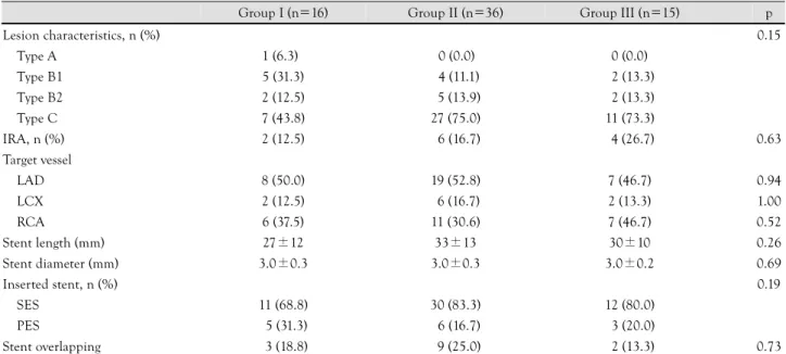
Distribution of target vessels was not significantly dif- ferent among groups, and the left anterior descending artery (LAD) was the most frequent target vessel. Mean stent length and diameter and proportions by type of inserted stents, i.e., SES or paclitaxel-eluting stent (PES), were not significantly different either (Table 2).
Clinical and angiographic characteristics by percu- taneous coronary intervention modality
Mean follow-up duration was 10±5 months in group
410·Treatment of In-Stent Restenosis After DES
I, 10±8 months in group II and 16±13 months in group III (p=0.06). Acute myocardial infarction frequen- cies were higher in group III (4, 26.7%) than in group I (2, 12.5%) or in group II (1, 2.8%) (p=0.02), and silent ischemia frequencies were higher in groups I (5, 31.2%) and II (13, 36.1%) than in group III (0, 0.0%) (p=0.02).
The latter observation might be related to higher fre- quencies of routine CAG follow-up in groups I and II than in group III. IVUS studies were done for 27 patients (40.3%), in which neointimal hyperplasia was the major cause of ISR. Stent fracture was seen in 1 (14.3%) case in
group I, 8 (57.1%) in group II, and 2 (33.3%) in group III, but the incidence rates were not significantly dif- ferent (p=0.20) (Table 3).
Group III patients had longer mean lesion length and narrower pre-procedure MLD and pre-procedure % DS;
however, this was related to grouping characteristics.
POBA, CBA and Re-DES were done for the following numbers of patients (and the following percentages by groups): POBA (5, 31.3%), CBA (3, 18.8%), and Re- DES (9, 56.3%) in group I; POBA (13, 36.1%), CBA (17, 47.2%), and Re-DES (6, 16.7%) in group II; and
Table 1. Baseline clinical characteristics
Group I (n=16) Group II (n=36) Group III (n=15) p
Age, years 58±8 60±9 64±10 0.25
Male sex (%) 19 (56.3) 29 (80.6) 10 (66.7) 0.18
Past medical history (%)
Diabetes 19 (56.3) 18 (50.0) 14 (26.7) 0.23
Hypertension 11 (68.8) 15 (41.7) 12 (80.0) 0.03
Smoking 15 (31.3) 16 (44.4) 15 (33.3) 0.59
Hyperlipidemia 11 (68.8) 23 (63.9) 10 (66.7) 0.94
Old MI 13 (18.8) 16 (16.7) 0 (0.0) 0.22
Stroke 13 (18.8) 2 (5.6) 13 (20.0) 0.19
LVEF (%) 55±13 55±12 53±12 0.75
Medication after DES, n (%)
Aspirin 16 (100.0) 36 (100.0) 15 (100.0)
Clopidogrel 15 (93.8) 35 (97.2) 15 (100.0) 0.59
Cilostazol 11 (68.8) 22 (61.1) 15 (33.3) 0.11
Beta-blocker 12 (75.0) 25 (71.5) 10 (66.7) 0.87
ACEI/ARB 17 (43.7) 27 (75.0) 12 (80.0) 0.07
Statin 10 (62.5) 17 (47.2) 15 (33.3) 0.27
MI: myocardial infarction, LVEF: left ventricular ejection fraction, DES: drug-eluting stent, ACEI: angiotensin converting enzyme inhibitor, ARB:
angiotensin receptor blocker
Table 2. Baseline angiographic characteristics of target lesions
Group I (n=16) Group II (n=36) Group III (n=15) p
Lesion characteristics, n (%) 0.15
Type A 1 (6.3) 20 (0.0) 10 (0.0)
Type B1 5 (31.3) 24 (11.1) 12 (13.3)
Type B2 2 (12.5) 25 (13.9) 12 (13.3)
Type C 7 (43.8) 27 (75.0) 11 (73.3)
IRA, n (%) 2 (12.5) 26 (16.7) 14 (26.7) 0.63
Target vessel
LAD 8 (50.0) 19 (52.8) 7 (46.7) 0.94
LCX 2 (12.5) 16 (16.7) 2 (13.3) 1.00
RCA 6 (37.5) 11 (30.6) 7 (46.7) 0.52
Stent length (mm) 27±12 33±13 30±10 0.26
Stent diameter (mm) 3.0±0.3 3.0±0.3 3.0±0.2 0.69
Inserted stent, n (%) 0.19
SES 11 (68.8) 30 (83.3) 12 (80.0)
PES 15 (31.3) 36 (16.7) 13 (20.0)
Stent overlapping 13 (18.8) 39 (25.0) 12 (13.3) 0.73
IRA: infarct related artery, LAD: left anterior descending artery, LCX: left circumflex artery, RCA: right coronary artery, SES: sirolimus- eluting stent, PES: paclitaxel-eluting stent
Ki-Hun Kim, et al.·411
POBA (6, 25.0%), CBA (4, 26.7%), and Re-DES (5, 33.3%) in group III. Frequencies of using treatment stra- tegies were not different among groups (p=0.07) (Table 4).
Angiographic and clinical outcomes at follow-up Angiographic follow-up was recommended nine mon- ths after repeated PCI with the different therapeutic modalities, and clinical follow-up was planned for 1 year. Anti-platelet medication (aspirin and clopidogrel) coverage was continued in all groups. Angiographic fol- low-up was performed in 10 (62.5%) patients in group I, 27 (75.0%) patients in group II, and 11 patients (73.3%)
in group III (p=0.64); mean follow-up periods were 12
±9 months in group I, 10±5 months in group II, and 8±3 months in group III (p=0.33). Restenosis was de- tected in 3 (30.0%) patients in group I, in 10 (37.0%) in group II, and in 6 (54.5%) in group III, with no stati- stically significant difference among proportions (p=0.48).
A total 19 MACEs developed: 4 (40.0%) in Group I, 6 (22.2%) in Group II and 9 (81.8%) in Group III. The rate of MACEs was significantly higher in group III than in groups I and II (p<0.01) (Table 5).
Comparison of angiographic and clinical outcomes according to therapeutic modality revealed that restenosis
Table 3. Clinical characteristics determining PCI modalities
Group I (n=16) Group II (n=36) Group III (n=15) p Diagnosis at first TLR, n (%)
Stable angina 7 (43.8) 18 (50.0) 9 (60.0) 0.68
Unstable angina 2 (12.5) 14 (11.1) 2 (13.3) 1.00
Acute MI 2 (12.5) 1 (2.8) 4 (26.7) 0.02
Silent ischemia 5 (31.2) 13 (36.1) 0 (0.0) 0.02
Follow up duration, months 10±5 10±8 16±13 0.06
IVUS, n (%) 7 (43.8) 14 (38.9) 6 (40.0) 0.95
Neointimal hyperplasia 6 (85.7) 16 (42.9) 4 (66.7) 0.20
Stent fracture 1 (14.3) 18 (57.1) 2 (33.3) 0.20
Stent recoil 0 (0.0) 10 (0.0) 0 (0.0)
TLR: target-lesion revascularization, MI: myocardial infarction, IVUS: intra vascular ultrasound, PCI: percutaneous coronary intervention
Table 4. Angiographic findings by PCI modality for ISR treatment
Group I (n=16) Group II (n=36) Group III (n=15) p
Lesion length (mm) 11.5±6.0 11.2±5.8 23.3±8.8 <0.01
RD (mm) 3.1±0.4 3.0±0.5 3.0±0.2 <0.56
Pre-procedure MLD (mm) 0.38±0.20 0.54±0.33 0.17±0.15 <0.01
Post-procedure MLD (mm) 2.63±0.69 2.65±0.47 2.71±0.32 <0.92
Pre-procedure DS (%) 88±9 81±11 94±5 <0.01
Post-procedure DS (%) 23±16 17±11 13±10 <0.31
Treatment strategies <0.07
POBA, n (%) 5 (31.3) 13 (36.1) 6 (25.0)
CBA, n (%) 3 (18.8) 17 (47.2) 4 (26.7)
Re-DES, n (%) 9 (56.3) 16 (16.7) 5 (33.3)
RD: reference diameter, MLD: minimal luminal diameter, DS: diameter stenosis, POBA: plain old balloon angioplasty, CBA: cutting balloon angioplasty, Re-DES: repeated drug-eluting stent implantation, PCI: percutaneous coronary intervention, ISR: instent restenosis
Table 5. Angiographic and clinical outcomes at follow-up
Group I (n=16) Group II (n=36) Group III (n=15) p
F/U rate of CAG, n (%) 10 (62.5) 27 (75.0) 11 (73.3) <0.64
F/U duration, months 12±9 10±5 8±3 <0.33
Restenosis rate, n (%) 3 (30.0) 10 (37.0) 6 (54.5) <0.48
MACE rate, n (%) 4 (40.0) 16 (22.2) 9 (81.8) <0.01
Death, n (%) 0 (0.0) 1 (3.7) 2 (18.2)
TLR, n (%) 3 (30.0) 15 (18.5) 6 (54.5)
TVR, n (%) 0 (0.0) 0 (0.0) 1 (9.1)
CABG, n (%) 1 (10.0) 0 (0.0) 0 (0.0)
MI, n (%) 0 (0.0) 0 (0.0) 0 (0.0)
F/U: follow up, CAG: coronary angiogram, MACE: major adverse cardiac event, TLR: target-lesion revascularization, TVR: target-vessel revascu- larization, CABG: coronary artery bypass graft, MI: myocardial infarction
412·Treatment of In-Stent Restenosis After DES
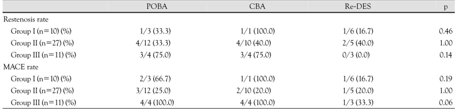
rates were not different according to therapeutic moda- lities within each group. Restenosis rates were as follows:
in group I, POBA (1/3, 33.3%), CBA (1/1, 100.0%), and Re-DES (1/6, 16.7%) (p=0.46); in group II, POBA (4/
12, 33.3%), CBA (4/10, 40.0%), and Re-DES (2/5, 40.0%) (p=1.00); and in group III, POBA (3/4, 75.0%), CBA (3/4, 75.0%), and Re-DES (0/3, 0.0%) (p=0.14).
There was no statistically significant difference in MACE rates between groups I and II. MACE rates were as fol- lows: in group I, POBA (2/3, 66.7%), CBA (1/1, 100.0%), and Re-DES (1/6, 16.7%) (p=0.19); and in group II, POBA (3/12, 25.0%), CBA (2/10, 20.0%), and Re- DES (1/5, 20.0%) (p=1.00). However, in group III, MACE rates were relatively higher for POBA (4/4, 100.0%) and CBA (4/4, 100.0%) than for Re-DES (1/3, 33.3%) (p=0.06), but these differences did not reach statistical significance (Table 6).
Discussion
The main findings of this study are as follows: 1) re- stenosis rates were not significantly different between focal (edge and body restenosis) and non-focal ISR; 2) restenosis rates were not significantly different among focal edge, focal body and non-focal ISR lesions regardless of the therapeutic modality chosen (POBA, CBA, and Re-DES); and 3) for non-focal ISR lesions, POBA and CBA strategies might be associated with higher MACE rates, and Re-DES might be a better choice for the le- sions.
Although ISR after DES implantation is much less frequent than after BMS, it is not less puzzling. Mole- cular mechanisms of arterial remodeling are less well understood and insight into the mechanisms of DES failure is still limited. It appears that the causes of res- tenosis after implantation of BMS and DES are funda- mentally the same. Restenosis is the arterial wall’s healing response to mechanical injury and includes two main processes: neointimal hyperplasia and vessel remodeling.1) Neointimal proliferation is the principal mechanism un- derlying ISR, and it is the result of endothelial damage after stent expansion.12-14) In our study, IVUS was per- formed in 27 cases (40.3%), and neointimal hyperplasia
was the main cause with stent fracture also being appa- rent in a total of 11 cases (40.7%). Although some reports have suggested that stent fracture might be another po- tential risk factor for restenosis, in our study the risk was not significantly elevated; however, there might have been some selection bias, and stent fracture might still be considered as another potential risk factor of ISR.15)16)
The patterns of angiographic restenosis after BMS implantation have been previously described, and it was shown that the Mehran ISR classification is an inde- pendent predictor of TLR, emphasizing the prognostic relevance of angiographic features after stent failure.
Mehran et al.17) showed that at 1-year follow-up in pa- tients undergoing percutaneous coronary intervention for BMS ISR, a significantly higher rate of TLR occur- red with more complex levels of ISR classification. Sim- ilarly, in the era of DES implantation, the incidence of TLR increases with the pattern of restenosis treated.11) In the present study, restenosis rates were not different among groups but MACE rates were numerically high- er in group III due to the high TLR rate. This result was comparable with that of a previous study.
Currently, there is a paucity of published data on the optimal management of DES restenosis. Some investi- gators have reported that repeated PCI with DES, BMS, brachytherapy, POBA, or CBA was safe and did not in- crease rates of vascular complications.2-7)18)19) Most reports on DES restenosis have indicated that the majority of cases, particularly those after SES implantation, are focal.
SES failure treated with traditional percutaneous coro- nary intervention yielded good outcomes at 1-year fol- low-up (a secondary failure rate of only 23%), perhaps due to the predominantly focal nature of the SES reste- notic lesion.8) Systematic use of SES to treat ISR was safe and effective in an unselected series of consecutive pati- ents treated in a real-world scenario, providing very low 9-month ischemia-driven TLR and MACE rates.20) On the basis of BMS-controlled RCT data, there is a definite therapeutic advantage associated with SES and PES use for the prevention of ISR. SES and PES continued to exceed the therapeutic potential of BMS, with a slight but consistent angiographic advantage being observed with SES.4)13)21) Furthermore, the use of DES for diffuse
Table 6. Angiographic and clinical outcomes by therapeutic modality
POBA CBA Re-DES p
Restenosis rate
Group I (n=10) (%) 1/3 (33.3) 1/1 (100.0) 1/6 (16.7) 0.46
Group II (n=27) (%) 4/12 (33.3) 4/10 (40.0) 2/5 (40.0) 1.00
Group III (n=11) (%) 3/4 (75.0) 3/4 (75.0) 0/3 (0.0) 0.14
MACE rate
Group I (n=10) (%) 2/3 (66.7) 1/1 (100.0) 1/6 (16.7) 0.19
Group II (n=27) (%) 3/12 (25.0) 2/10 (20.0) 1/5 (20.0) 1.00
Group III (n=11) (%) 4/4 (100.0) 4/4 (100.0) 1/3 (33.3) 0.06
POBA: plain old balloon angioplasty, CBA: cutting balloon angioplasty, Re-DES: repeated drug-eluting stent implantation
Ki-Hun Kim, et al.·413
ISR is feasible and safe, and is associated with acceptable early and mid-term results.9)10)20-22) On the other hand, Albiero et al.18) suggested that CBA and traditional PT- CA are equally effective in preventing ISR recurrence.
In our study, restenosis rates according to therapeu- tic modality were not significantly different within each group, and MACE rates were not different between the focal edge and focal body lesion groups. But, even if dif- ferences did not reach statistical significance due to re- latively small sample sizes, MACE rates were relatively higher for POBA and CBA than for Re-DES for non- focal lesions. Based on our results, it appears that all PCI options are associated with favorable outcomes when used for focal restenotic lesions, while Re-DES appears to be associated with more favorable clinical outcomes for non-focal lesions.
Limitations of this study is that: the data were collected in a retrospective and nonrandomized manner; the sam- ple size was small; there was a relatively low rate of an- giographic follow-up. A large scale randomized clinical trial is warranted to determine the optimal strategy for the treatment of ISR after DES implantation.
In conclusion, treatment of DES ISR should be indi- vidualized according to restenosis pattern, with all PCI strategies appearing appropriate for focal ISR patterns, and Re-DES appearing to be a better choice for non-fo- cal ISR patterns.
REFERENCES
1) Costa MA, Simon DI. Molecular basis of restenosis and drug- eluting stents. Circulation 2005;111:2257-73.
2) Costa MA. Treatment of drug-eluting stent restenosis. Am Heart J 2007;153:447-9.
3) Solinas E, Dangas G, Kirtane AJ, et al. Angiographic patterns of drug-eluting stent restenosis and one-year outcomes after treat- ment with repeated percutaneous coronary intervention. Am J Cardiol 2008;102:311-5.
4) Kim YH, Lee BK, Park DW, et al. Comparison with conventi- onal therapies of repeated sirolimus-eluting stent implantation for the treatment of drug-eluting coronary stent restenosis. Am J Cardiol 2006;98:1451-4.
5) Steinberg DH, Pinto Slottow TL, Buch AN, et al. Impact of in- stent restenosis on death and myocardial infarction. Am J Car- diol 2007;100:1109-13.
6) Lee SR, Jeong MH, Lim SY, et al. Comparison of the clinical ef- fect of cutting balloon angioplasty and drug-eluting stent for treat- ing the focal type of in-stent restenosis. Korean Circ J 2006;36:
279-84.
7) Cosgrave J, Melzi G, Corbett S, et al. Repeated drug-eluting stent
implantation for drug-eluting stent restenosis: the same or a dif- ferent stent. Am Heart J 2007;153:354-9.
8) Moussa ID, Moses JW, Kuntz RE, et al. The fate of patients with clinical recurrence after sirolimus-eluting stent implantation (a two-year follow-up analysis from the SIRIUS trial). Am J Cardiol 2006;97:1582-4.
9) Airoldi F, Briguori C, Iakovou I, et al. Comparison of sirolimus versus paclitaxel eluting stents for treatment of coronary in-stent restenosis. Am J Cardiol 2006;97:1182-7.
10) Lee HG, Chun KJ, Cho KI, et al. Impact of drug-eluting stents on clinical outcomes in patients with diffuse coronary lesions. Korean Circ J 2008;38:612-7.
11) Cosgrave J, Melzi G, Biondi-Zoccai GG, et al. Drug-eluting stent restenosis the pattern predicts the outcome. J Am Coll Cardiol 2006;47:2399-404.
12) Sukhija R, Aronow WS, Sureddi R, et al. Predictors of in-stent restenosis and patient outcome after percutaneous coronary in- tervention in patients with diabetes mellitus. Am J Cardiol 2007;
100:777-80.
13) Eisenberg MJ, Konnyu KJ. Review of randomized clinical trials of drug-eluting stents for the prevention of in-stent restenosis.
Am J Cardiol 2006;98:375-82.
14) Mitra AK, Agrawal DK. In stent restenosis: bane of the stent era.
J Clin Pathol 2006;59:232-9.
15) Yang TH, Kim DI, Park SG, et al. Clinical characteristics of stent fracture after sirolimus-eluting stent implantation. Int J Cardiol 2009;131:212-6.
16) Aoki J, Nakazawa G, Tanabe K, et al. Incidence and clinical im- pact of coronary stent fracture after sirolimus-eluting stent im- plantation. Catheter Cardiovasc Interv 2007;69:380-6.
17) Mehran R, Dangas G, Abizaid AS, et al. Angiographic patterns of in-stent restenosis: classification and implications for long-term outcome. Circulation 1999;100:1872-8.
18) Albiero R, Silber S, Di Mario C, et al. Cutting balloon versus conventional balloon angioplasty for the treatment of in-stent re- stenosis: results of the restenosis cutting balloon evaluation trial (RESCUT). J Am Coll Cardiol 2004;43:943-9.
19) Airoldi F, Rogacka R, Briguori C, et al. Comparison of clinical and angiographic outcome of sirolimus-eluting stent implantation versus cutting balloon angioplasty for coronary in-stent resteno- sis. Am J Cardiol 2004;94:1297-300.
20) Liistro F, Fineschi M, Angioli P, et al. Effectiveness and safety of sirolimus stent implantation for coronary in-stent restenosis: the TRUE (Tuscany Registry of Sirolimus for Unselected In-Stent Re- stenosis) Registry. J Am Coll Cardiol 2006;48:270-5.
21) Alfonso F, Perez-Vizcayno MJ, Hernandez R, et al. A randomized comparison of sirolimus-eluting stent with balloon angioplasty in patients with in-stent restenosis: results of the Restenosis In- trastent: Balloon Angioplasty Versus Elective Sirolimus-Eluting Stenting (RIBS-II) trial. J Am Coll Cardiol 2006;47:2152-60.
22) Kastrati A, Mehilli J, von Beckerath N, et al. Sirolimus-eluting stent or paclitaxel-eluting stent vs balloon angioplasty for pre- vention of recurrences in patients with coronary in-stent restenosis:
a randomized controlled trial. JAMA 2005;293:165-71.