Switching-on of serotonergic calcium signaling in activated hepatic stellate cells
Kyu-Sang Park, Pyo-Jin Sin, Dong Hyeon Lee, Seung-Kuy Cha, Min-Jeong Kim, Na-Hyun Kim, Soon-Koo Baik, Seong-Woo Jeong, In Deok Kong
Kyu-Sang Park, Pyo-Jin Sin, Seung-Kuy Cha, Min-Jeong Kim, Seong-Woo Jeong, In Deok Kong, Department of Physiology and Institute of Lifelong Health, Yonsei University Wonju Col- lege of Medicine, Wonju, Gangwon-Do, 220-701, South Korea Dong Hyeon Lee, Department of Physiology, College of Medi- cine, Pochon CHA University, Seongnam, Gyeonggi-Do, 463-712, South Korea
Na-Hyun Kim, Department of Basic Nursing Science, Keimyung University College of Nursing, Daegu, 700-712, South Korea Soon-Koo Baik, Department of Internal Medicine, Yonsei Univer- sity Wonju College of Medicine, Wonju, Gangwon-Do, 220-701, South Korea
Author contributions: Park KS, Sin PJ and Kong ID designed the project; Park KS, Sin PJ, Lee DH, Cha SK, Kim MJ and Kim NH performed the experiments; Park KS, Sin PJ and Kong ID wrote the manuscript; Baik SK and Jeong SW contributed to the discussion of the data and the revision of the manuscript.
Supported by Grants from the Korean National Research Foun- dation (2010-0014617); the Myung Sun Kim Memorial Founda- tion (2009); and the Yonsei University Faculty Research Grant (2004)
Correspondence to: In Deok Kong, Professor, Department of Physiology and Institute of Lifelong Health, Yonsei University Wonju College of Medicine, 162 Ilsan-Dong, Wonju, Gangwon- Do, 220-701, South Korea. [email protected]
Telephone: +82-33-7410292 Fax: +82-33-7456461 Received: September 6, 2010 Revised: October 11, 2010 Accepted: October 18, 2010
Published online: January 14, 2011
Abstract
AIM: To investigate serotonergic Ca2+
signaling and the expression of 5-hydroxytryptamine (5-HT) receptors, as well as Ca
2+transporting proteins, in hepatic stellate cells (HSCs).
METHODS: The intracellular Ca2+
concentration ([Ca
2+]
i) of isolated rat HSCs was measured with a fluorescence microscopic imaging system. Quantitative PCR was per-
formed to determine the transcriptional levels of 5-HT receptors and endoplasmic reticulum (ER) proteins in- volved in Ca
2+storage and release in cultured rat HSCs.
RESULTS: Distinct from quiescent cells, activated HSCs
exhibited [Ca
2+]
itransients following treatment with 5-HT, which was abolished by U-73122, a phospholipase C inhibitor. Upregulation of 5-HT
2Aand 5-HT
2Breceptors, but not 5-HT
3, was prominent during trans-differenti- ation of HSCs. Pretreatment with ritanserin, a 5-HT
2antagonist, inhibited [Ca
2+]
ichanges upon application of 5-HT. Expression of type 1 inositol-5’-triphosphate receptor and type 2 sarcoplasmic/endoplasmic reticulum Ca
2+ATPase were also increased during activation of HSCs and serve as the major isotypes for ER Ca
2+stor- age and release in activated HSCs. Ca
2+binding chap- erone proteins of the ER, including calreticulin, calnexin and calsequestrin, were up-regulated following activa- tion of HSCs.
CONCLUSION: The appearance of 5-HT-induced [Ca2+
]
iresponse accompanied by upregulation of metabotropic 5-HT
2receptors and Ca
2+transporting/chaperone ER proteins may participate in the activating process of HSCs.
© 2011 Baishideng. All rights reserved.
Key words: Hepatic stellate cells; 5-hydroxytryptamine;
Intracellular Ca
2+transient; Sarcoplasmic/endoplasmic reticulum Ca
2+ATPase; Inositol-5’-triphosphate recep- tor; Endoplasmic reticulum chaperone
Peer reviewer: Bronislaw L Slomiany, PhD, Professor, Re- search Center, C-875, UMDNJ-NJ Dental School, 110 Bergen Street, PO Box 1709, Newark, NJ 07103-2400, United States Park KS, Sin PJ, Lee DH, Cha SK, Kim MJ, Kim NH, Baik SK, Jeong SW, Kong ID. Switching-on of serotonergic calcium sig- naling in activated hepatic stellate cells. World J Gastroenterol
ORIGINAL ARTICLE
© 2011 Baishideng. All rights reserved.
doi:10.3748/wjg.v17.i2.164
2011; 17(2): 164-173 Available from: URL: http://www.wjg- net.com/1007-9327/full/v17/i2/164.htm DOI: http://dx.doi.
org/10.3748/wjg.v17.i2.164
INTRODUCTION
Hepatic stellate cells (HSCs), also known as “Ito cells”
or “fat-storing cells”, localize between hepatocytes and sinusoids (space of Disse) in mammalian livers. In their healthy state, HSCs control retinoid homeostasis, sinusoi- dal blood flow, macromolecule transport, and potentially act as antigen-presenting cells in the liver
[1,2]. However, in response to hepatic injury, HSCs undergo gross morpho- logical and functional changes, transforming to a myofi- broblast-like phenotype in a process called “activation”
or “trans-differentiation”
[3,4]. Manifestations of activated HSCs include: (1) the expression of contractile cytoskele- tal proteins such as α-smooth muscle actin (α-SMA)
[5,6]; (2) enhanced extracellular matrix synthesis
[7,8]; (3) increased cell size and proliferation
[9]; (4) decreased size of lipid droplets
[8,10]; and (5) well developed endoplasmic reticulum (ER), Golgi bodies, and compacted microfilaments
[11,12]. In particular, the deposition of cross-linked collagen during the activation process may result in cirrhotic changes ac- companied by life-threatening hepatic dysfunction.
Serotonin [5-hydroxytryptamine (5-HT)] is a neurotrans- mitter that also acts as a multifunctional hormone in vari- ous tissues
[13], where it modulates proliferation and dif- ferentiation of muscle, neurons, and mammary glands
[14-16]. Serotonin released from platelets at sites of injury plays an important role in liver regeneration and fibrosis
[17]. It has been reported that patients with cirrhosis of the liver and portal hypertension have increased plasma serotonin levels
[18]. The expression levels of 5-HT
2Aand 5-HT
2Bare increased in the liver after hepatectomy as well as in activated HSCs
[2,17]. Moreover, 5-HT
2receptor antagonists suppress cell proliferation and expression of key fibro- genic factors in activated HSCs
[2,19]. Among the mamma- lian 5-HT receptors (5-HT
1to 5-HT
7), the 5-HT
2receptor family is coupled to the G
q/11protein and increases intra- cellular Ca
2+concentration ([Ca
2+]
i) mobilized from ER reservoirs
[20].
As the major intracellular calcium storage site, the ER possesses various kinds of calcium regulatory proteins that participate in: (1) pumping Ca
2+into the ER lumen, such as the sarcoplasmic/endoplasmic reticulum Ca
2+ATPase (SERCA); (2) releasing Ca
2+into the cytosol, such as IP
3or ryanodine receptors; and (3) buffering Ca
2+, such as calre- ticulin and calnexin, which are also known as chaperones.
ER Ca
2+homeostasis is maintained by a balance between Ca
2+release and replenishment
[21]. The free Ca
2+concentra- tion in the ER ([Ca
2+]
ER) ranges from 60-400 μmol/L, and disturbances in [Ca
2+]
ERhomeostasis can affect many of the functions of the ER including protein synthesis, secre- tion
[22], protein folding
[23], and sensitivity of cells to apop- tosis
[24]. Further, [Ca
2+]
ERhomeostasis might be critically required for the activation process of HSCs in order to
keep up with accelerated protein synthesis. However, until now, the compensatory changes in ER protein expression involved in Ca
2+homeostasis and chaperone function have not been clearly elucidated.
[Ca
2+]
imay be important for the activation of HSCs, primarily because [Ca
2+]
iregulates the transcription of genes critical for cell function
[25], and secondly because contractile elements such as α-SMA respond sensitively to [Ca
2+]
i[26]. We hypothesized that serotonin, acting as an au- tocrine or paracrine mediator, can elicit a Ca
2+signal, and this signal might be involved in the activation of HSCs.
Moreover, there may be an alteration in the ER function of HSCs such as Ca
2+release and protein folding. In this study, we isolated and cultured rat HSCs on plastic dishes in vitro, which has been widely accepted as an appropriate model for the study of activated HSCs
[8,27]. Appearance of [Ca
2+]
itransients induced by 5-HT and the upregula- tion of 5-HT
2receptors and ER proteins were observed during HSC activation. These observed changes may par- ticipate in an activation signal as well as adaptive changes during the trans-differentiation of HSCs.
MATERIALS AND METHODS
Isolation of rat HSCs
HSCs were isolated from male Sprague-Dawley rats (150- 250 g) by means of a collagenase/pronase perfusion and Nycodenz-gradient centrifugation, as previously de- scribed
[28,29]. HSCs were cultured with DMEM containing fetal bovine serum (10%) and antibiotics-antimycotics (Invitrogen, Carlsbad, CA, USA) in a humidified incuba- tor (5% CO
2, 37 ℃ ). The purity of HSCs was > 95% as assessed by their typical microscopic morphology and positive immunocytochemical staining for desmin at 24 to 48 h after seeding.
Quantitative reverse transcription-polymerase chain reaction analysis
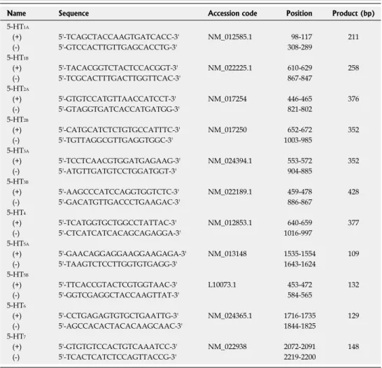
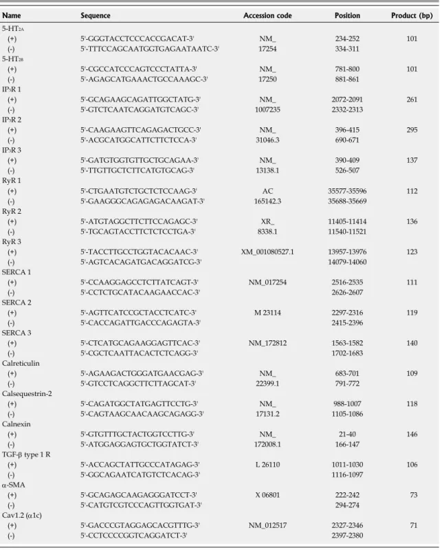
Total cellular RNA was isolated and purified from HSCs at different culture periods, and reverse transcription (RT) was performed with random hexamers. Quantitative real time PCR using SYBR Green PCR Master mix (Applied Biosystems, Foster City, CA, USA) was performed on an ABI PRISM 7900HT Sequence Detection System (Applied Biosystems). Sequence specific oligonucleotide primers for the genes of interest were designed based on rat sequenc- es deposited in the GenBank database (Tables 1 and 2), and the amplification program included the activation of AmpliTaq Gold at 95 ℃ for 10 min, followed by 45 cycles of a two-step PCR reaction with denaturation at 95 ℃ for 15 s and annealing/extension at 60 ℃ for 1 min. The con- stitutively expressed housekeeping gene glyceraldehydes- 3-phosphate dehydrogenase (GAPDH) was selected as an endogenous control to correct for potential variation in RNA loading and efficiency of amplification reactions.
Fluorescent [Ca
2+]
imeasurement
HSCs at 3 d or 2 wk after isolation were seeded on glass
coverslips and loaded with fura-2/AM (5 μmol/L) in a dark room for 30 to 60 min at room temperature. Dye- loaded cells were then washed and transferred to a per- fusion chamber on a fluorescence microscope (IX-70, Olympus, Tokyo, Japan). The HSCs were alternately excited at 340 and 380 nm by a monochromatic light source (LAMDA DG-4; Sutter, Novato, CA, USA), and fluorescence images were captured at 510 nm with an intensified CCD camera (Cascade; Roper, Duluth, GA, USA). Images were analyzed using the Metafluor 6.1 soft- ware package (Universal Imaging Corporation, Downing- town, PA, USA).
Immunocytochemistry
HSCs cultured on coverslips were fixed in 4% paraformal- dehyde and immunocytochemical staining was performed using an antibody for α-SMA (Sigma Chemical Co., St Louis, MO, USA). After incubating with a biotinylated secondary antibody, an avidin-conjugated peroxidase com- plex was added to the slides and 3-amino-9-ethylcarbazole (AEC) was used as the chromogen.
Electrophysiology
Whole-cell membrane currents were recorded using the gramicidin-perforated patch-clamp technique as described
previously
[28]. All experiments were performed at room temperature (20-24 ℃ ). The internal solution for the per- forated patch clamp contained (in mmol/L): 140 KCl, 5 EGTA, 10 HEPES, 0.5 CaCl
2, 5 NaCl, and gramicidin (50 μg/mL) (pH 7.2). The external solution contained (in mmol/L): 135 NaCl, 5.4 KCl, 1.8 CaCl
2, 1 MgCl
2, 5 HEPES, and 10 glucose (pH 7.4).
Statistical analysis
Quantitative data are expressed as the mean ± SE. Statisti- cal comparisons were made by the two-tailed Student’s t-test and ANOVA. Differences with P < 0.05 were con- sidered to be significant. PCR from each cDNA sample was done in triplicate and n indicates the number of ex- periments. For quantitative comparisons, the expression level of each gene was normalized to that of GAPDH and presented as relative expression ratio (target/GAP- DH) by applying the formula 2
-ΔΔCt[30].
RESULTS
Serotonergic signaling and receptor expression during HSC activation
We isolated HSCs using density gradient-based separa- tion with Nycodenz. Most of the harvested cells (> 95%)
Table 1 Primers for reverse transcription-polymerase chain reaction
Name Sequence Accession code Position Product (bp)
5-HT1A
(+) 5'-TCAGCTACCAAGTGATCACC-3' NM_012585.1 98-117 211
(-) 5'-GTCCACTTGTTGAGCACCTG-3' 308-289
5-HT1B
(+) 5'-TACACGGTCTACTCCACGGT-3' NM_022225.1 610-629 258
(-) 5'-TCGCACTTTGACTTGGTTCAC-3' 867-847
5-HT2A
(+) 5'-GTGTCCATGTTAACCATCCT-3' NM_017254 446-465 376
(-) 5'-GTAGGTGATCACCATGATGG-3' 821-802
5-HT2B
(+) 5'-CATGCATCTCTGTGCCATTTC-3' NM_017250 652-672 352
(-) 5'-TGTTAGGCGTTGAGGTGGC-3' 1003-985
5-HT3A
(+) 5'-TCCTCAACGTGGATGAGAAG-3' NM_024394.1 553-572 352
(-) 5'-ATGTTGATGTCCTGGATGGT-3' 904-885
5-HT3B
(+) 5'-AAGCCCATCCAGGTGGTCTC-3' NM_022189.1 459-478 428
(-) 5'-GACATGTTGACCCTGAAGAC-3' 886-867
5-HT4
(+) 5'-TCATGGTGCTGGCCTATTAC-3' NM_012853.1 640-659 377
(-) 5'-CTCATCATCACAGCAGAGGA-3' 1016-997
5-HT5A
(+) 5'-GAACAGGAGGAAGGAAGAGA-3' NM_013148 1535-1554 109
(-) 5'-TAAGTCTCCTTGGTGTGAGG-3' 1643-1624
5-HT5B
(+) 5'-TTCACCGTACTCGTGGTAAC-3' L10073.1 453-472 132
(-) 5'-GGTCGAGGCTACCAAGTTAT-3' 584-565
5-HT6
(+) 5'-CCTGAGAGTGTGCTGAATTG-3' NM_024365.1 1716-1735 129
(-) 5'-AGCCACACTACACAAGCAAC-3' 1844-1825
5-HT7
(+) 5'-GTGTGTCCACTGTCAAATCC-3' NM_022938 2072-2091 148
(-) 5'-TCACTCATCTCCAGTTACCG-3' 2219-2200
5-HT: 5-hydroxytryptamine.
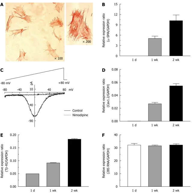
exhibited positive intra-cytoplasmic staining for desmin and glial fibrillary acidic proteins (GFAP). Expression of HSC trans-differentiation markers was tested at 1 d, 1 wk and 2 wk after isolation. In activated HSCs (2 wk after isolation), bundles of α-SMA were clearly observed as cytoskeletal fibers in immunocytochemical staining (Figure 1A), which was not evident in quiescent cells. In a voltage-clamp mode, nimodipine (10 μmol/L)-sensitive L-type Ca
2+currents were recorded only for activated
HSCs (Figure 1C). The expression level of α-SMA and the L-type Ca
2+channel (Cav1.2) were proportional to the activation period elicited by culturing cells on plastic dishes (Figure 1B and D). Transforming growth factor-β1 (TGF-β1), an abundant isoform of TGF in both normal and cirrhotic liver, is known as the main profibrogenic cytokine
[31]. We observed that the type Ⅰ receptor for TGF-β1 (Tβ-RI) was also upregulated during activation (Figure 1E), while the expression of 28S RNA as well as
Table 2 Primers for quantitative reverse transcription-polymerase chain reaction
Name Sequence Accession code Position Product (bp)
5-HT2A
(+) 5'-GGGTACCTCCCACCGACAT-3' NM_ 234-252 101
(-) 5'-TTTCCAGCAATGGTGAGAATAATC-3' 17254 334-311
5-HT2B
(+) 5'-CGCCATCCCAGTCCCTATTA-3' NM_ 781-800 101
(-) 5'-AGAGCATGAAACTGCCAAAGC-3' 17250 881-861
IP3R 1
(+) 5'-GCAGAAGCAGATTGGCTATG-3' NM_ 2072-2091 261
(-) 5'-GTCTCAATCAGGATGTCAGC-3' 1007235 2332-2313
IP3R 2
(+) 5'-CAAGAAGTTCAGAGACTGCC-3' NM_ 396-415 295
(-) 5'-ACGCATGGCATTCTTCTCCA-3' 31046.3 690-671
IP3R 3
(+) 5'-GATGTGGTGTTGCTGCAGAA-3' NM_ 390-409 137
(-) 5'-TTGTTGCTCTTCATGTGCAG-3' 13138.1 526-507
RyR 1
(+) 5'-CTGAATGTCTGCTCTCCAAG-3' AC 35577-35596 112
(-) 5'-GAAGGGCAGAGAGACAAGAT-3' 165142.3 35688-35669
RyR 2
(+) 5'-ATGTAGGCTTCTTCCAGAGC-3' XR_ 11405-11414 136
(-) 5'-TGCAGTACCTTCTCTCCTGA-3' 8338.1 11540-11521
RyR 3
(+) 5'-TACCTTGCCTGGTACACAAC-3' XM_001080527.1 13957-13976 123
(-) 5'-AGTCACAGATGACAGGATCG-3' 14079-14060
SERCA 1
(+) 5'-CCAAGGAGCCTCTTATCAGT-3' NM_017254 2516-2535 111
(-) 5'-CCTCTGCATACAAGAACCAC-3' 2626-2607
SERCA 2
(+) 5'-AGTTCATCCGCTACCTCATC-3' M 23114 2297-2316 119
(-) 5'-CACCAGATTGACCCAGAGTA-3' 2415-2396
SERCA 3
(+) 5'-CTCATGCAGAAGGAGTTCAC-3' NM_172812 1563-1582 140
(-) 5'-CGCTCAATTACACTCTCAGG-3' 1702-1683
Calreticulin
(+) 5'-AGAAGACTGGGATGAACGAG-3' NM_ 683-701 109
(-) 5'-GTCCTCAGGCTTCTTAGCAT-3' 22399.1 791-772
Calsequestrin-2
(+) 5'-CAGATGGCTATGAGTTCCTG-3' NM_ 988-1007 118
(-) 5'-CAGTAAGCAACAAGCAGAGG-3' 17131.2 1105-1086
Calnexin
(+) 5'-GTGTTTGCTACTGGTCCTTG-3' NM_ 21-40 146
(-) 5'-ATGGAGGAGTGCTGGTATCT-3' 172008.1 166-147
TGF-β type 1 R
(+) 5'-ACCAGCTATTGCCCATAGAG-3' L 26110 1011-1030 106
(-) 5'-GGCAGAATCATGTCTCACAG-3' 1116-1097
α-SMA
(+) 5'-GCAGAGCAAGAGGGATCCT-3' X 06801 222-242 73
(-) 5'-CATGTCGTCCCAGTTGGTGAT-3' 294-274
Cav1.2 (α1c)
(+) 5'-GACCCGTAGGAGCACGTTTG-3' NM_012517 2327-2346 71
(-) 5'-CCTCCCCGGTCAGGATCT-3' 2397-2380
5-HT: 5-hydroxytryptamine; SERCA: Sarcoplasmic/endoplasmic reticulum Ca2+ ATPase; α-SMA: α-smooth muscle actin; RyR: Ryanodine receptor; TGF: Transforming growth factor.
GAPDH was not changed during the activation process of HSCs (Figure 1F).
Serotonergic signaling has been suggested as a candi- date for triggering activation of HSCs
[2,17]. We focused on [Ca
2+]
isignaling in HSCs, which has been emphasized by previous work as having an important role in the activa- tion process
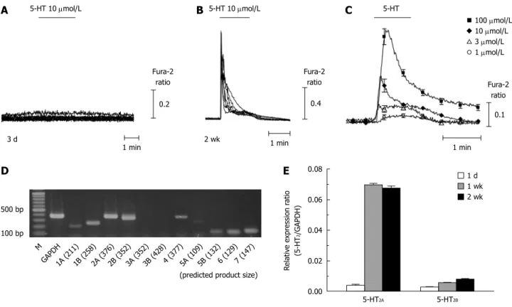
[26,32]. As shown in Figure 2A and B, strong [Ca
2+]
itransients followed by a slow plateau increase were recorded in response to 5-HT (10 μmol/L) application only from most of the activated HSCs (2 wk after isola- tion; 81 cells out of 92 cells), but not from quiescent cells (3 d after isolation; 0 out of 11 cells). The 5-HT-induced [Ca
2+]
iincrease was dose-dependent in activated HSCs (Figure 2C). Consistent with a previous report
[33], ATP also evoked [Ca
2+]
itransients in activated HSCs while ace- tylcholine did not (Figure 3).
Among the 5-HT receptors, 5-HT
2is known to re- lease Ca
2+from the ER while 5-HT
3acts as a ligand-gated cation channel
[20]. We estimated the steady-state mRNA levels of 5-HT receptor isotypes (5-HT
1to 5-HT
7) using reverse transcription-polymerase chain reaction (RT-PCR) and found that the 5-HT
2Aand 5-HT
2Breceptors, but not 5-HT
3, were abundantly transcribed (Figure 2D). Consis- tent with the observed changes in [Ca
2+]
i, the expression of 5-HT
2Awas increased by about 17-fold after 2 wk of isolation (5-HT
2A/GAPDH; from 0.004 at 1 d to 0.067 at 2 wk). 5-HT
2Bwas also found to be upregulated in acti- vated HSCs (from 0.003 to 0.008) using quantitative RT- PCR (Figure 2E).
It has been recognized that 5-HT
2receptors are coupled with the G
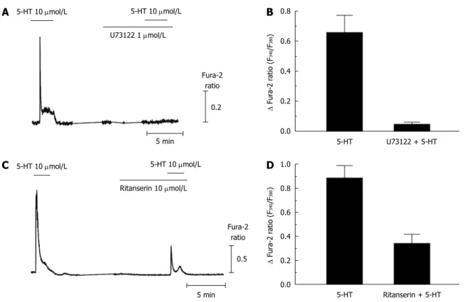
q/11-phospholipase C pathway. Figure 4A and B show that the 5-HT-induced [Ca
2+]
ichanges were abolished
× 100
× 200
15 12 9 6 3 0 Relative expression ratio (α-SMA/GAPDH)
1 d 1 wk 2 wk 0.08
0.06
0.04
0.02
0.00 Relative expression ratio (Cav1.2/GAPDH)
1 d 1 wk 2 wk 40
30
20
10
0 Relative expression ratio (28S RNA/GAPDH)
1 d 1 wk 2 wk 0.20
0.15
0.10
0.05
0.00 Relative expression ratio (Tβ-RI/GAPDH)
1 d 1 wk 2 wk -80 -40 40 8010
-40
-90
mV +80 mV -80 mV
Control Nimodipine
A B
D C
F E
Figure 1 Expression of α-smooth muscle actin, L-type calcium channels and type 1 transforming growth factor-β receptors in activated rat hepatic stel- late cells. A: Immunocytochemical staining for α-smooth muscle actin (α-SMA) was performed on hepatic stellate cells (HSCs) cultured for 1 wk; C: Whole cell Ca2+
currents in a voltage-clamp mode were recorded from 2 wk-cultured HSCs, and were completely blocked by nimodipine (10 μmol/L); Changes in the transcript levels of α-SMA (B), the α1c subunit of the L-type Ca2+ channel (Cav1.2) (D), the type 1 receptor of transforming growth factor-β (Tβ-RI) (E), and 28S RNA (F) during HSC culturing (1 d, 1 wk and 2 wk) were measured by quantitative real-time reverse transcription-polymerase chain reaction analysis. Expression levels were normalized to glyceraldehyde-3-phosphate dehydrogenase (GAPDH) and expressed as a relative expression ratio (target/GAPDH). Data are presented as the mean ± SE (n = 3).
pA
by pretreatment with 1 μmol/L U73122, a phospholipase C inhibitor (0.05 ± 0.05 peak changes of Fura-2 ratio from 0.66 ± 0.12, n = 13). We also observed that [Ca
2+]
itransients induced by 5-HT were not altered in extracel- lular Ca
2+-free conditions (data not shown). These results suggest that 5-HT activates phospholipase C to produce IP
3, which induces Ca
2+release from ER in activated HSCs. To confirm the receptor subtype, we tested block- ing effects of a universal 5-HT
2antagonist, ritanserin, which does not discriminate among 5HT
2isotypes. 5-HT- induced [Ca
2+]
iresponses were attenuated by pretreatment
with 10 μmol/L ritanserin by 46.3% (0.34 ± 0.08 from 0.89
± 0.10, n = 11).
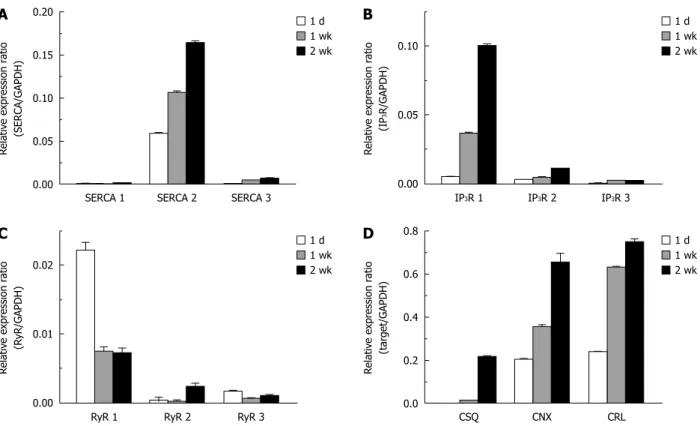
Upregulation of calcium transporting and binding proteins in the ER
In mammalian cells, there are three major subtypes of the sarcoplasmic/endoplasmic reticulum Ca
2+ATPase (SERCA1, 2, and 3) which pump Ca
2+into the ER lu- men. We observed SERCA2 to be the dominant subtype in HSCs. SERCA2, especially SERCA2b, is considered to be a house-keeping protein expressed constitutively
5-HT 10 μmol/L
Fura-2 ratio
0.2
1 min 3 d
5-HT 10 μmol/L
Fura-2 ratio
0.4
1 min 2 wk
5-HT
Fura-2 ratio
0.1
1 min
100 μmol/L 10 μmol/L 3 μmol/L 1 μmol/L
0.08
0.06
0.04
0.02
0.00 500 bp
100 bp M
1A (211) GAPDH 1B (258)
2A (376) 2B (352)
3A (352) 3B (428)
4 (377)5A (109) 5B (132)
6 (129)7 (147)
(predicted product size) Relative expression ratio 2(5-HT/GAPDH)
1 d 1 wk 2 wk
5-HT2A 5-HT2B
C B
A
D E
Figure 2 5-hydroxytryptamine-induced intracellular Ca2+ concentration changes and the expression of 5-hydroxytryptamine2 receptors in quiescent and activated hepatic stellate cells. A, B: 5-hydroxytryptamine (5-HT)-induced intracellular Ca2+ concentration ([Ca2+]i) transients were recorded from hepatic stellate cells (HSCs) at 3 d (A) and 2 wk (B) after isolation; C: Averages of [Ca2+]i changes (from 13-40 cells/each trace) in response to 5-HT (1-100 μmol/L) application to 2 wk- cultured HSCs are shown; D: Steady-state mRNA levels of the 5-HT receptor isotypes in 2 wk-cultured HSCs were compared using reverse transcription-polymerase chain reaction (RT-PCR); E: Using quantitative RT-PCR, the transcriptional changes in 5-HT2 receptors among 1 d-, 1 wk- and 2 wk-cultured HSCs were compared.
Expression levels were normalized to GAPDH and expressed as a relative expression ratio (target/GAPDH, n = 3). Data are presented as the mean ± SE.
0.6
0.4
0.2
0.0
Δ Fura-2 ratio (F340/F380)
(28)
(28)
(16) ATP 5-HT ACh
B
5-HT 10 μmol/L
Fura-2 ratio (F340/F380)
0.2 1 min ATP 100 μmol/L
A
Figure 3 Comparison of intracellular Ca2+ concentration responses to various metabotropic receptor agonists in activated hepatic stellate cells. Intracellular Ca2+ concentration changes following application of ATP (100 μmol/L), 5-hydroxytryptamine (5-HT) (10 μmol/L), or acetylcholine (ACh, 10 μmol/L) were measured in 2 wk- cultured hepatic stellate cells (n = 3-6, 16-28 cells). Data are presented as the mean ± SE.
Figure 4 5-hydroxytryptamine-induced intracellular Ca2+ concentration transients via metabotropic 5-hydroxytryptamine2 receptor in activated hepatic stellate cells. A, B: 5-hydroxytryptamine (5-HT)-induced intracellular Ca2+ concentration ([Ca2+]i) transients were completely abolished by pretreatment with U73122 (1 μmol/L), a phospholipase C blocker (n = 3, 11 cells); C, D: Ritanserin (10 μmol/L), a 5-HT2 antagonist, inhibited the [Ca2+]i responses to 5-HT in activated hepatic stellate cells (2 wk-cultured cells; n = 3, 13 cells). Data are presented as the mean ± SE.
0.8
0.6
0.4
0.2
0.0
Δ Fura-2 ratio (F340/F380)
5-HT U73122 + 5-HT
B
1.0 0.8 0.6 0.4 0.2 0.0
Δ Fura-2 ratio (F340/F380)
5-HT 10 μmol/L
Fura-2 ratio
0.5
5 min 5-HT 10 μmol/L Ritanserin 10 μmol/L
C
5-HT Ritanserin + 5-HT
D
5-HT 10 μmol/L
Fura-2 ratio
0.2
5 min 5-HT 10 μmol/L U73122 1 μmol/L
A
in most kinds of cells; however, in HSCs, the expression of SERCA2 tends to increase during activation. Specifi- cally, the relative expression ratio of SERCA2 (SERCA2/
GAPDH) at 1 d after isolation was 0.058, and increased to 0.106 after 1 wk in culture and 0.164 after 2 wk in culture in vitro (Figure 5A). The expression of SERCA3 was also increased during culture (SERCA3/GAPDH; 0.4 × 10
-3at 1 d and 6.9 × 10
-3at 2 wk).
Among the three isoforms (types 1 through 3) of the IP
3receptor, the type 1 IP
3receptor was the main subtype expressed in activated HSCs. We observed that the expres- sion of the type 1 IP
3receptor increased by about 7-fold (IP
3R 1/GAPDH = 0.037) after 1 wk of culture, and 20-fold (0.100) after 2 wk of culture compared to (0.005) levels 1 d after isolation (Figure 5B). In contrast, the ex- pression level of ryanodine receptors, which are a family of Ca
2+-releasing channel proteins expressed in the ER, either did not change or was decreased during the activa- tion of HSCs (Figure 5C).
We investigated whether Ca
2+binding chaperones of the ER could be up-regulated following the activation process of HSCs. There were similar increases in the ex- pression levels of calreticulin (calreticulin/GAPDH; from 0.204 at 1 d to 0.655 at 2 wk), calnexin (calnexin/GAP- DH; from 0.240 at 1 d to 0.750 at 2 wk), and calsequestrin in HSCs. In the case of calsequestrin, the expression level in HSCs at 1 d after isolation was undetectable, but was markedly increased (calsequestrin/GAPDH; 0.217) after 2 wk of culturing (Figure 5D).
DISCUSSION
Trans-differentiation of HSCs is accompanied by marked increases in protein synthesis, including collagen, elastin, and glycoproteins
[7]. It is well known that Ca
2+homeosta- sis in the ER is critical for the synthesis, folding, and se- cretion of protein
[22,23]. In HSCs, the depletion of ER Ca
2+stores inhibits protein synthesis and increases intracellular degradation of collagen
[34]. Maintaining a high Ca
2+gradi- ent across the ER membrane (around 1000-fold) is ac- complished by active Ca
2+transport by SERCAs. Among the three different isoforms of SERCAs, SERCA2 is con- sidered to be a house-keeping protein expressed in the ER of most cell types, including HSCs
[34]. We observed that SERCA2 was the main isotype in quiescent and activated HSCs (Figure 5A). During activation, the expression of SERCA2 (and also SERCA3) was increased, which likely helped to maintain appropriate ER Ca
2+concentrations.
Chaperone proteins in the ER facilitate the folding
of newly synthesized proteins and glycoproteins. In par-
ticular, calreticulin and calnexin are important chaperones
involved in a “quality control” system for protein synthe-
sis
[35]. In addition, these chaperones act as Ca
2+binding
proteins in the ER. Overexpression of calreticulin increas-
es the total amount of Ca
2+in intracellular stores, whereas
calreticulin-deficient cells have reduced ER Ca
2+storage
capacity
[36]. Impaired collagen synthesis has been observed
in cells derived from mice possessing genetic defects in
ER chaperone proteins
[37]. In this study, we observed for
the first time that the expression of ER Ca
2+binding pro- teins was markedly increased during the activation process of HSCs, which might be an important adaptive change for trans-differentiation.
Upon stimulation from the extracellular space, ER Ca
2+is the main source for releasing Ca
2+and is respon- sible for enabling biologic signaling mediated by Ca
2+. In addition, Ca
2+release from the ER stimulates store-oper- ated Ca
2+entry into the cytosol, which eventually increases the refilling of the ER Ca
2+reservoir. It has been shown that cytosolic Ca
2+signaling is important for proliferation and differentiation of HSCs
[25]. Similar to myofibroblast- like cells, activated HSCs can have a contractile response to [Ca
2+]
ichanges, which may increase vascular resistance leading to portal hypertension in vivo
[32]. During trans- differentiation, the expression of L-type calcium channels increases, which may contribute to cytosolic Ca
2+signaling in HSCs
[26,38]. In the present study, we observed that 5-HT increased [Ca
2+]
ionly in activated HSCs via a serotonergic receptor. Until now, 5-HT-induced [Ca
2+]
ichanges have not been reported in HSCs. Physiologic concentrations of 5-HT in plasma are known to be less than 100 nmol/L, but those in cirrhotic patients are significantly elevated (3-4 fold) compared to controls
[39]. Moreover, intrahepatic neighboring cells secrete 5-HT to act as an autocrine/
paracrine regulator
[40]. Thus, we hypothesize that local 5-HT concentration close to the releasing cells might be higher than the plasma level and repetitive exposure may
have additive effects on [Ca
2+]
i-mediated changes in the process of HSC activation.
We observed that 5-HT elicited a [Ca
2+]
iresponse via the metabotropic 5-HT
2receptor in activated HSCs. This was demonstrated by the findings that 5-HT-induced [Ca
2+]
itransients were (1) completely blocked by a PLC inhibitor;
(2) not altered by nominally Ca
2+free conditions; and (3) reduced by a 5-HT
2blocker. 5-HT
2Ais known to mediate mitogenic effects in fibroblasts
[41], while 5-HT
2Bis involved in the development of the heart and enteric nervous system
[42]. However, we did not discriminate whether the 5-HT
2Aand/or 5-HT
2Breceptor mediated the serotonergic Ca
2+signaling in activated HSCs. We also observed that the type Ⅰ IP
3receptor (IP
3R 1) is the main isoform expressed in activated HSCs, which is consistent with a recent report by Kruglov et al
[32]. The expression level of IP
3R 1 was in- creased during the activation process (Figure 5B).
Various ligands for G
q/11-coupled metabotropic recep- tors could be important extracellular stimuli, as they gen- erate IP
3by activating phospholipase-C. Interestingly, it has been reported that the expression of the P2Y metabo- tropic purinoceptor (P2Y6) is rapidly upregulated follow- ing activation of HSCs, with a similar increase in ATP- induced [Ca
2+]
itransients
[33]. The same study also reported that extracellular UDP increases the transcription of procollagen in activated HSCs via activation of the P2Y receptor, and this effect is partially inhibited by a P2Y receptor blocker. These results add further support to the
0.8
0.6
0.4
0.2
0.0 Relative expression ratio (target/GAPDH)
1 d 1 wk 2 wk
CSQ CRL
D
CNX 0.20
0.15
0.10
0.05
0.00 Relative expression ratio (SERCA/GAPDH)
1 d 1 wk 2 wk
SERCA 1 SERCA 2
A
SERCA 3
0.10
0.05
0.00 Relative expression ratio (IP3R/GAPDH)
1 d 1 wk 2 wk
IP3R 1 IP3R 3
B
IP3R 2
0.02
0.01
0.00 Relative expression ratio (RyR/GAPDH)
1 d 1 wk 2 wk
RyR 1 RyR 3
C
RyR 2
Figure 5 Up-regulation of endoplasmic reticulum Ca2+ transporting and binding proteins in activated hepatic stellate cells. Changes in the expression level of 3 isoforms of the sarcoplasmic/endoplasmic reticulum Ca2+ ATPase (SERCA) (A), inositol triphosphate receptor (IP3R) (B), ryanodine receptor (RyR) (C) and Ca2+
binding chaperones (D) during the culture periods (1 d, 1 wk and 2 wk) were measured by quantitative real-time reverse transcription-polymerase chain reaction analysis. Expression levels were normalized to GAPDH and expressed as a relative expression ratio (target/GAPDH). Data are presented as the mean ± SE (n = 3).
CSQ: Calsequestrin, CNX: Calnexin, and CRL: Calreticulin.
hypothesis that Ca
2+signaling released from ER stores is associated with HSCs undergoing the process of activa- tion. We also observed that ATP increased [Ca
2+]
i, which might be mediated by the metabotropic P2Y receptor (Figure 3). However, acetylcholine did not induce calcium changes, indicating that muscarinic acetylcholine receptors do not functionally exist in activated HSCs, even in the presence of machinery for ER Ca
2+release.
In this study, we observed the pronounced increase in serotonergic [Ca
2+]
iresponse related to the upregula- tion of metabotropic 5-HT
2receptors, type 1 inositol-5’- triphosphate receptor, type 2 sarcoplasmic/endoplasmic reticulum Ca
2+ATPase, and Ca
2+binding ER chaperone proteins following trans-differentiation of HSCs. These changes may be involved in the pathophysiologic (profi- brotic) process of rat HSCs as well as being a compensa- tory mechanism for maintaining ER Ca
2+homeostasis and protein synthesis/maturation. Switching on and off of the serotonergic signaling pathway might be implicated in potential treatment for portal hypertension. Yet, the biological relevance of a 5-HT-induced [Ca
2+]
itransient in HSCs remains to be clarified. Moreover, it is not obvious whether simply switching-off this serotonergic signal- ing is an ideal target for developing treatments for liver cirrhosis. While there is evidence to suggest that 5-HT
2antagonists reduce proliferation and increase cell death of isolated HSCs
[2,19], a recent study found that fibrotic changes induced by CCl
4are not ameliorated by a 5-HT
2antagonist
[29,43]. Further studies to elucidate the detailed role of serotonergic signaling in HSCs are needed in order to develop therapeutic approaches to hepatic fibrosis.
COMMENTS
Background
Hepatic stellate cells (HSCs) are known to initiate hepatic fibrosis by trans- differentiating into myofibroblast-like cells. Changes in intracellular Ca2+ concen- tration ([Ca2+]i) have been suggested as a stimulus for the activation of HSCs.
Research frontiers
Recent data showed that activated HSCs responded to 5-hydroxytryptamine (5-HT) in a profibrogenic manner, which can be suppressed by 5-HT2 antago- nists. In this study, the authors demonstrated that 5-HT generated [Ca2+]i tran- sients released from endoplasmic reticulum (ER) in trans-differentiated HSCs, which was consistent with the upregulation of 5-HT2 receptors.
Innovations and breakthroughs
Serotonergic [Ca2+]i signaling has not been reported in HSCs, until now. It is also a novel finding that the expression of ER Ca2+ binding proteins was mark- edly increased during the activation process of HSCs.
Applications
The identification of [Ca2+]i signaling and the expressional changes of Ca2+ han- dling proteins in the process of HSC activation could help us to understand the pathophysiology and develop therapeutic approaches to hepatic fibrosis.
Terminology
IP3 receptor and sarcoplasmic/endoplasmic reticulum Ca2+ ATPase are ER proteins involved in Ca2+ release from, and refilling into, ER. Calsequestrin, cal- nexin, and calreticulin are ER Ca2+ binding chaperone proteins. Upregulation of all these proteins is important not only for [Ca2+]i signaling but also for maintain- ing ER Ca2+ levels needed for protein synthesis/maturation.
Peer review
The manuscript by Park et al reports the results of investigations on the seroto- nergic Ca2+ signaling, and the expression of 5-HT receptors and Ca2+ transporting proteins in rat HSCs. By employing reverse transcription-polymerase chain reac-
tion, and fluorescent (fura-2) and electrophysiological techniques, as well as im- munocytochemistry, the authors conclude that the increase in serotonergic [Ca2+]i responses accompanied by the upregulation in 5-HT2 receptors and Ca-transport proteins attests to their role in HSC activation. It is worthy of publication.
REFERENCES
1 Gressner AM, Weiskirchen R. Modern pathogenetic concepts of liver fibrosis suggest stellate cells and TGF-beta as major players and therapeutic targets. J Cell Mol Med 2006; 10: 76-99 2 Ruddell RG, Oakley F, Hussain Z, Yeung I, Bryan-Lluka LJ,
Ramm GA, Mann DA. A role for serotonin (5-HT) in hepatic stellate cell function and liver fibrosis. Am J Pathol 2006; 169:
861-876
3 Gressner AM. Transdifferentiation of hepatic stellate cells (Ito cells) to myofibroblasts: a key event in hepatic fibrogenesis.
Kidney Int Suppl 1996; 54: S39-S45
4 Friedman SL. Molecular regulation of hepatic fibrosis, an integrated cellular response to tissue injury. J Biol Chem 2000;
275: 2247-2250
5 Ramadori G, Veit T, Schwögler S, Dienes HP, Knittel T, Rie- der H, Meyer zum Büschenfelde KH. Expression of the gene of the alpha-smooth muscle-actin isoform in rat liver and in rat fat-storing (ITO) cells. Virchows Arch B Cell Pathol Incl Mol Pathol 1990; 59: 349-357
6 Rockey DC, Housset CN, Friedman SL. Activation-depen- dent contractility of rat hepatic lipocytes in culture and in vivo. J Clin Invest 1993; 92: 1795-1804
7 Ogawa K, Suzuki J, Mukai H, Mori M. Sequential changes of extracellular matrix and proliferation of Ito cells with en- hanced expression of desmin and actin in focal hepatic injury.
Am J Pathol 1986; 125: 611-619
8 Friedman SL, Rockey DC, McGuire RF, Maher JJ, Boyles JK, Yamasaki G. Isolated hepatic lipocytes and Kupffer cells from normal human liver: morphological and functional character- istics in primary culture. Hepatology 1992; 15: 234-243 9 Senoo H, Imai K, Matano Y, Sato M. Molecular mechanisms
in the reversible regulation of morphology, proliferation and collagen metabolism in hepatic stellate cells by the three-di- mensional structure of the extracellular matrix. J Gastroenterol Hepatol 1998; 13 Suppl: S19-S32
10 Ramm GA, Britton RS, O'Neill R, Blaner WS, Bacon BR.
Vitamin A-poor lipocytes: a novel desmin-negative lipocyte subpopulation, which can be activated to myofibroblasts. Am J Physiol 1995; 269: G532-G541
11 Minato Y, Hasumura Y, Takeuchi J. The role of fat-storing cells in Disse space fibrogenesis in alcoholic liver disease.
Hepatology 1983; 3: 559-566
12 Wanless IR, Belgiorno J, Huet PM. Hepatic sinusoidal fi- brosis induced by cholesterol and stilbestrol in the rabbit: 1.
Morphology and inhibition of fibrogenesis by dipyridamole.
Hepatology 1996; 24: 855-864
13 Veenstra-VanderWeele J, Anderson GM, Cook EH Jr. Phar- macogenetics and the serotonin system: initial studies and future directions. Eur J Pharmacol 2000; 410: 165-181
14 Fanburg BL, Lee SL. A new role for an old molecule: sero- tonin as a mitogen. Am J Physiol 1997; 272: L795-L806 15 Vitalis T, Parnavelas JG. The role of serotonin in early corti-
cal development. Dev Neurosci 2003; 25: 245-256
16 Matsuda M, Imaoka T, Vomachka AJ, Gudelsky GA, Hou Z, Mistry M, Bailey JP, Nieport KM, Walther DJ, Bader M, Horse- man ND. Serotonin regulates mammary gland development via an autocrine-paracrine loop. Dev Cell 2004; 6: 193-203 17 Lesurtel M, Graf R, Aleil B, Walther DJ, Tian Y, Jochum W,
Gachet C, Bader M, Clavien PA. Platelet-derived serotonin mediates liver regeneration. Science 2006; 312: 104-107 18 Beaudry P, Hadengue A, Callebert J, Gaudin C, Soliman H,
Moreau R, Launay JM, Lebrec D. Blood and plasma 5-hy- droxytryptamine levels in patients with cirrhosis. Hepatology 1994; 20: 800-803
COMMENTS
19 Li T, Weng SG, Leng XS, Peng JR, Wei YH, Mou DC, Wang WX. Effects of 5-hydroxytamine and its antagonists on he- patic stellate cells. Hepatobiliary Pancreat Dis Int 2006; 5: 96-100 20 Raymond JR, Mukhin YV, Gelasco A, Turner J, Collinsworth G, Gettys TW, Grewal JS, Garnovskaya MN. Multiplicity of mechanisms of serotonin receptor signal transduction. Phar- macol Ther 2001; 92: 179-212
21 Putney JW Jr, McKay RR. Capacitative calcium entry chan- nels. Bioessays 1999; 21: 38-46
22 Sambrook JF. The involvement of calcium in transport of secretory proteins from the endoplasmic reticulum. Cell 1990;
61: 197-199
23 Corbett EF, Oikawa K, Francois P, Tessier DC, Kay C, Bergeron JJ, Thomas DY, Krause KH, Michalak M. Ca2+
regulation of interactions between endoplasmic reticulum chaperones. J Biol Chem 1999; 274: 6203-6211
24 Demaurex N, Distelhorst C. Cell biology. Apoptosis--the cal- cium connection. Science 2003; 300: 65-67
25 Gallo EM, Canté-Barrett K, Crabtree GR. Lymphocyte calci- um signaling from membrane to nucleus. Nat Immunol 2006; 7:
25-32
26 Bataller R, Gasull X, Ginès P, Hellemans K, Görbig MN, Nicolás JM, Sancho-Bru P, De Las Heras D, Gual A, Geerts A, Arroyo V, Rodés J. In vitro and in vivo activation of rat hepatic stellate cells results in de novo expression of L-type voltage-operated calcium channels. Hepatology 2001; 33:
956-962
27 Rockey DC, Boyles JK, Gabbiani G, Friedman SL. Rat hepatic lipocytes express smooth muscle actin upon activation in vivo and in culture. J Submicrosc Cytol Pathol 1992; 24: 193-203 28 Lee DH, Kong ID, Lee JW, Park KS. Changes in inward recti- fier K+ channels in hepatic stellate cells during primary cul- ture. Yonsei Med J 2008; 49: 459-471
29 Baik SK, Jo HS, Suk KT, Kim JM, Lee BJ, Choi YJ, Kim HS, Lee DK, Kwon SO, Lee KI, Cha SK, Park KS, Kong ID. [In- hibitory effect of angiotensin II receptor antagonist on the contraction and growth of hepatic stellate cells] Korean J Gas- troenterol 2003; 42: 134-141
30 Livak KJ, Schmittgen TD. Analysis of relative gene expres- sion data using real-time quantitative PCR and the 2(-Delta Delta C(T)) Method. Methods 2001; 25: 402-408
31 Breitkopf K, Haas S, Wiercinska E, Singer MV, Dooley S.
Anti-TGF-beta strategies for the treatment of chronic liver disease. Alcohol Clin Exp Res 2005; 29: 121S-131S
32 Kruglov EA, Correa PR, Arora G, Yu J, Nathanson MH, Dranoff JA. Molecular basis for calcium signaling in hepatic
stellate cells. Am J Physiol Gastrointest Liver Physiol 2007; 292:
G975-G982
33 Dranoff JA, Ogawa M, Kruglov EA, Gaça MD, Sévigny J, Robson SC, Wells RG. Expression of P2Y nucleotide receptors and ectonucleotidases in quiescent and activated rat hepatic stellate cells. Am J Physiol Gastrointest Liver Physiol 2004; 287:
G417-G424
34 Stefanovic B, Stefanovic L, Schnabl B, Bataller R, Brenner DA. TRAM2 protein interacts with endoplasmic reticulum Ca2+ pump Serca2b and is necessary for collagen type I syn- thesis. Mol Cell Biol 2004; 24: 1758-1768
35 Gelebart P, Opas M, Michalak M. Calreticulin, a Ca2+-bind- ing chaperone of the endoplasmic reticulum. Int J Biochem Cell Biol 2005; 37: 260-266
36 Nakamura K, Zuppini A, Arnaudeau S, Lynch J, Ahsan I, Krause R, Papp S, De Smedt H, Parys JB, Muller-Esterl W, Lew DP, Krause KH, Demaurex N, Opas M, Michalak M.
Functional specialization of calreticulin domains. J Cell Biol 2001; 154: 961-972
37 Nagai N, Hosokawa M, Itohara S, Adachi E, Matsushita T, Hosokawa N, Nagata K. Embryonic lethality of molecular chaperone hsp47 knockout mice is associated with defects in collagen biosynthesis. J Cell Biol 2000; 150: 1499-1506
38 Oide H, Tateyama M, Wang XE, Hirose M, Itatsu T, Wata- nabe S, Ochi R, Sato N. Activated stellate (Ito) cells possess voltage-activated calcium current. Biochim Biophys Acta 1999;
1418: 158-164
39 Culafic DM, Mirkovic DS, Vukcevic MD, Rudic JS. Plasma and platelet serotonin levels in patients with liver cirrhosis.
World J Gastroenterol 2007; 13: 5750-5753
40 Marzioni M, Glaser S, Francis H, Marucci L, Benedetti A, Alvaro D, Taffetani S, Ueno Y, Roskams T, Phinizy JL, Venter J, Fava G, Lesage GD, Alpini G. Autocrine/paracrine regula- tion of the growth of the biliary tree by the neuroendocrine hormone serotonin. Gastroenterology 2005; 128: 121-137 41 Julius D, Huang KN, Livelli TJ, Axel R, Jessell TM. The 5HT2
receptor defines a family of structurally distinct but function- ally conserved serotonin receptors. Proc Natl Acad Sci USA 1990; 87: 928-932
42 Nebigil CG, Choi DS, Dierich A, Hickel P, Le Meur M, Mes- saddeq N, Launay JM, Maroteaux L. Serotonin 2B receptor is required for heart development. Proc Natl Acad Sci USA 2000;
97: 9508-9513
43 Hauso O, Gustafsson BI, Nordrum IS, Waldum HL. The ef- fect of terguride in carbon tetrachloride-induced liver fibrosis in rat. Exp Biol Med (Maywood) 2008; 233: 1385-1388
S- Editor Sun H L- Editor Logan S E- Editor Zheng XM