Toll-like receptor 4 mediated
inflammatory signaling by bacterial
lipopolysaccharide in human hepatic
stellate cells
Paik, Yong Han
Department of Medical Science
Toll-like receptor 4 mediated
inflammatory signaling by bacterial
lipopolysaccharide in human hepatic
stellate cells
Directed By Professor Chon, Chae Yoon
The Doctoral Dissertation submitted to
the Department of Medical Science,
the Graduate School, Yonsei University
in partial fulfillment of the requirements for the
degree of Doctor of Philosophy of Medical Science
Paik, Yong Han
This certifies that the Doctoral Dissertation of
‘Paik, Yong Han’ is approved.
(Supervisory committee, Chairman)
(Supervisory committee)
(Supervisory committee)
(Supervisory committee)
(Supervisory committee)
The Graduate School
Yonsei University
Acknowledgements
I am very delighted to acknowledge a number of people who have
enabled me to complete this thesis. First of all, I have to express my
sincere appreciation to Prof. Chae Yoon Chon, a thesis director, who
encouraged and supported me to complete this thesis successfully. He
tought me sincerity, patience and consistency in the devotion to
research.
I would like to thank specially to readers as supervisory committee
of this thesis, Prof. Kwang-Hyub Han, who ceaselessly encouraged me
and offered important suggestions and advices. I also want to express
my deep gratitude to Prof. Kwan Sik Lee, Prof. Bong Ki Lee and Prof.
Young Nyun Park, who gave me kind comments and review. And I
deeply thank to Prof. Kyung Whan Kim, a director of BK21 Medical
Science, for his encouragement and kind support.
I am particulary grateful to Prof. David Brenner who invited me to
University of North Carolina at Chapel Hill and kindly supported me to
do research about hepatic fibrosis in a highly academic atmosphere.
I will never forget the absolute support and love from my parents,
brother and sister and I really thank to my wife, Ji Eun and two sons,
Jun Yub and Jun Suk for their love. Finally I would like to devote this
thesis for the honor of God.
i
Table of contents
I. Introduction... 1
II. Materials and Methods ... 5
1. Reagents ... 5
2. Isolation and culture of human HSCs ... 6
3. Animals... 7
4. Reverse transcription-polymerase chain reaction
(RT-PCR) analysis ... 7
5. Western blot Analysis ... 9
6. Kinase assays... 10
7. Recombinant adenoviral infection of HSCs ... 10
8. NF-
κB responsive luciferase assay... 11
9. Immunofluorescent staining of p65 ... 12
10. Electrophoretic mobility shift assay (EMSA)... 12
11. RNase protection assay ... 13
12. IL-8 and MCP-1 enzyme-linked immunosorbent
assay ... 14
13. Detection of surface ICAM-1 and VCAM-1
expression by flow cytometry ... 14
ii
III. Results ... 15
1. Detection of LPS receptors in human HSCs... 15
2. LPS activates NF-
κB in culture-activated human
HSCs... 16
3. LPS-induced NF-
κB activation is serum-dependent
and is mediated by TLR4 ... 21
4. LPS increases NF-
κB DNA binding activity in
activated human HSCs ... 24
5. LPS activates JNK in activated human HSCs... 28
6. LPS induces the production of inflammatory
chemokines including IL-8 and MCP-1 in
activated HSCs ... 28
7. LPS-induced IL-8 production depends on NF-
κB
and JNK activation in HSCs ... 31
8. LPS upregulates the cell surface expression of
adhesion molecules including ICAM-1 and
VCAM-1 in activated HSCs ... 31
IV. Discussion ... 35
V. Conclusions ... 41
References ... 44
- iii -
List of figures
Figure 1. Expression of LPS receptors in human HSCs ... 17
Figure 2. LPS stimulates IKK activity, I
κBα
phosphorylation and I
κBα degradation ... 19
Figure 3. LPS-induced NF-
κB transcriptional activity is
inhibited by Polymyxin B ... 20
Figure 4.
Lipid A-induced NF-
κB activity depends on
the presence of serum and is blocked by anti-TLR4
blocking Ab (HTA 125) or Polymyxin B ... 23
Figure 5. LPS-induced p65 nuclear translocation is
blocked by HTA 125 or Polymyxin B ... 26
Figure 6.
LPS stimulates NF-
κB DNA-binding activity in
activated human HSC... 27
Figure 7. LPS activates JNK in activated human HSCs ... 29
Figure 8. LPS induces the secretion of IL-8 and MCP-1
in activated human HSCs... 30
Figure 9.
LPS-induced IL-8 secretion is dependent on NF-
κB
and JNK activity in activated human HSCs...33
Figure10. LPS-induced upregulation of ICAM-1 and
1
Abstract
Toll-like receptor 4 mediated inflammatory signaling
by bacterial lipopolysaccharide in human hepatic
stellate cells
Paik, Yong Han
Department of Medical Science
The Graduate School, Yonsei University
<Directed by Professor Chon, Chae Yoon>
Bacterial lipopolysaccharide (LPS) stimulates Kupffer cells and participates in the pathogenesis of alcohol-induced liver injury. However, it is unknown whether LPS directly affects hepatic stellate cells (HSCs), the main fibrogenic cell type in the injured liver. This study characterizes LPS-induced signal transduction and pro-inflammatory gene expression in activated human HSCs. Human HSCs were isolated and activated in culture. Expressions of CD14, Toll-like receptor (TLR) 4, and MD2 mRNA were
assessed by RT-PCR. IκBα and phospho-c-Jun were assessed by
2
kinase (JNK) activity were measured by in vitro kinase assay using
a GST-IκB(1-54) or GST-c-Jun substrates, respectively.
LPS-induced NF-κB transcriptional activation was assessed by a
luciferase reporter gene assay in response to various concentrations of purified LPS (1-1000 ng/ml). Nuclear
translocation of NF-κB was assessed by immunofluorescent
staining for p65 and electrophoretic mobility shift assay (EMSA). IL-8 and MCP-1 expression were assessed by an RNase protection assay and enzyme-linked immunosorbent assay (ELISA). ICAM-1 and VCAM-1 expression were assessed by flow cytometry. Culture-activated HSCs and HSCs isolated from patients with hepatitis C virus-induced cirrhosis express LPS-associated signaling molecules including CD14, TLR4, and MD2. Stimulation of culture-activated HSCs with LPS results in a
rapid and marked activation of NF-κB. Lipid A induces NF-κB
activation in a similar manner. Both LPS- and lipid A-induced
NF-κB activation is blocked by preincubation with either
anti-TLR4 blocking antibody (HTA125) or Polymyxin B. Lipid A
3
sufficient mice) but not from C3H/HeJ (TLR4-deficient mice). LPS also activates c-Jun N-terminal Kinase (JNK). LPS upregulates gene expression and secretion of IL-8 and MCP-1. LPS-induced
IL-8 secretion is completely inhibited by the IκB super-repressor
(Ad5IκB) and partially inhibited by a specific JNK inhibitor,
SP600125. LPS also upregulates cell surface expression of ICAM-1 and VCAM-1. Human activated HSCs utilize components
of TLR4 signal transduction cascade to stimulate NF-κB and JNK,
and upregulate chemokines and adhesion molecules. Thus, HSCs are a potential mediator of LPS-induced liver injury.
Key Words: lipopolysaccharide, toll-like receptor, hepatic inflammation, hepatic stellate cell, hepatic fibrosis
4
Toll-like receptor 4 mediated inflammatory signaling
by bacterial lipopolysaccharide in human hepatic
stellate cells
<Directed by Professor
Chon, Chae Yoon
>Paik, Yong Han
Department of Medical Science The Graduate School, Yonsei University
I. Introduction
Lipopolysaccharide (LPS), a major constituent of the outer
membrane of gram-negative bacteria, potently stimulates host
innate immune responses.1 LPS-induced activation of
monocytes/macrophages leads to secretion of a number of
proinflammatory cytokines such as TNF-α, IL-1, and IL-6.2
Although upregulation of these cytokines is an important host defense mechanism to eliminate bacteria from infected site, their
excessive production may results in fatal septic shock.1 The
5
endogenous LPS. LPS is absorbed from the gut and transported in the portal vein to the liver where it is rapidly cleared by the
Kupffer cells.3 Several lines of evidence indicate that LPS plays a
role in chronic liver diseases. Serum LPS levels are significantly
elevated in patients with chronic hepatitis and cirrhosis.4
Moreover, LPS has been implicated in experimental liver injury
induced by alcohol, CCl4, galactosamine, and choline deficiency.5
An emerging concept is that LPS-induced Kupffer cell activation mediates hepatic injury in these experimental models. Acute or
chronic treatment with ethanol increases gut permeability to LPS,6
resulting in increased level of LPS in serum, which activates Kupffer cells to produce proinflammatory mediators such as TNF-α.5,7
Hepatic stellate cells (HSCs) mediate fibrosis and inflammation in the injured liver. HSCs regulate leukocyte trafficking and activation through secretion of chemokines such as monocyte
chemotactic protein-1 (MCP-1)8,9 and interleukin (IL)-8.10 HSCs
express CD40, which activates NF-κB and JNK and upregulates
chemokine secretion. Therefore, cross-talk between HSCs and
6
chemokines including MCP-1 and macrophage inflammatory
protein-2 (MIP-2) in activated rat HSCs.12,13 Cytokines such as
TNF-α or IL-1βinduce NF-κB and upregulate adhesion
molecules such as ICAM-1 in activated HSCs.14 However, the
direct effect of LPS on the proinflammatory response in human HSCs has not been studied.
Recent studies have begun to clarify the molecular basis for LPS intracellular signaling. Activation of LPS-responsive cells, such as
monocytes and macrophages, occurs after LPS interacts with
circulating LPS-binding protein (LBP).15 After binding with LBP,
LPS-induced cell activation depends on the presence of three
proteins: CD14, TLR4, and MD2 comprising the LPS receptor
complex.16 CD14 is a 55-kDa-sized glycophosphatidylinositol-
linked protein expressed on the surface of macrophages and
monocytes.17 However, because CD14 lacks a transmembrane
domain, it cannot transduce a signal intracellularly.17 The human
TLRs are mammalian homologues of the Drosophila Toll protein,
which plays a critical role in the establishment of dorsoventral
polarity and the antifungal response in adult flies.18 Human TLR
7
recognition of pathogen-associated molecular patterns.19 TLR2
and TLR4 confer responsivenessto bacterial products.20,21 Recent
evidence suggests that TLR4 is involved in LPS signaling,22
whereas TLR2 responds to peptidoglycans, lipoteichoic acids and
lipoptroteins.23,24 Human MD2 is a novel accessory molecule
expressed on the cell surface that associates with TLR4 and
confers LPS responsiveness.25 LPS is brought into close proximity
to TLR4 only when CD14 and TLR4 are co-expressed with MD2.16
Activation of TLR4 by LPS triggers several crucial intracellular signaling pathways, including stress-activated mitogen activated protein kinases (MAPKs), c-Jun N-terminal kinase (JNK) and p38,
as well asNF-κB by utilizing components of the IL-1 pathway,
such as MyD88, IRAK, and TNF receptor-associated factor
(TRAF)-6 in monocyte, macrophage, and endothelial cell lines.26
Interestingly, alcohol-induced liver injury and
TNF-αexpression are reduced in C3H/HeJ mice compared to
C3H/OuJ mice.27 However, the molecular mechanism of LPS signal
transduction in HSCs is unknown.
This study characterized TLR4-mediated LPS signaling associated with proinflammatory gene expression in activated
8
HSC. The activated human HSCs expressed LPS recognizing receptors such as CD14, TLR4, and MD2. Low concentration of
LPS induced activation of NF-κB and JNK and expression of
chemokines and adhesion molecules in activated human HSCs. The direct regulation of HSC gene expression by LPS represents a novel mechanism for hepatic injury and fibrosis.
II. Materials and Methods
1. Reagents
LPS from E. Coli serotype O127:B8 (phenol extracted and then
chromatographically purified by gel filtration, protein content <1%) was purchased from Sigma (St.Louis, MO, USA). Synthetic
lipid A from Salmonella Minnesota R595 LPS was obtained from
ICN Biomedicals (Costa Mesa, CA, USA). Human recombinant
TNF-αand IL-1βwere purchased from RD Systems
(Minneapolis, MN, USA). Polymyxin B and phorbol myristate acetate (PMA) were purchased from Sigma. Mouse anti-human TLR4 blocking Ab (HTA 125) was obtained from eBioscience (San
9
PharMigen (San Diego, CA, USA). SP600125, a selective JNK inhibitor, was obtained from Celgene Inc. (San Diego, CA, USA).
2. Isolation and culture of human HSCs
HSCs were isolated by a two-step collagenase perfusion from surgical specimens of six nontumorous normal portion of human livers with metastatic carcinoma or two HCV-induced human
cirrhotic livers as described previously.11 All tissues were
obtained by qualified medical staff, with donor consent and the approval of the Institutional Ethical Committee. HSC purity was assessed microscopically and by using the autofluorescence property of the stored retinoids in the HSC. Cell viability was determined by Trypan blue dye exclusion. More than 96% of the cells were identified as HSCs and viable. Isolated HSCs were seeded on uncoated plastic tissue culture dishes and cultured in DMEM (Life Technologies, Grand Island, NY, USA) supplemented with 10% heat inactivated FCS and standard antibiotics in 95% air
and 5% CO2 humidified atmosphere at 37°C. After 2 days in culture,
HSCs had a quiescent phenotype, and after 14 days in culture, the
10
culture-activated human HSCs isolated from normal portion of
human liver from passage 3 to 9 were used in all
experiments. The purity of activated HSCs were assessed by immunohistochemistry using mouse anti-human smooth muscle α-actin Ab (DAKO, Carpinteria, CA, USA). Activated HSCs were >99% pure. We used HSCs isolated from the same patient in each different assays to avoid individual variations. The human onocytic cell line THP-1 (from American Type Culture Collection) were cultured in RPMI medium with 10% FCS. THP-1 cells were
differentiated by adding 10ng/ml of PMA for 18h.
3. Animals
C3H/HeJ and C3H/OuJ mice were purchased from The Jackson
Laboratory (Bar Harbor, Maine, USA). HSCs were isolated by in
situ perfusion of the livers with collagenase and pronase, followed
by arabinogalactan gradient ultracentrifugation and then cultured
in DMEM with 10% FCS.14 All animal procedures were performed
11
4. Reverse transcription-polymerase chain reaction (RT-PCR) analysis
Total RNA was isolated from quiescent or in vitro
culture-activated human HSCs (from normal portion of human
liver), in vivo-activated human HSCs (from HCV-induced cirrhotic
liver) and differentiated THP-1 cells by the TRIzol method (Invitrogen, Carlsbad, CA, USA), according to the manufacturer’s instructions. One microgram of RNA was reverse transcribed
using dT15-oligonucleotide and Moloney murine leukemia virus
reverse transcriptase (Perkin-Elmer/Applied Biosystems, Foster
City, CA, USA) in 25µl. One microliter of the reverse transcriptase
reaction was subjected to PCR to measure the mRNA of CD14,
TLR4, MD2, and β-actin. PCR amplification was performed with
Taq polymerase (Qiagen, Valencia, CA, USA) for 35 cycles at
92 °C for 45 s, 56 °C for 45 s, and 72°Cfor 2 min. PCR primers
for CD14 were 5'-GGTGCCGCTGTGTAGGAAAGA sense and 5'-GGTCCTCGAGCGTCAGTTCCT antisense. PCR primers for
TLR4 were 5'-TGTCCCTGAACCCTATGAAC sense and
5'-GCCTTTTGAGAGATTTGAGT antisense. PCR primers for
12
GGTTGGTGTAGGATGAC AAACTCC antisense. PCR primers for β-actin were 5'- CCAACCGCGAGAAGATGACC sense and 5´- GATCTTCATGAGGTAGTCAGT antisense.
5. Western blot analysis
Whole cell extracts were prepared from activated human HSCs by using Triton lysis buffer containing protease and phosphatase
inhibitors as described.11One hundred
µg of protein (for CD14 and
TLR4) or 20 µg of protein (for IκBα, phospho-c-Jun, and
phospho-IκBα) were electrophoresed on 10%
SDS-polyacrylamide gels. The gels were then blotted onto the nitrocellulose membrane. For immunoreaction of CD14, TLR4,
IκBα, phospho-c-Jun, and phospho-IκBα, rabbit anti-human CD14
(Santa Cruz Biotechnology, Santa Cruz, CA, USA), rabbit
anti-human TLR4 Ab (eBioscience, San Diego, CA, USA), rabbit
anti-human IκBα, mouse anti-human phospho-c-Jun antibody
(Santa Cruz Biotechnology) and rabbit anti-human phospho-IκBα
(Cell Signaling Technology, Beverly, MA, USA), all diluted 1:1000 were used, respectively, and the enhanced chemiluminescence
13
Piscataway, NJ, USA) was used as described.11
6. Kinase assays
Activated HSCs were lysed in Triton lysis buffer containing protease and phosphatase inhibitors after treatment with LPS (100
ng/ml), TNF-α (10 ng/ml), IL-1β (5 ng/ml) for various times. IKK
and JNK kinase assays were performed as previously described.11
For IKK assays, 100 µg protein was immunoprecipitated with 2 µl
anti-IKKγantibody (Santa Cruz Biotechnology) for 2 h, followed
by 20 µl protein A/G agarose (Santa Cruz Biotechnology) for 1 h.
The kinase reaction was performed for 30 min at 30°C using
GST-IκBα (amino acid 1-54). For JNK assays, 25 µg protein was
incubated with 1µl GST-c-Jun bound to reduced glutathione beads,
washed, and subjected to a kinase reaction for 30 min at 30°C. Supernatant from the kinase reactions was analysed on 10% SDS-acrylamide gel. Coomassie staining was used to demonstrate equal protein loading. Phosphate incorporation was determined by autoradiography and quantitated by phosphoimager analysis
14
7. Recombinant adenoviral infection of HSCs
Recombinant adenoviral vectors expressing either a luciferase
reporter gene driven by NF-κB transcriptional activation
(Ad5NF-κBLuc),29 a dominant negative mutant form (S32A/S36A)
of IκB(Ad5IκB),30 or a green fluorescent protein (Ad5GFP)31
were used forfunctional studies. Infection of activated HSCs by
adenovirus was evaluated using different multiplicities of infection (MOI) of Ad5GFP and subsequent checking GFP expression by fluorescent microscopy. Increasing MOI efficiently infected HSCs in a dose-dependent manner, Based on this data, the lowest MOI required to achieve 100% transduction rate (MOI=500) was used
for Ad5NF-κBLuc infection. Incubation of Polymyxin B did not
affect the infection of HSCs by Ad5GFP (data not shown). HSCs
were infected with Ad5NF-κBLuc (MOI 500) and/or Ad5IκB (MOI
1000), Ad5GFP (MOI 1000) for 12 h in DMEM containing 0.5%
FCS.28 After infection the medium was changed to fresh medium
with 0.5% FCS and the culture was continued for an additional 8 h before performing the individual experiments.
15
8. NF-κB responsive luciferase assay
The luciferase assay system with Luciferase Cell Lysis Buffer (BD Pharmigen, San Diego, CA, USA) was used to measure
NF-κB-mediated transcriptional induction according to the
manufacturer's protocol. All measurements of luciferase activity
(relative light units)were normalized to the protein concentration.
9. Immunofluorescent staining of p65
Twenty thousand human HSCs were plated onto uncoated 6-well plate, and treated as described. After 1 h of LPS (100
ng/ml) treatment or 15 min of IL-1β (5 ng/ml) treatment, the cells
were fixed in ice-cold methanol for 10 min, washed 3 times with PBS, and blocked with 10% normal goat serum in PBS. For the detection of p65, the cells were incubated with rabbit anti-human p65 (Rockland, Gilbertsville, PA, USA) primary antibody at 1:100 in blocking solution for 1 h, and rhodamine-conjugated goat anti-rabbit secondary antibody (Pierce, Rockford, IL, USA) at 1:100 for 1 h. The cells were visualized on an Olympus microscope (Olympus, Melville, NY, USA) using a digital imaging system (Molecular Dynamics, Sunnyvale, CA, USA).
16
10. Electrophoretic mobility shift assay (EMSA)
Activated HSCs were stimulated with LPS (100 ng/ml) for 1 h or
with IL-1β for 15min. Nuclear extracts were prepared as
described.28 Eight micrograms of protein was incubated with a
radiolabeled probe containing the NF-κB consensus site (top:
5’-GCAGAGGGGACTTTCCGAGA-3’;bottom: 5’-GTCTCGGAAA GTCCCCTCTG-3’), separated by electrophoresis and analyzed by
autoradiography as described previously.14 For supershift analysis
and competition assays, nuclear extracts were preincubated with antibodies to p65, p50, c-Rel or CBF-1 (Santa Cruz) or 100-fold excess of unlabeled oligonucleotide.
11. RNase protection assay
Total RNA was isolated from human activated HSCs culturing in DMEM containing 0.5% FCS after treatment of LPS (10 ng/ml) or
IL-1β (5 ng/ml) for 18 h with or without pretreatment of Polymyxin
B (10 IU/ml) for 2 h by the TRIzol method (Invitrogen), according to the manufacturer’s instructions. RNase protection assays were
17
determined by a using human cytokine multiprobe template set (hCK-5; PharMingen, San Diego, CA, USA). Thirty µg of total RNA
was hybridized with 105 cpm of riboprobe. Twenty
µg of yeast tRNA was hybridized as a negative control. The protected riboprobes were visualized by autoradiography and quantitated by phosphoimager analysis (Molecular Dynamics, Sunnyvale, CA, USA).
12. IL-8 and MCP-1 enzyme-linked immunosorbent assay IL-8 and MCP-1 secretion into culture media from activated human HSCs were measured by ELISA according to the manufacturer’s instruction (R D Systems).
13. Detection of surface ICAM-1 and VCAM-1 expression by flow cytometry
Cultured HSCs were resuspended in PBS containing 2mM EDTA and then incubated with 10% normal goat serum in PBS for 10 min on ice to block Fc receptors. HSCs were washed twice and incubated with the mouse anti-human ICAM-1 Ab, mouse anti-human VCAM-1 Ab (Santa Cruz Biotechnology), or isotype
18
mouse anti-human IgG1αAb for 30 min on ice. Cells were washed
twice and incubated with FITC-conjugated goat anti-mouse Ab (Pierce Biotechnology) for 30 min on ice. Cells were again washed
twice and analysed using a FACScanTM (Becton Dickinson, San
Jose, CA, USA) and SummitTM software (Cytomation, Inc., Fort
Collins, CO, USA).
14. Statsitics
Data were analyzed using Mann-Whitney U-test. Data were considered to be statistically significant with p= <0.05.
III. Results
1. Detection of LPS Receptors in Human HSCs
The expression of the LPS receptor molecules, CD14, TLR4, and MD2 in human HSCs was first assessed by RT-PCR. In quiescent HSCs, low levels of mRNA encoding TLR4 were detected, whereas mRNA for CD14 and MD2 was not detected.
19
vitro culture-activated HSCs (from normal portion of human liver)
and in vivo activated HSCs (from HCV-induced cirrhotic liver)
(Figure 1A). Activation of HSCs in vivo was confirmed by DNA
microarray analysis. Compared to quiescent HSCs in vivo
activated HSCs showed upregulation of α1(I) collagen and smooth
muscle α-actin, that are known markers of HSC activation14 (data
not shown). In the presence of PMA, THP-1 cells differentiate toward a more monocyte-like phenotype and coordinately up-regulate the expressions of mRNAs for CD14, TLR4, and
MD2,33 serving as a positive control. Human CD14 and TLR4
proteins were expressed in both activated HSCs and THP-1 cells, as assessed by Western blotting (Figure 1B). LPS stimulation for 6 h did not affect the expression of CD14 and TLR4 in culture-activated HSCs or THP-1 cells.
2. LPS activates NF-κB in culture-activated human HSCs
We examined the LPS signal transduction pathway leading to
NF-κB activation in culture-activated human HSCs. IκB kinase
20
LPS stimulation (Figure 2A). TNF-α(10 ng/ml) and IL-1β(5
ng/ml) were used as positive controls. Figure 2B (upper panel)
shows decreased IκBαsteady-state levels at 60-120 min and
A B CD14 β−actin MD2 TLR4 438bp 454bp 236bp 422bp No Tx LPS No Tx LPS 6 h 6 h Activated HSC THP-1 (in vitro) TLR4 (100kDa) CD14 (53kDa) Quie scen t HSC Activ ated (in vitro ) HSC Activ ated (in vivo ) HSC TH P-1 H2O actin (48kDa)
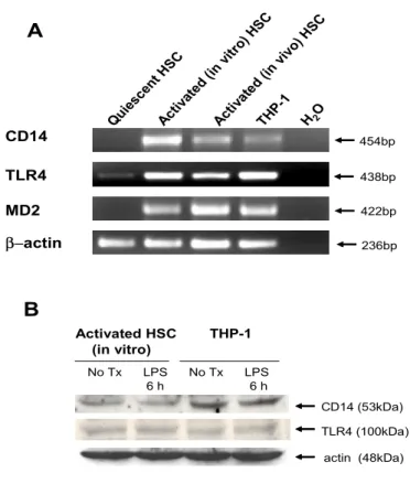
Figure 1. Expression of LPS receptors in human HSCs. (A) CD14, TLR4, and MD2 mRNA expression was assessed by RT-PCR. Total RNA was obtained from quiescent human HSCs, in vitro culture-activated human HSCs (from normal portion of human liver), in vivo activated human HSCs (from HCV-induced cirrhotic liver), or differentiated THP-1 cells. Data represent HSCs isolated from three normal portion of human livers and two human cirrhotic livers. THP-1 cells were differentiated by
treatment with 10 ng/ml of PMA for 18h, and were used as a positive control. (B) CD14
and TLR4 protein expression was assessed by Western blotting in culture-activated HSCs and PMA-treated THP-1 cells with or without LPS (100 ng/ml) for 6 h. Actin expression was measured to assess equal protein loading. (No Tx), No treatment.
21
resynthesis at 240 min following LPS stimulation. A proteosome
inhibitor, MG-132, was used to assess IκBαphosphorylation,
since phosphorylated IκBα is ubiquinated and rapidly degraded by
a proteosome complex.28 After preincubation with MG-132
(10µM) for 30 min, phosphorylated IκBα was detected from 60 min
following LPS stimulation (Figure 2B, middle panel). Western
blotting of α-tubulin was shown as a control for equal loading of
samples (Figure 2B, low panel). These data demonstrated that LPS
activated IKK that correlated with increased endogenous
IκBα phosphorylation and degradation in activated human HSCs.
An NF-κB-driven luciferase reporter assay showed that LPS
induced NF-κB transcriptional activity in a time- (Figure 3A) and
dose- dependent (Figure 3B) manner in activated HSCs. Polymyxin B is a cationic cyclic polypeptide antibiotic that binds
the lipid A moiety of LPS, inactivating its biological function.34
Preincubation of Polymyxin B (10 IU/ml) for 30 min before
stimulation completely inhibited LPS-induced NF-κB activation,
but did not inhibit IL-1β-induced NF-κB activation in HSCs
22 0 15 30 60 120 240 min IκBα (37 kDa) α−tubulin (55 kDa) phospho-IκBα (41 kDa) + MG-132 0 5 15 30 60 120 240 5 15 5 15 min LPS 100 ng/ml TNF-α IL-1β GST-IκBα (1-54) Fold Increase 1 6 7 21 111 91 25 239 317 432 720 A B IP: ΙΚΚγ Coomassie staining - MG-132
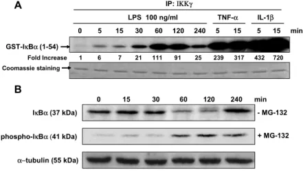
Figure 2. LPS stimulates IKK activity, IκBα phosphorylation and IκBα degradation. (A) Time
course of activation of IKK in response to LPS in activated human HSCs. Cells were cultured in DMEM containing 10% FBS. HSCs were stimulated with LPS (100 ng/ml) for the
indicated times. TNF-α (10 ng/ml) and IL-1β (5 ng/ml) were used as positive controls. An in
vitro kinase assay for IKK was performed using GST-IκBα (amino acid 1-54) as a substrate. Coomassie staining was used to demonstrate equal protein loading. (B) HSCs were stimulated with LPS (100 ng/ml) in the presence or absence of MG-132 (10 ng/ml). A
proteosome inhibitor, MG-132, was used to assess IκBα phosphorylation. IκBα steady-state
level and IκBα phosphorylation were assessed by Western blotting. A representative of
23 0 1 2 4 8 16 hrs 0 5 10 15 20 25 0 5 10 15 20 25 30 35 40 0 0.1 1 10 100 1000 10 5 ng/ml IL-1β TNFα LPS LPS 100ng/ml N F -κ B Fo ld In cr ea se 0 5 10 15 20 25 30 N F -κ B F ol d In cr ea se N F -κ B Fo ld In cr ea se
A
C
B
No Tx LPS PMB IL-1β PMB + + LPS IL-1β*
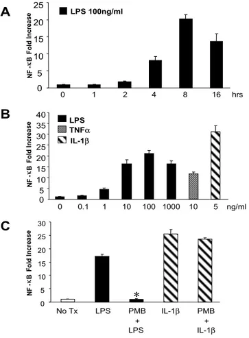
Figure 3. LPS-induced NF-κB transcriptional activity is inhibited by Polymyxin B. Activated human
HSCs were infected with the Ad5NF-κBLuc (MOI 500) for 12 h in DMEM containing 0.5% FCS. At
20 h post-infection, HSCs were stimulated with (A) LPS (100 ng/ml) for the indicated times (0-16 h),
or (B) with the indicated concentrations of LPS, TNF-α, and IL-1β for 8 h or (C) with LPS (100
ng/ml), or IL-1β (5 ng/ml) for 8 h with or without pretreatment of Polymyxin B (10 IU/ml) for 30 min.
Cells were lysed, and NF-κB-mediated luciferase activity was quantified. Data represent the mean
± SD of 3 independent experiments and are expressed as fold-increase over unstimulated cells. All measurements of luciferase activity were normalized to the protein concentration. *: P<0.01, when compared with HSCs which were treated with LPS. (PMB), Polymyxin B.
24
3. LPS-induced NF-κB activation is serum-dependent and
is mediated by TLR4
Lipid A is the LPS component that mediates its biological
effects.35 However, contamination of LPS with highly bioactive
"endotoxin proteins" can also activate NF-κB through TLR2.36 To
rule out this possibility, we stimulated HSCs with synthetic lipid A, which is free of contaminating proteins. In the presence of serum,
lipid A activated NF-κB in HSCs in a dose-dependent manner
(Figure 4A). These results demonstrate that LPS-induced NF-κB
activation in HSCs is dependent on the presence of serum, a
source of LBP that is required for LPS to act through TLR4.5,15
Polymyxin B eliminated lipid A-induced NF-κB activation in
human HSCs, confirming this response was due to LPS but not to contaminants (Figure 4B).
To examine whether LPS acts directly through TLR4, we stimulated HSCs with lipid A after preincubation with anti-TLR4
blocking Ab (HTA 125). Preincubation of HTA 125 for 30 min
significantly reduced lipid A-induced NF-κB activation in HSCs,
whereas isotype antibody (mouse IgG2α) had no effect (Figure 4B).
25 A B 0 2 4 6 8 10 N F-κ B Fo ld In cr ea se
No Tx Lipid A PMB HTA 125 IgG2α Ab + + + Lipid A Lipid A Lipid A
§ ‡ No Tx Lipid A Lipid A 100ng/ml 1µg/ml N F-κ B Fo ld In cr ea se 0 5 10 15 20 25 30 10% FCS 0% FCS * † 0 5 10 15 20 25 N F-κ B Fo ld In cr ea se No Tx Lipid A Lipid A mTNF-α 100ng/ml 1µg/ml 10ng/ml C ** ** C3H/OuJ C3H/HeJ
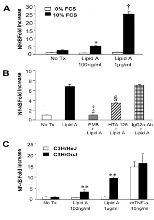
Figure 4. Lipid A-induced NF-κB activity depends on the presence of serum and is blocked by
anti-TLR4 blocking Ab (HTA 125) or Polymyxin B. Activated human HSCs were infected with the
Ad5NF-κBLuc (MOI 500) for 12 h. At 20 h post-infection, HSCs were treated with (A) lipid A for 8
h with 10% FCS or without serum, or (B) with 1µg/ml of lipid A with 10% FCS for 8 h with or
without preincubation of Polymyxin B (10 IU/ml), HTA 125 (20µg/ml), or IgG2Ab (20 µg/ml) for 30
min. Activated HSCs from C3H/HeJ or C3H/OuJ mice were infected with the Ad5NF-κBLuc (MOI
500) for 12 h. At 20 h post-infection, HSCs were treated with lipid A or mouse TNF-α (10 ng/ml)
for 8 h with 10% FCS (C). Luciferase assays were performed as described in “Materials and Methods”. Data represent the mean ± SD of 3 independent experiments and are expressed as fold increase over unstimulated cells. All measurements of luciferase activity were normalized to the protein concentration. *, †: p<0.01 when compared with HSCs stimulated with Lipid A without serum; ‡, §: p<0.01 when compared with HSCs stimulated with Lipid A only; **:p<0.01when
26
(Lpsd/d) that confers hyporesponsiveness to LPS. In contrastto
C3H/HeJ mice, substrain C3H/OuJ (Lpsn homozygotes) that
diverged from the same stock as C3H/HeJ mice,exhibits vigorous
responses to LPS. We compared lipid A-induced NF-κB
activation in activated HSCs derived from TLR4-sufficient C3H/OuJ mice and TLR4-deficient C3H/HeJ mice. Lipid A
induced NF-κB activation in HSCs from C3H/OuJ mice, but failed
to induce NF-κB activation in HSCs from C3H/HeJ mice (Figure
4C). These results provide further evidence that TLR4 mediates
LPS-induced NF-κB activation in HSCs.
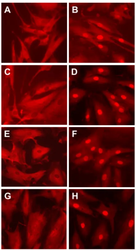
Untreated HSCs cultured with 10% FCS showed a cytoplasmic
staining of p65 as demonstrated by immunofluorescent p65 staining (Figure 5A), whereas LPS treatment induced p65 nuclear translocation in HSCs (Figure 5B). However, no p65 translocation was found in HSCs cultured without serum following LPS
stimulation (Figure 5C). IL-1β was used as a positive control
(Figure 5D). Polymyxin B blocked LPS-induced, but not
IL-1β-induced p65 nuclear translocation (Figure 5E-F).
27
nuclear translocation (Figure 5G), but preincubation of isotype Ab
(mouse IgG2α) did not (Figure 5H). These results demonstrate that
LPS-induced NF-κB activation is serum dependent and TLR4 is
the major receptor for LPS-induced NF-κB activation.
4. LPS increases NF-κB DNA binding activity in activated
human HSCs
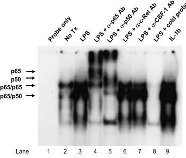
In activated HSCs, at least three NF-κB DNA binding complexes
have been observed: complex 1 (p65:p65 homodimer), 2 (p65:p50 heterodimer), and 3 (novel component) based on their relative
electrophoretic mobilities in response TNF-α or IL-1β.37 LPS or
IL-1β stimulation increased NF-κB DNA binding activity in
activated HSC, as demonstrated by EMSA (Figure 6). Supershift EMSA experiments were performed to identify constituent
proteins of the NF-κB DNA binding activities. We also did a
supershift assay with antibody against recombination signal
binding protein Jκ(RBP-Jκ) also designated CBF1, which is a
transcriptional factor that binds κB binding site of IL-6 promotor
resulting in transcriptional repression.38 The supershift complexes
28
supershifts were obtained with anti-c-Rel, or anti-CBF1 (Figure 6). A D G E C B H F
Figure 5. LPS-induced p65 nuclear translocation is blocked by HTA 125 or Polymyxin B. Immunofluorescent staining was used to assess the p65 nuclear translocation in HSCs. Cells were cultured in DMEM with 10% FBS (A, B, D-H) or in DMEM without serum (C). After 1 h of
stimulation with LPS (10 ng/ml) or 15 min stimulation with IL-1β (5 ng/ml), cells were fixed and then
stained with anti-p65 primary antibody, and then a rhodamine-conjugated secondary antibody.
Cells were treated as follows: (A) DMEM, (B, C) LPS (10 ng/ml), (D) IL-1β (5 ng/ml), (E) LPS
(10 ng/ml) after preincubation of polymyxin B (10 IU/ml) for 30 min, (F) IL-1β (5 ng/ml) after
preincubation of polymyxin B (10 IU/ml) for 30 min, (G) LPS (10 ng/ml) after preincubation of HTA
29
Figure 6. LPS stimulates NF-κB DNA-binding activity in activated human HSC.
Culture-activated HSCs were incubated with DMEM alone (Lane 2), LPS (100 ng/ml)
for 1 h (Lane 3) or IL-1β (5 ng/ml) for 15 min (Lane 9). Nuclear extracts (8µg) were
assayed for NF-κB binding activity by EMSA using a radiolabeled consensus NF-κB
site as a probe. Lane 1, probe without nuclear extract. Lane 4-7, Nuclear extracts
from LPS-treated HSCs were incubated with p65, p50, c-Rel or RBP-Jκantibodies,
respectively, and then incubated with radiolabeled probe. Antibody supershift, produced by binding of the p65 and p50 antibody are identified by arrow. Lane 8 is the same as lane 3 with the addition of 100-fold molar excess cold oligonucleotide as competitor. p65 p50 Prob e on ly No T x LPS LPS + α-p 65 A b LPS + α-p 50 A b LPS + α-c -Rel Ab LPS + α-C BF-1 Ab LPS + co ld p robe IL-1 b p65/p65 p65/p50 Lane 1 2 3 4 5 6 7 8 9
30
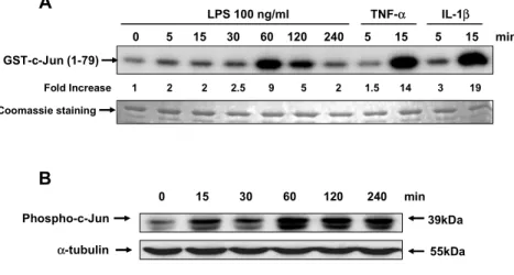
5. LPS activates JNK in activated human HSCs
The JNK/AP-1 pathway is an important regulator of inflammation and host defense response. LPS increased JNK activity, which peaked 9-fold at 60 min after stimulation (Figure
7A). TNF-α and IL-1βincreased JNK activity 14- and 19-fold at
15 min after stimulation, respectively. LPS induced
phosphorylation of c-Jun, a JNK substrate, a maximum at 60 min after stimulation (Figure 7B), correlating with the time course of JNK activation.
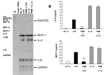
6. LPS induces the production of inflammatory chemokines including IL-8 and MCP-1 in activated HSCs
As shown by RNase protection assay (Figure 8A), LPS induced mRNA expression of IL-8 (10-fold) and MCP-1 (2.4-fold) in HSCs. Preincubation of Polymyxin B for 30 min before stimulation
completely inhibited LPS-induced, but not IL-1β-induced
increase in IL-8 and MCP-1 mRNA levels (Fig. 8A). Both LPS and
IL-1β stimulated IL-8 and MCP-1 secretion. Polymyxin B blocked
LPS-induced, but not IL-1β-induced chemokine secretion (Figure
31
Figure 7. LPS activates JNK in activated human HSCs. HSCs cultured in DMEM
containing 10% FCS were stimulated with LPS (100 ng/ml), TNF-α (10 ng/ml), and
IL-1β (5 ng/ml) for the indicated times. (A) An in vitro kinase assays for JNK was
performed using GST-c-Jun as a substrate. Coomassie staining was used to demonstrate equal protein loading. (B) HSCs cultured in DMEM containing 10% FCS were stimulated with LPS (100 ng/ml) for the indicated times. Western blotting was performed using Ab specific for phosphorylated c-Jun as described in “Materials and Methods”. A representative of three independent experiments is shown.
0 5 15 30 60 120 240 5 15 5 15 min LPS 100 ng/ml TNF-α IL-1β Fold Increase 1 2 2 2.5 9 5 2 1.5 14 3 19 GST-c-Jun (1-79) 0 15 30 60 120 240 min B A Phospho-c-Jun 39kDa α-tubulin 55kDa Coomassie staining
32 Y e ast t-RN A No T x LPS PM B + LPS IL -1 β PM B + I L -1β MCP-1 IL-8 L32 GAPDH Ltn RANTES IP-10 MIP-1β MIP-1α MCP-1 lL-8 I-309 L32 GAPDH RANTES No Tx LPS PMB IL-1β PMB + + LPS IL-1β IL -8 ( n g /m l) MC P-1 (n g /ml ) A B M 0 10 20 30 40 50 60 70 80 0 2 4 6 8 10 12 14 16 18 20 * † No Tx LPS PMB IL-1β PMB + + LPS IL-1β
Figure 8. LPS induces the secretion of IL-8 and MCP-1 in activated human HSCs. (A) RNase protection assay was performed to assess mRNA expression of chemokines in HSC. Total RNA was isolated from HSCs culturing in DMEM containing 0.5% FCS
after stimulation with LPS (10 ng/ml) or IL-1β (5 ng/ml) for 18 h with or without
pretreatment of Polymyxin B (10 IU/ml) for 30 min. Chemokine mRNA levels were determined by a using human cytokine multiprobe template set (hCK-5; PharMingen,
San Diego, CA). Thirty µg of total RNA was hybridized with 105 cpm of riboprobe.
Twenty µg of yeast tRNA was hybridized as a negative control. Lane M contains the
radiolabeled markers. Migration of the protected bands is indicated. (B) Secreted IL-8 and MCP-1 were quantified by ELISA. HSCs culturing 0.5% FCS containing DMEM
were stimulated with LPS (10 ng/ml) or IL-1β (5 ng/ml) for 24 h with or without
pretreatment of Polymyxin B (10 IU/ml) for 30 min. Data represent the mean ± SD of 2 experiments in triplicates. *: p<0.01, when compared with HSCs stimulated with LPS; †: p<0.01, when compared with HSCs stimulated with LPS. (PMB), Polymyxin B.
33
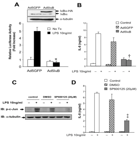
7. LPS-induced IL-8 production depends on NF-κB and
JNK activation in HSCs
The IκB super-repressor is expressed in Ad5IκB infected HSCs
(Figure 9A). This IκB super-repressor (Ad5IκB) completely
inhibited LPS-induced NF-κB activation in activated HSCs (Figure
9A). ELISA showed that LPS-induced IL-8 secretion was nearly
completely blocked by Ad5IκB (Figure 9B). Activated HSCs were
preincubated with DMSO vehicle or 20µM of SP600125, a
selective JNK inhibitor39for 2 h and then subsequently stimulated
with LPS for 1 h. c-Jun phosphorylation was induced in control and DMSO-treated HSCs by LPS stimulation, but inhibited by SP600125 (Figure 9C). IL-8 secretion was induced in control and DMSO treated HSCs by LPS stimulation, but inhibited about 70% by SP600125 (Figure 9D).
8. LPS upregulates the cell surface expression of adhesion molecules including ICAM-1 and VCAM-1 in activated HSCs
To assess the expression of the adhesion molecules ICAM-1 and VCAM-1, flow cytometry analysis was performed. ICAM-1
34
and VCAM-1 proteins were expressed on the activated HSCs (Figure 10A-B). LPS significantly upregulated the cell surface expression of ICAM-1 and VCAM-1, an effect that was blocked by polymyxin B (Figure 10A-B).
35 A C R el ati ve L uci fe rase A cti vi ty (F ol d I ncr ease) Ad5IκB Ad5GFP Ad5IκB Ad5GFP Control LPS 10ng/ml – + – + – + IL -8 (n g/ ml ) SP600125 (20µM) DMSO Control IL -8 (n g/ ml ) LPS 10ng/ml – + – + – + LPS 10ng/ml – + – + – + IB: p-c-Jun control DMSO SP600125 (20µM) IB: α-tubulin B D 0 1 2 3 4 5 6 LPS 10ng/ml No Tx 0 1 2 3 4 5 6 7 8 0 2 4 6 8 10 12 * † ‡ Ad5IκB Ad5GFP IκBα-HA IκBα α-tubulin
Figure 9. LPS-induced IL-8 secretion is dependent on NF-κB and JNK activity in activated human
HSCs. (A) HSCs were co-infected with Ad5NF-κBLuc virus (MOI 500) together with Ad5IκB or
Ad5GFP virus (MOI 1000) for 20 h. Infection of Ad5IκB was confirmed by Western blotting of
IκBα expression. The hemagglutinin (HA)-tagged IκBα has higher molecular weight than the
endogenous IκBα (upper panel). At 20 h post-infection, cells were stimulated with LPS (10 ng/ml)
and 10% FCS for 8 h. Cells were lysed, and NF-κB-mediated luciferase activity in each sample
was quantified (low panel). (B) HSCs were either left uninfected (control) or infected with Ad5IκB
or Ad5GFP virus at a MOI of 1000 particles/cell for 12 h. Cells were stimulated with LPS (10 ng/ml) with 0.5% FCS for 24 h. Secreted IL-8 was quantified by ELISA. (C) HSCs were either left
untreated as control, or preincubated with DMSO or SP600125 (20 µM) for 2 h and then stimulated
with LPS (10 ng/ml) with 10% FCS for 1 h. Whole cell extracts (20 µg) were assessed for
phospho-c-Jun expression using Western Blotting. (D) Human HSCs were either left untreated as
control, or preincubated with DMSO or SP600125 (20 µM) for 2 h and then stimulated with LPS
(10 ng/ml) with 0.5% FCS for 24 h. Secreted IL-8 was quantified by ELISA from culture supernatant of HSCs. The values in all data represent the mean ± S.D. from two experiments in triplicates. *: P<0.01 when compared with HSCs which were infected with Ad5GFP and stimulated with LPS; †: P<0.01 when compared with HSCs which were infected with Ad5GFP and stimulated with LPS; ‡: P<0.01 when compared with HSCs which were preincubated with DMSO and stimulated with LPS.
36
Figure 10. LPS-induced upregulation of ICAM-1 and VCAM-1 is inhibited by Polymyxin B. HSCs were stimulated for 24 h with 10 ng/ml LPS, stained for ICAM-1 (A) or VCAM-1 (B), and analysed by flow cytometry. Thin solid line, Isotype-matched control Ab; Thick solid line, ICAM-1 or VCAM-1 expression on untreated HSCs; Thickest solid line, ICAM-1 or VCAM-1 expression on LPS-stimulated HSCs; dotted line, ICAM-1 or VCAM-1 expression on LPS-stimulated HSCs pretreated with polymyxin B (10 IU/ml) 30 min before each stimulation. Figure is representative of four independent experiments. (FL), fluorescence.
A
B
FL1-FITC
37
IV. Discussion
There is increasing evidence for a role of portal bacteremia in hepatic inflammation in particular in alcoholic liver disease. The critical cell in the inflammatory process is the Kupffer cell which can be directly activated by bacterial products such as LPS and produce a wide variety of cytokines and reactive oxygen
species.5,7 However, the mechanism by which this inflammatory
response can progress to fibrosis is largely unknown. One thesis
is that the products of Kupffer cells including TGF-βand
reactive oxygen species activate HSCs and therefore there is an indirect effect. Alternatively, the bacterial products can directly stimulate the HSCs or the two pathways can be synergistic. The underlying hypothesis of the present study was that there is a direct role for bacterial products on HSC signal transduction. This is largely been validated in our present studies in which an intact TLR4 pathway was demonstrated in activated human HSCs.
Our study demonstrated that three critical components of LPS signaling receptor complex CD14, TLR4, and MD2 are expressed
38
in both in vitro- and in vivo- activated human HSCs. Meanwhile, in
quiescent HSCs, low levels of mRNA for TLR4 were detected, but mRNA for CD14 and MD2 was not detected. These results suggest that activation of HSCs is associated with up-regulation of LPS receptors. Activation of HSC results in the acquisition of myofibroblast-like properties, including cell proliferation,
pro-inflammatory properties and increased collagen synthesis.40
This activation process is associated with a dramatic up-regulation of specific cell surface receptors, including
receptors for PDGF, TGF-β1, and vasoactive substances.41
Therefore, the finding that LPS receptors are up-regulated in activated HSCs suggests that receptors mediating the inflammatory actions of HSCs are co-ordinantly up-regulated.
To assess LPS signaling inhuman HSCs, we investigated the
activation of NF-κB and JNK. NF-κB is a critical transcriptional
activator involved in the rapid inductionof a number of cytokines
involved inthe inflammatory response.42 We demonstrated that
LPS directly induced NF-κB activation in a time- and
dose-dependent manner in activated human HSCs. NF-κB binding
39
stimulation, as demonstrated by EMSA. Supershift EMSA
experiments demonstrated that p65:p50 and p65:p65 NF-κB
dimers are major NF-κB DNA binding complexes induced by LPS
in activated HSCs. These findings are consistent with previous
reports showing p65 is major NF-κB component in response to
TNF-α or IL-1β in activated HSC.14,28 Activation of
LPS-responsive cells, such as monocytes and macrophages,
occurs after LPS interacts with circulating LBP.15 Analysis of
LBP-deficient mice showed that LBP was essential for the rapid induction of an inflammatory response by low concentrations of
LPS or Gram-negative bacteria.43 As expected, LPS-induced
NF-κB activation in activated human HSCs was dependent on the
presence of serum, a source of LBP.
Preincubation with anti-TLR4 blocking Ab (HTA125) before
lipid A stimulation significantly decreased NF-κB activation.
Immunofluorescent staining also showed that TLR4 bloking Ab (HTA125) inhibited LPS-induced p65 nuclear translocation in HSCs. These results demonstrate that LPS-induced signaling in human HSCs requires TLR4. C3H/HeJ mice are characterized by
40
mutation within the coding region of the Tlr4 gene, resulting in a
nonconservative substitution of a highly conserved proline by
histidine at codon 712.22 Lipid A failed to induce
NF-κB activation in HSCs derived from C3H/HeJ mice, but not from its
corresponding control mice, confirming LPS-induced
proinflammatory pathway is mediated by TLR4 in HSCs. Activated JNKs play an essential role in the activation of
transcriptional factors, such as c-Jun, ATF-2, and Elk-1.45 In
macrophages, activated JNKs mediate the expressionof cytokines
such as TNF- , IL-1, and IL-6,46 chemokines such as RANTES,47
all of which often lead to inflammatory responses. We demonstrated that LPS is an effective inducer of JNK activation in
activated human HSCs. The kinetics of LPS-induced NF-κB and
JNK activation in human HSCs were delayed (maximal 60 min)
compared with TNF-α or IL-1β (maximal 15 min), suggesting that
the assemblage/recruitment of signaling proteins is slower in endotoxin than in cytokine-stimulated cells.
Chemokines such as MCP-1 and IL-8 may perpetuate liver fibrosis due to their proinflammatory properties. MCP-1 expression is elevated in patients with chronic viral hepatitis and
41
in experimental liver injury models.8,9 In cultured HSC,
proinflammatory cytokines including TNF-α, IL-1βand IFN-γ
potently stimulate MCP-1 expression.8,12 Activated HSCs promote
hepatic inflammation by production of potent neutrophil
chemoattractant such as cytokine-induced neutrophil
chemoattractant (CINC), the rat homolog to human IL-8.48
Similarly, serum and hepatic levels of IL-8 correlate with severity
of chronic viral hepatitis and liver cirrhosis.10 LPS induces the
expression of MCP-112 and MIP-213 in activated HSCs in rat. Our
study demonstrated that purified LPS induced both MCP-1 and IL-8 mRNA and protein expression in activated human HSCs. HSCs also contribute to hepatic inflammation by upregulating expression on their cell surface of leukocytes adhesion molecules
including ICAM-1,49 and VCAM-150 in response to
TNF-αor
INF-γ. In this report, we demonstrated that LPS upregulated the
expression of ICAM-1 and VCAM-1 in HSCs. Thus, LPS may participate in hepatic inflammation by aiding with neutrophil transmigration out of the hepatic sinusoid and into the liver parenchyma during LPS-induced liver injury.
42
molecules (ICAM-1, VCAM-1) genes are regulated by NF-κB.42 In
HSCs, NF-κB is upregulated upon cell activation, and is further
induced by cytokines such as TNF-α, IL-1β resulting in the
induction of inflammatory chemokines and cell adhesion
molecules.14 We showed that LPS-induced IL-8 secretion was
blocked by Ad5IκB, which provides an exogenous nondegradable
IκB, blocking NF-κB activation. In HSC, JNK is upregulated upon
cell activation, is required for activation of HSC, stimulates HSC
proliferation, and decreases collagen α1(I) expression.39 However,
the role of JNK in the inflammatory process in HSCs is unclear. We demonstrated that LPS-induced IL-8 production is partially inhibited by selective inhibition of JNK activation. This study provides a rationale for novel therapies for LPS-induced hepatic
inflammation by targeting NF-κB or JNK.
Interestingly, the expression of chemokines such as MCP-1 and MIP-2 induced by LPS was reported as being dependent on the activation state of HSCs in rats. Only fully activated HSCs responded directly to LPS with the release of chemokines in rat.12,13 This observation may be related to a changes in the expression of LPS receptors between activated and quiescent
43
HSCs. On the other hand, it may be due to an unidentified
postreceptor defect of the NF-κB signaling pathway in quiescent
HSC like TNF-α or IL-1β- induced response.14 Further studies
are needed to clarify the exact role of LPS in quiescent HSCs.
In conclusion, our study characterized LPS-induced
proinflammatory signaling in activated human HSCs. LPS directly
acts through TLR4 and then activates NF-κB and JNK to induce
proinflammatory chemokines and adhesion molecules in activated human HSCs. Therefore, HSCs in addition to Kupffer cells may be a target for LPS-induced liver injury, and provide a direct link between inflammatory and fibrotic liver injury.
V. Conclusions
Bacterial LPS stimulates Kupffer cells and participates in the pathogenesis of alcohol-induced liver injury. However, it is unknown whether LPS directly affects HSCs, the main fibrogenic cell type in the injured liver. This study characterizes LPS-induced signal transduction and pro-inflammatory gene
44
expression in activated human HSCs.
The results as below were obtained:
1. Culture-activated HSCs and HSCs isolated from patients with hepatitis C virus-induced cirrhosis express LPS-associated signaling molecules including CD14, TLR4, and MD2.
2. Stimulation of culture-activated HSCs with LPS results in a
rapid and marked activation of NF-κB, as assessed by in vitro
kinase assays for IκB kinase (IKK), IκBα steady state levels, p65
nuclear translocation, NF-κB-dependent luciferase reporter
gene assays and electrophoretic mobility shift assays. Lipid A
induces NF-κB activation in a similar manner.
3. Both LPS- and lipid A-induced NF-κB activation is blocked by
preincubation with either anti-TLR4 blocking antibody (HTA125) or Polymyxin B.
4. Lipid A induces NF-κB activation in HSCs from
TLR4-sufficient (C3H/OuJ) mice but not from TLR4-deficient (C3H/HeJ) mice.
5. LPS also activates c-Jun N-terminal Kinase (JNK), as
45
6. LPS upregulates IL-8 and MCP-1 gene expression and secretion. LPS-induced IL-8 secretion is completely inhibited by the IB super-repressor (Ad5IB) and partially inhibited by a specific JNK inhibitor, SP600125.
7. LPS also upregulates cell surface expression of ICAM-1 and VCAM-1.
In conclusion, human activated HSCs utilize components of TLR4 signal transduction cascade to stimulate NF-B and JNK and upregulate chemokines and adhesion molecules. Thus HSCs are a potential mediator of LPS-induced liver injury.
46
References
1. Ulevitch RJ, Tobias PS. Receptor-dependent mechanisms of cell stimulation by bacterial endotoxin. Annu Rev Immunol 1995; 13:437-457.
2. Schletter J, Heine H, Ulmer AJ, Rietschel ET. Molecular mechanisms of endotoxin activity. Arch Microbiol 1995; 164:383-389.
3. Nolan JP. The role of endotoxin in liver injury. Gastroenterology 1975;69:1346-1356.
4. Lumsden AB, Henderson JM, Kutner MH. Endotoxin levels measured by a chromogenic assay in portal, hepatic and peripheral venous blood in patients with cirrhosis. Hepatology 1988;8:232-236.
5. Su GL. Lipopolysaccharides in liver injury: molecular mechanisms of Kupffer cell activation. Am J Physiol Gastrointest Liver Physiol 2002;283:G256-G265.
47
6. Enomoto N, Ikejima K, Yamashina S, Hirose M, Shimizu H, Kitamura T et al. Kupffer cell sensitization by alcohol involves increased permeability to gut-derived endotoxin. Alcohol Clin Exp Res 2001;25:1-4.
7. Thurman RG. II. Alcoholic liver injury involves activation of Kupffer cells by endotoxin. Am J Physiol 1998;275:1-11.
8. Marra F, Valente AJ, Pinzani M, Abboud HE. Cultured human liver fat-storing cells produce monocyte chemotactic protein-1. Regulation by proinflammatory cytokines. J Clin Invest 1993;92:1674-1680.
9. Czaja MJ, Geerts A, Xu J, Schmiedeberg P, Ju Y. Monocyte chemoattractant protein 1 (MCP-1) expression occurs in toxic rat liver injury and human liver disease. J Leukoc Biol 1994;55:120-126.
10. Masumoto T, Ohkubo K, Yamamoto K, Ninomiya T, Abe M, Akbar SM et al. Serum IL-8 levels and localization of IL-8 in liver
from patients with chronic viral hepatitis.
48
11. Schwabe RF, Schnabl B, Kweon YO, Brenner DA. CD40 activates NF-kappa B and c-Jun N-terminal kinase and enhances chemokine secretion on activated human hepatic stellate cells. J Immunol 2001;166:6812-6819.
12. Sprenger H, Kaufmann A, Garn H, Lahme B, Gemsa D, Gressner AM. Differential expression of monocyte chemotactic protein-1 (MCP-1) in transforming rat hepatic stellate cells. J Hepatol 1999;30:88-94.
13. Sprenger H, Kaufmann A, Garn H, Lahme B, Gemsa D, Gressner AM. Induction of neutrophil-attracting chemokines in transforming rat hepatic stellate cells. Gastroenterology 1997; 113:277-285.
14. Hellerbrand C, Jobin C, Licato LL, Sartor RB, Brenner DA. Cytokines induce NF-kappaB in activated but not in quiescent rat hepatic stellate cells. Am J Physiol 1998;275:G269-G278.
15. Schumann RR, Leong SR, Flaggs GW, Gray PW, Wright SD, Mathison JC et al. Structure and function of lipopolysaccharide binding protein. Science 1990;249:1429-1431.
49
16. da Silva CJ, Soldau K, Christen U, Tobias PS, Ulevitch RJ. Lipopolysaccharide is in close proximity to each of the proteins in its membrane receptor complex transfer from CD14 to TLR4 and MD-2. J Biol Chem 2001;276:21129-21135.
17. Haziot A, Chen S, Ferrero E, Low MG, Silber R, Goyert SM. The monocyte differentiation antigen, CD14, is anchored to the cell membrane by a phosphatidylinositol linkage. J Immunol 1988;141:547-552.
18. Lemaitre B, Nicolas E, Michaut L, Reichhart JM, Hoffmann JA. The dorsoventral regulatory gene cassette spatzle/Toll/cactus controls the potent antifungal response in Drosophila adults. Cell 1996;86:973-983.
19. Rock FL, Hardiman G, Timans JC, Kastelein RA, Bazan JF. A family of human receptors structurally related to Drosophila Toll. Proc Natl Acad Sci U S A 1998;95:588-593.
20. Kirschning CJ, Wesche H, Merrill AT, Rothe M. Human toll-like
receptor 2 confers responsiveness to bacterial
50
21. Chow JC, Young DW, Golenbock DT, Christ WJ, Gusovsky F. Toll-like receptor-4 mediates lipopolysaccharide-induced signal transduction. J Biol Chem 1999;274:10689-10692.
22. Poltorak A, He X, Smirnova I, Liu MY, Huffel CV, Du X et al. Defective LPS signaling in C3H/HeJ and C57BL/10ScCr mice: mutations in Tlr4 gene. Science 1998;282:2085-2088.
23. Yoshimura A, Lien E, Ingalls RR, Tuomanen E, Dziarski R, Golenbock D. Cutting edge: recognition of Gram-positive bacterial cell wall components by the innate immune system occurs via Toll-like receptor 2. J Immunol 1999;163:1-5.
24. Schwandner R, Dziarski R, Wesche H, Rothe M, Kirschning CJ. Peptidoglycan- and lipoteichoic acid-induced cell activation is mediated by toll-like receptor 2. J Biol Chem 1999; 274:17406-17409.
25. Shimazu R, Akashi S, Ogata H, Nagai Y, Fukudome K, Miyake K et al. MD-2, a molecule that confers lipopolysaccharide responsiveness on Toll- like receptor 4. J Exp Med 1999; 189:1777-1782.
51
26. Zhang FX, Kirschning CJ, Mancinelli R, Xu XP, Jin Y, Faure E et al. Bacterial lipopolysaccharide activates nuclear factor-kappaB through interleukin-1 signaling mediators in cultured human dermal endothelial cells and mononuclear phagocytes. J Biol Chem 1999;274:7611-7614.
27. Uesugi T, Froh M, Arteel GE, Bradford BU, Thurman RG. Toll-like receptor 4 is involved in the mechanism of early
alcohol-induced liver injury in mice. Hepatology
2001;34:101-108.
28. Hellerbrand C, Jobin C, Iimuro Y, Licato L, Sartor RB, Brenner DA. Inhibition of NFkappaB in activated rat hepatic stellate cells by proteasome inhibitors and an IkappaB super-repressor. Hepatology 1998;27:1285-1295.
29. Sanlioglu S, Williams CM, Samavati L, Butler NS, Wang G,
McCray PB, Jr. et al. Lipopolysaccharide induces
Rac1-dependent reactive oxygen species formation and coordinates tumor necrosis factor-alpha secretion through IKK regulation of NF-kappa B. J Biol Chem 2001;276:30188-30198.
52
30. Iimuro Y, Nishiura T, Hellerbrand C, Behrns KE, Schoonhoven R, Grisham JW et al. NFkappaB prevents apoptosis and liver dysfunction during liver regeneration. J Clin Invest 1998; 101:802-811.
31. Moriyoshi K, Richards LJ, Akazawa C, O'Leary DD, Nakanishi S. Labeling neural cells using adenoviral gene transfer of membrane-targeted GFP. Neuron 1996;16:255-260.
32. Lang A, Schoonhoven R, Tuvia S, Brenner DA, Rippe RA. Nuclear factor kappaB in proliferation, activation, and apoptosis in rat hepatic stellate cells. J Hepatol 2000;33:49-58.
33. Zarember KA, Godowski PJ. Tissue expression of human Toll-like receptors and differential regulation of Toll-like receptor mRNAs in leukocytes in response to microbes, their products, and cytokines. J Immunol 2002;168:554-561.
34. Morrison DC, Jacobs DM. Binding of polymyxin B to the lipid A portion of bacterial lipopolysaccharides. Immunochemistry 1976;13:813-818.
53
35. Rietschel ET, Kirikae T, Schade FU, Mamat U, Schmidt G, Loppnow H et al. Bacterial endotoxin: molecular relationships of structure to activity and function. FASEB J 1994;8:217-225.
36. Hirschfeld M, Ma Y, Weis JH, Vogel SN, Weis JJ. Cutting edge: repurification of lipopolysaccharide eliminates signaling through both human and murine toll-like receptor 2. J Immunol 2000;165:618-622.
37. Elsharkawy AM, Wright MC, Hay RT, Arthur MJ, Hughes T, Bahr MJ et al. Persistent activation of nuclear factor-kappaB in cultured rat hepatic stellate cells involves the induction of potentially novel Rel-like factors and prolonged changes in the expression of IkappaB family proteins. Hepatology 1999; 30:761-769.
38. Vales LD, Friedl EM. Binding of C/EBP and RBP (CBF1) to Overlapping Sites Regulates Interleukin-6 Gene Expression. J Biol Chem 2002;277:42438-42446.