• 검색 결과가 없습니다.

The Suppressive Effect of Pueraria lobata Root Extract and Its Biotransformed Preparation against Skin Wrinkle Formation

N/A
N/A
Protected

Academic year: 2021

Share "The Suppressive Effect of Pueraria lobata Root Extract and Its Biotransformed Preparation against Skin Wrinkle Formation"

Copied!
8
0
0

로드 중.... (전체 텍스트 보기)

전체 글

(1)

Introduction

Skin aging involves the structural and physiological alterations and progressive changes in each skin layer as well as changes in skin appearance. Skin aging is a complex biological process influenced by both intrinsic (genetics, cellular metabolism, hormone and metabolic processes) and extrinsic (chronic light exposure, pollution, ionizing radiation, chemicals, toxins) factors (Baumann, 2007; Ganceviciene et al., 2012; Lephart, 2016). Skin health and beauty is considered one of the principal factors representing overall “well-being”

and the perception of “health” in humans (Ganceviciene et

al., 2012). Sometimes, whitening agents such as corticosteroids, tretinoin (all-trans retinoic acid) and hydroquinone those medically applied for effectively lighten the skin tone of hyperpigmented lesions, may cause a variety of side-effect (Desmedt et al., 2016). Thus an increasing interest in herbs, many studies focused on discovering novel natural skin- whitening agents that are currently underway (Smit et al., 2009). Wrinkle formation involves degradation of the skin’s three primary structural constituents, collagen, elastin and glycosaminoglycans (e.g., hyaluronic acid) and these three components are known to decline with age or exposure to ultraviolet light). Thus, prevention of wrinkle development has assumed a fundamental status in anti-ageing skin care.

Isoflavones are a subclass of flavonoids, which include over 6000 identified family members and is a phenolic

The Suppressive Effect of Pueraria lobata Root Extract and Its Biotransformed Preparation against Skin Wrinkle Formation

Hyun Jung Koo

1†

, SungRyul Lee

2†

, Se Chan Kang

3

, Jung Eun Kwon

3

, Da Eun Lee

3

, Eui-Su Choung

4

, Jong-Sub Lee

4

, Jin Woo Lee

5

, Yuna Park

5

, Dong Soo Sim

5

and Eun-Hwa Sohn

5

*

1

Department of Medicinal and Industrial Crops, Korea National College of Agriculture and Fisheries, Jeonju 54874, Korea

2

Department of Integrated Biomedical Science, Cardiovascular and Metabolic Disease Center, Inje University, Busan 47392, Korea

3

Department of Oriental Medicine Biotechnology, Kyung Hee University, Yongin 17104, Korea

4

DanjoungBio Co., Ltd., Wonju 26303, Korea

5

Department of Herbal Medicine Resources, Kangwon National University, Samcheok 25949, Korea

Abstract - EP was obtained through 20% ethanol extraction of Pueraria lobata root, and the fermented form of EP, FEP, was prepared from the EP after incubating with Lactobacillus rhamnosus vitaP1. There was no significant toxicity by EP and FEP up to 1000 ㎍/㎖ in NIH-3T3, HaCaT, and B16F10 cells. In addition to antioxidant potentials of EP and FEP determined by DPPH and ABST assays, we confirmed increase of procollagen type I and elastin synthesis by supplementation of the EP and FEP at the concentration of 50 ㎍/㎖ using ELISA kits. The protein expression levels of matrix metalloprotease (MMP)-1, -3, and -9, those are involved in the degradation of collagen or other skin matrix proteins, were remarkably suppressed while their inhibitory protein metallopeptidase inhibitor 1 (TIMP-1) was greatly up-regulated by supplementation of the EP and FEP at a concentration of 50 ㎍/㎖. Taken together, both EP and FEP supplementation could be involved in the suppression of the skin wrinkle formation through inhibiting degradation of collagen and stimulating the synthesis of collagen and elastin. The results showed that the anti-wrinkle potential of the EP and FEP will be a promising candidate for developing cosmeceutical compounds or products.

Key words - Antioxidant, Fermentation, Pueraria lobate, Wrinkle

*Corresponding author. E-mail : [email protected] Tel. +82-33-540-3322

These authors contributed equally to this work.

ⓒ 2017 by The Plant Resources Society of Korea

Original Research Article

(2)

compound is rich in soy beans, soy foods, legumes and fungus (Woo et al., 2003; Yu et al., 2016). Recently, it has been found that root part of Pueraria thunbergiana possesses abundant flavonoid (Shin et al., 2000; Han et al., 2015).

Besides their antioxidant capacities, antimicrobial, and anti-inflammatory activities, isoflavones possess a weak estrogenic (agonistic) activity through its binding to estrogen receptor (Yu et al., 2016). Recent efforts are aimed at isoflavone biotransformation to increase the health benefits of isoflavones like as an equol (Gao et al., 2016). Moreover, in the topical application of isoflavones on skin whitening, transformation of isoflavones like metabolism by microbiota in the intestine will be necessary since isoflavone itself has less potential bioavailability. In this experiment, we prepared the ethanol extract of the root part of Pueraria lobata (EP) and fermented extract (FEP) after biotransformation of the EP with Lactobacillus rhamnosus vitaP1. Then, we investigated the anti-wrinkle effect of the EP and FEP, fermented form of EP, based on the production of procollagen type I, elastin, and the suppression of matrix metalloprotease (MMP)-1 (collagenase-1), -3 (stromelysin-1), and -9 (gelatinase). In addition, antioxidant capacity of EP and FEP were determined by DPPH and ABTS method.

Materials and Methods

Chemicals and reagents

2,2-diphenyl-1-picryl-hydrazyl-hydrate (DPPH), β-Phycoerythrin, mushroom tyrosinase, and α-melanocyte stimulating hormone (α-MSH) were purchased from Sigma (St Louis, MO, USA).

Unless indicated otherwise, all other chemicals were obtained from Sigma.

Preparation of EPL and FEPL

Pueraria lobata (Wild.) Ohwi roots were purchased from Daegu oriental medicine market and identified by Dr. T.H.

Park of Daegu University (Daegu, Korea). Roots of P. lobata were chopped into small pieces and extracted three times in 20% ethanol for 24 hours each at room temperature (yield 20.5%). The extract was subsequently filtered to remove any particulates and was concentrated under vacuum at 50°C.

Then, the concentrated crude 20% EtOH extract of P. lobata

(EP) was lyophilized to obtain a powder and stored at –20°C for further experiments (20.5% yield). FEP was prepared after fermentation of 2% PE by incubation with L. rhamnosus vitaP1 for 48 hours at 37°C with shaking (250 rpm). Then fermented of EP (FEP) was re-extracted with ethyl acetate hexane (EtOAc). EP and FEP were dissolved in phosphate buffer saline (PBS) (Gibco, USA).

Cell culture

NIH-3T3 (mouse embryo fibroblast cell), HaCaT (im- mortalized human keratinocyte cell), and B16F10 (mouse melanoma cell) were obtained from Korean cell line bank (Seoul, Korea) and were maintained in Dulbecco’s modified Eagle’s medium (DMEM) (Gibco, USA) containing 10%

fetal bovine serum (Gibco, USA), 1% penicillin (Gibco, USA) in a humidified atmosphere of 5% CO

2

at 37°C.

Cell cytotoxicity

Cell cytotoxicity was measured by quantitative colorimetric assay with a 3-(4,5-dimethylthiazol-2-yl)-2,5-diphenyltetrazolium bromide (MTT), which measures the mitochondrial activity of living cells. Exponentially growing NIH-3T3, HaCaT, and B16F10 cells were seeded at 1×10

4

cells/well in 96-well tissue culture plates and treated with different dose of EP and FEP (1, 10, 100, 500, 1000 ㎍/㎖) for 24 hours. After incubation of MTT (150 ㎍/㎖) for 4 hours, the formazan crystals were dissolved in dimethyl sulfoxide (DMSO), and then measured the optical density at 540 ㎚ using a Molecular Devices microplate reader (Menlo Park, CA). Cytotoxicity was expressed as a percent of untreated control cells.

DPPH scavenging activity

Different concentrations of EP and FEP were adjusted at

100 ㎕ with reaction mixture and then reacted with l00 ㎕ of

0.4 mM DPPH (2,2-diphenyl-1-picrylhydrazyl) solution in

99% EtOH. After vigorous shaking, reaction mixtures were

allowed to reach a steady state at room temperature for 30

minutes. Decolorization of DPPH was determined by measuring

the absorbance at 540 ㎚ with using a Molecular Devices

microplate reader.

(3)

ABTS assay

The 2,2’-azino-bis (3-ethylbenzthiazoline-6-sulfonic acid (ABTS) antioxidant assay was generated by adding 7.4 mM ABTS to 2.4 mM potassium persulfate and the mixture was left at room temperature for overnight in dark. The ABTS radical cation solution was diluted with distilled water to obtain an absorbance of approximately 1.1 ± 0.02 (1.09) at 734 ㎚. 400 ㎕ EP and FEP extract at various concentrations was added to 800 ㎕ of ABTS reagent. Reaction was conducted at room temperature for 10 minutes in dark, and then absorbance was measured at 734 ㎚ with using UV/VIS spectrophotometer (Shimadzu, Japan).

Effect of EP and FEP on the production of procollagen type I and elastin

NIH-3T3 cells were inoculated into 24-well plate (1×10

5

cells/well) and were cultured for 24 hours. The media were switched into serum-free DMEM in the presence or absence of EP or FEP (50 ㎍/㎖) for 24 hours. After collecting supernatants, procollagen type I and elastin contents were determined by a procollagen type I C-peptide assay kit (Takara Bio, Japan) and FASTINTM assay (Biocolor, Belfast, Ireland), respectively.

Effect of EP and FEP on the secretion of metallopeptidase inhibitor 1, matrix metalloprotease (MMP)-1, -3, and -9

To determine the secreted forms metallopeptidase inhibitor-1 (TIMP-1), matrix metalloprotease (MMP)-1, -3, and -9 in NIH-3T3 cells were switched into serum free-medium and then EP and FEP (50 ㎍/㎖) were treated for 24 hours. After collecting the culture medium, TIMP-1, MMP-1, -3, -9 levels were measured by each ELISA assay kits (Amesham Pharmacia biotech, Centenial Ave., USA) according to manufacturer’s protocol. Culture supernatants were four-fold diluted with assay buffer solution. Equal volume (100 ㎕) of diluted culture medium and peroxidase conjugate (100 ㎕) were mixed and then incubated for 2 hours at room temperature.

Then 100 ㎕ of TIMP-1 was added and reacted for 30 minutes. The absorbance of each samples were measured at 450 nm using a Molecular Devices microplate reader.

Statistical Analyses

Each experiment was repeated three or four times, and the results of a representative experiment are shown. The results are expressed as means ± SEM and were analyzed using one-Way ANOVA followed by Turkey’s method (Systat Software Inc., San Jose, CA, USA). A statistical probability of p < 0.05 was considered significant.

Results

Cell cytotoxicity of EPL and FEPL

To exclude the possible cell cytotoxicity of the EP and FEP, MTT reduction assay was conducted in HaCaT, B16F10, and NIH-3T3 cells. Both EP and FEP up to 1000 ㎍/㎖ did not show any significant cell cytotoxicity on keratinocytes (Fig. 1A), melanocytes (Fig. 1B), and embryonic fibroblast cells (Fig. 1C).

Antioxidant potential of EP and FEP

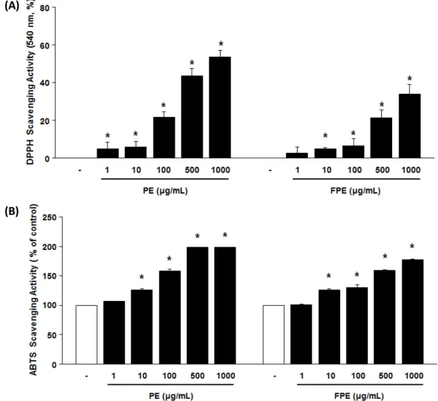
Antioxidant capacities of the EP and FEP were determined by DPPH (Fig. 2A) and ABTS-based (Fig. 2B) assays. When determined the antioxidant capacities of the EP and FEP based on their free radical scavenging activity, EP showed remarkable antioxidant capacity but in the same amount of FEP the antioxidant capacity was less than that of EP (Fig.

2A). ABTS scavenging activity represents the compound’s cationic radical scavenging potential. As shown in Fig. 2B, ABST scavenging activity of the EP and FEP showed the similar results as shown in DPPH assay. These results suggest that both EP and FEP have an antioxidant potential, which has been helpful in reducing oxidative stress involved in skin aging.

Effect of EP and FEP on the production (or secretion) of procollagen type I and Elastin

Collagen and elastin are important proteins involved in forming skin matrix. Reduction in collagen and elastin may be associated with a wrinkle formation due to destruction of skin architecture (Contet-Audonneau et al., 1999).

Procollagen type I is cleaved at its carboxy-terminus to

produce mature collagen type I, so the content of procollagen

type I is a good indicator of collagen biosynthesis (Díez et al.,

(4)

1996). As shown in Fig. 3A, EP (50 ㎍/㎖) and FEP (50 ㎍/

㎖) increased the production of procollagen type I by 2.2 fold and 2.4 fold. Test with equol used as a positive control showed 2.2 fold increase of procollagen type I synthesis at same concentration (Fig. 3A). Stimulation of elastin in skin cells will be a good contributor involved in reduction of wrinkle formation. EP and FEP could increase the production of elastin in NIH-3T3 cells by 1.9 and 2.1 fold, compared to untreated control (Fig. 3B). FEP was more potent than that of equol which showed 1.9 fold increase of elastin synthesis.

Effect of EP and FEP on the production (or secretion) of MMP-1, -2, and -3

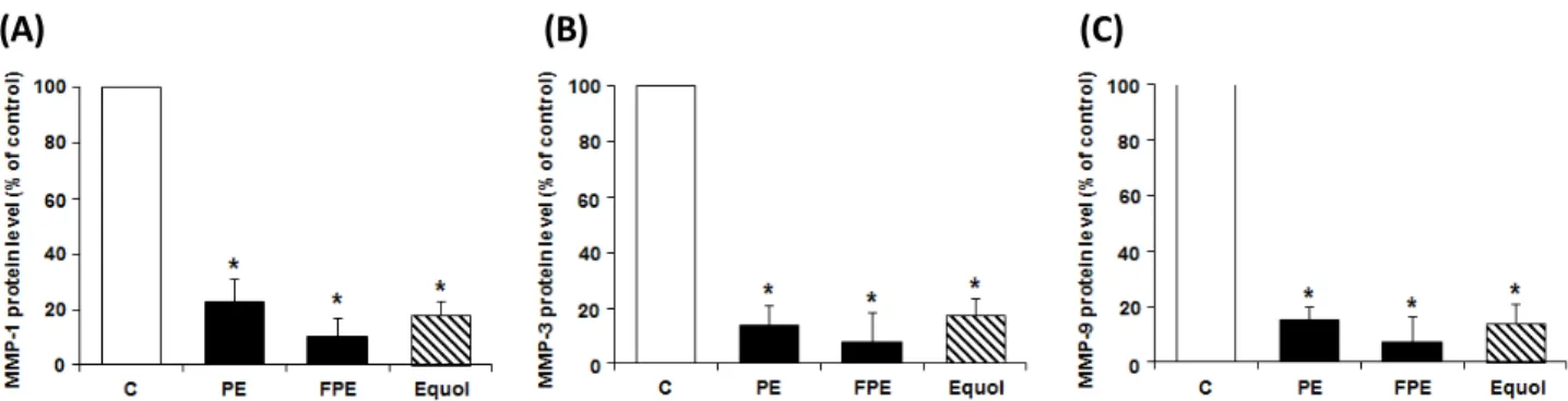
As a causal promotion of wrinkle formation, it has been suggested that suppression of expression or activities of matrix metalloproteases (MMPs) will be another approach to alleviate wrinkle formation (Pittayapruek et al., 2016). When determined the inhibitory effect of EP and FEP on the expression of MMP-1 (collagenase-1), -3 and -9 by ELISA assay (Fig. 4), the supplementation of EP could remarkably suppress the expression level of MMP-1, -3 and -9. In addition, FEP showed more potent activity than that of EP but statistical significance was not observed.

(A)

(B)

(C)

Fig. 1. Cytotoxic effects of PE ( Pueraria lobata extracts) and Fermented PE (FPE) in (A) keratniocytes (HaCaT), (B) melanocytes

(B16F10), and (C) fibroblast cells (NIH-3T3). Cells were treated with indicated concentrations of extracts. Cell viability was

measured using the MTT assay. The viability of untreated control cells was defined as 100%. (a) Cells (2×10

4

cells/well) treated with

PE and FPE (1 ㎍/㎖ to 1000 ㎍/㎖) for 24 hrs. The results are mean ± S.E. of quintuplicates from a representative experiment

(*p<0.05; significantly different from the control).

(5)

(A)

(B)

Fig. 2. The antioxidant activity of PE ( Pueraria lobata extracts) and Fermented PE (FPE) using (A) DPPH radical scavenging activity assay and (B) ABTS radical scavenging activity assay. Each bar indicates the mean S.D. of determinations, n=4. Statistical analysis was performed by the one-way ANOVA (p<0.05).

(A) (B)

Fig. 3. Effects of PE ( Pueraria lobata extracts) and Fermented PE (FPE) on the production of (A) type I procollagen and (B) elastin

levels by ELISA assay in fibroblast NIH-3T3 cells. The results are mean ± S.E. of quintuplicates from a representative experiment

(*p<0.05; significantly different from the control).

(6)

Effect of EP and FEP on the production of TIMP-1 In addition to down regulation in protein expression of MMPs, the strategy involved in inhibition of their activities will be another choice in suppression of wrinkle formation.

TIMP-1 is an endogenous MMPs inhibitor and behaves as a growth factor-like molecule in keratinocytes and fibroblasts (Hornebeck, 2003). When the effect of EP and FEP on the protein expression of TIMP-1 was determined by ELISA assay, supplementation of EP and FEP could significantly increase the protein expression level of TIMP-1, by 5.4 and 6.5 times more compared to untreated control (Fig. 5). These results suggest that the EP and FEP could not only suppress the protein expression levels of MMP-1, -3, -9 (Fig. 4) but also inhibit the activities of MMP-1, -3, and -9 through

upregulation of TIMP-1.

Discussion

In this report, we found that EP supplementation could involve in alleviation of wrinkle formation through suppression of protein expression level of MMP-1, -3, and -9 in conjunction with up-regulation of TIMP-1. In addition, augmentation of protein expression level of procollagen type I and elastin by EP supplementation will be another beneficial effect against wrinkle formation.

Pueraria lobata is a species of climbing plant belonging to the Leguminosae family and contains various phenolic compounds and isoflavonoids, including daidzin, daidzein, genistin, and puerarin, used for alleviating problems with liver damage, bone loss (Kim et al., 2015), obesity (Lee et al., 2015; Jung et al., 2017), allergy (Jeong et al., 2013), inflammation (Choo et al., 2002) and metabolic disorders such as obesity (Jung et al., 2017). The extract of Pueraria lobata (EP) has been used as an alternative herb for alleviating postmenopausal symptoms (Woo et al., 2003). Many studies have been conducted to find a natural material that has high biologic functions for human skin without any side effects (Smit et al., 2009; Lee et al., 2012; Kim et al., 2015; Chen et al., 2016). Recently, it has been found that anti-melanogenic effects of aerial part of Pueraria thunbergiana in vitro and in vivo (Han et al., 2015). In addition to intervention regarding anti-melanogenesis, prevention of wrinkle formation is another concern in skin-aging and beauty. It is noteworthy that looking better is one of important factors in well-being

(A) (B) (C)

Fig. 4. Inhibitory effects of PE ( Pueraria lobata extracts) and Fermented PE (FPE) on the expression of (A) MMP-1, (B) MMP-3, and (C) MMP-9 secretion by NIH-3T3 cells. The levels of MMPs were measured by ELISA assay. Data are presented as mean ± S.E. of quintuplicates from a representative experiment (*p<0.05; significantly different from the control).

Fig. 5. Effects of PE ( Pueraria lobata extracts) and Fermented

PE (FPE) on the expression of TIMP-1 in NIH-3T3 cells. The

level of TIMP-1 was measured by ELISA method. Data are

presented as mean ± S.E. of quintuplicates from a

representative experiment (*p<0.05; significantly different

from the control).

(7)

and quality life. In addition, maintenance of well performing skin barrier is also important point for keeping a good health, since destruction of skin barrier will cause vulnerability to infection, or penetration of environmental pollutants (Ganceviciene et al., 2012). Wrinkle formation is one of cutaneous signs of aging due to alterations of collagen structure, composition, function, synthesis, and degradation (Ramos-e-Silva and Carneiro, 2001).

Therefore, the blockage of collagen-degrading MMPs or stimulation of collagen synthesis, or both, can be a good target. In this view, the good starting point would be EP and FEP from natural products. Like other extracts isolated form plants, EP contains antioxidant potential that was confirmed by DPPH (Fig. 2A) and ABST (Fig. 2B) assay. However, it is unclear why FEP was less potent than that of EP even after fermentation process. Interestingly, EP and FEP could suppress the wrinkle formation via increase of collagen and elastin and suppression of MMP-1, -3, and -9 protein levels (Fig. 4). In addition, up-regulation of TMIP-1 by supplementation of EP and FEP further suppressed activities of MMPs (Fig. 5). This result is valuable since more active isoflavone metabolites could be obtained from relatively small starting quantity. It is speculated that L. rhamnosus vitaP1 could biotransform from isoflavones of EP into more active forms of isoflavones including dihydrodaidzein (DHD), dihydrogenistein (DHG) or equol, which have more potent estrogenic activity, biological effect and bioavailability.

Besides the inhibitory effect of EP and FEP on the collagen degrading process by MMPs, additional protective effects of the EP and FEP were shown in the view of increase production in collagen (Fig. 3A) and elastin (Fig. 3B), those involve the healthy skin architecture and are diminished with aging or exposure to sunlight (Lephart, 2016; Makrantonaki et al., 2016). However, the elucidation of the underlying molecular mechanism involved in down-regulation of MMP-1, -3, and -9 and up-regulation of procollagen type I, elastin and TIMP-1 expression should be under more extensive studies.

Especially, the mechanistic insight of robustly increased expression of TIMP-1 by the supplementation of EP and FEP should be paid more attentions.

Taken together, supplementation of EP and FEP could involve in the suppression of parameters associated with

wrinkle formation through suppression of MMP-1, -3, and -9 expression and up-regulation of TIMP-1. In addition, acceleration of collagen and elastin synthesis by EP and FEP supplementation may prove to have potential values as anti-wrinkle cosmetic additives.

Acknowledgment

This work was carried out with the support of “Cooperative Research Program for Agriculture Science & Technology Development (Project No. PJ01128102)” Rural Development Administration, and 2015 Research Grant from Kangwon National University (No. 201510101), Korea.

References

Baumann, L. 2007. Skin ageing and its treatment. The Journal of Pathology 211:241-251.

Chen, Y.H., L .Huang, Z.H. Wen, C. Zhang, C.H. Liang, S.T.

Lai, L.Z. Luo, Y.Y. Wang and G.H. Wang. 2016. Skin whitening capability of shikimic acid pathway compound.

Eur. Rev. Med. Pharmacol. Sci. 20:1214-1220.

Choo, M.K., E.K. Park, H.K. Yoon and D.H. Kim. 2002.

Antithrombotic and antiallergic activities of daidzein, a metabolite of puerarin and daidzin produced by human intestinal microflora. Biol. Pharm. Bull. 25:1328-1332.

Contet-Audonneau, J.L., C. Jeanmaire and G. Pauly. 1999. A histological study of human wrinkle structures: comparison between sun-exposed areas of the face, with or without wrinkles, and sun-protected areas. Br. J. Dermatol. 140:

1038-1047.

Desmedt, B., P. Courselle, J.O. De Beer, V. Rogiers, M.

Grosber, E. Deconinck and K. De Paepe. 2016. Overview of skin whitening agents with an insight into the illegal cosmetic market in Europe. J. Eur. Acad. Dermatol.

Venereol. 30:943-950.

Díez, J., A. Panizo, M.J. Gil, I. Monreal, M. Hernández and J.

Pardo Mindán. 1996. Serum markers of collagen type I metabolism in spontaneously hypertensive rats: relation to myocardial fibrosis. Circulation 93(5):1026-1032.

Ganceviciene, R., A.I. Liakou, A. Theodoridis, E. Makrantonaki and C.C. Zouboulis. 2012. Skin anti-aging strategies.

Dermatoendocrinol 4:308-319.

(8)

Gao, Y.N., Q.H. Hao, H.L. Zhang, B. Zhou, X.M. Yu and X.L.

Wang. 2016. Reduction of soy isoflavones by use of Escherichia coli whole-cell biocatalyst expressing isoflavone reductase under aerobic conditions. Lett. Appl. Microbiol.

63:111-116.

Han, E., B. Chang, D. Kim, H. Cho and S. Kim. 2015.

Melanogenesis inhibitory effect of aerial part of Pueraria thunbergiana in vitro and in vivo. Arch Dermatol Res.

307:57-72.

Hornebeck, W. 2003. Down-regulation of tissue inhibitor of matrix metalloprotease-1 (TIMP-1) in aged human skin contributes to matrix degradation and impaired cell growth and survival. Pathol. Biol. 51:569-573.

Jeong, Y.J., Y.J. Jang and S.C. Kang. 2013. Constituent alterations of Puerariae radix and lacquer tree mixture extract and their anti-allergic effects. Korean J. Plant Res.

26(1):103-110 (in Korean).

Jung, H.W., A.N. Kang, S.Y. Kang, Y.K. Park and M.Y. Song.

2017. The root extract of Pueraria lobata and its main compound, puerarin, prevent obesity by increasing the energy metabolism in skeletal muscle. Nutrients 9(1):pii:

E33.

Kim, D.Y., K.J. Won, D.I. Hwang, S.W. Yoon, S.J. Lee, J.H.

Park, M.S. Yoon, B. Kim and H. M. Lee. 2015. Potential skin regeneration activity and chemical composition of absolute from Pueraria thunbergiana flower. Nat. Prod. Commun.

10:2009-2012.

Lee, C.M., M.S. Yoon and Y.C. Kim. 2015. Effects of Pueraria lobata root ethanol extract on adipogenesis and lipogenesis during 3T3-L1 differentiation into adipocytes. Toxicol. Res.

31:191-201.

Lee, H.S., M.R. Kim, Y. Park, H.J. Park, U.J. Chang, S.Y. Kim and H.J. Suh. 2012. Fermenting red ginseng enhances its safety and efficacy as a novel skin care anti-aging ingredient:

in vitro and animal study. J. Med. Food 15:1015-1023.

Lephart, E.D. 2016. Skin aging and oxidative stress: Equol's anti-aging effects via biochemical and molecular mechanisms.

Ageing Res. Rev. 31:36-54.

Makrantonaki, E., G.P. Pfeifer and C.C. Zouboulis. 2016.

Intrinsic factors, genes, and skin aging. Der Hautarzt 67:

103-106.

Pittayapruek, P., J. Meephansan, O. Prapapan, M. Komine and M. Ohtsuki. 2016. Role of matrix metalloproteinases in photoaging and photocarcinogenesis. Int. J. Mol. Sci. 17(6):

pii: E868.

Ramos-e-Silva, M. and S.C. Carneiro. 2001. Cosmetics for the elderly. Clin. Dermatol. 19:413-423.

Shin S.C., S.K. Kang and M.J. Jang. 2000. A study on the screening of antimicrobial activity and the isoflavonoids in Korean arrowrrot. Korean J. Plant Res. 13(1):25-28 (in Korean).

Smit, N., J. Vicanova and S. Pavel. 2009. The hunt for natural skin whitening agents. Int. J. Mol. Sci. 10:5326-5349.

Woo, J., E. Lau, S.C. Ho, F. Cheng, C. Chan, A.S. Chan, C.J.

Haines, T.Y. Chan, M. Li and A. Sham. 2003. Comparison of Pueraria lobata with hormone replacement therapy in treating the adverse health consequences of menopause.

Menopause 10:352-361.

Yu, J., X. Bi, B. Yu and D. Chen. 2016. Isoflavones: Anti- inflammatory benefit and possible caveats. Nutrients 8(6):

pii:E361.

(Received 7 April 2017 ; Revised 19 June 2017 ; Accepted 21 June 2017)

수치

Fig. 1. Cytotoxic effects of PE ( Pueraria lobata extracts) and Fermented PE (FPE) in (A) keratniocytes (HaCaT), (B) melanocytes  (B16F10), and (C) fibroblast cells (NIH-3T3)
Fig. 2. The antioxidant activity of PE ( Pueraria lobata extracts) and Fermented PE (FPE) using (A) DPPH radical scavenging activity  assay and (B) ABTS radical scavenging activity assay
Fig. 4. Inhibitory effects of PE ( Pueraria lobata extracts) and Fermented PE (FPE) on the expression of (A) MMP-1, (B) MMP-3, and  (C) MMP-9 secretion by NIH-3T3 cells

참조

관련 문서

Effects of Anthriscus sylvestris Hoffmann aqueous layer (ASAL) on cell apoptosis and PARP activity in NIH/3T3 fibroblast and KB, FaDu oral cancer cells by

Frequency of conidial germination (A) and appressorium formation (B) of Colletotrichum orbiculare and callose formation of plant cells (C) at the

The effects of the acceleration voltage, the probe current and the travel speed on the formation, the ingredient variation and the surface roughness of the deposited

In terms of the mould design, the effects of runner system design and the mould temperature on filling characteristics, the weldline formation, the

In order to achieve this goal first, PE teachers learning the difference between commitment and satisfaction of junior classes depending on the type of

이하선의 실질 속에서 하악경의 후내측에서 나와 하악지의 내측면을 따라 앞으로 간다. (귀밑샘 부위에서 갈라져 나와

1 John Owen, Justification by Faith Alone, in The Works of John Owen, ed. John Bolt, trans. Scott Clark, &#34;Do This and Live: Christ's Active Obedience as the

Inhibitory effect of Quercetin and Aronia extract on the MITF, Tyrosinase, TRP-1, TRP-2 and Actin in expression B16F10 cells.. Inhibitory effect of C3G on the Tyrosinase,