Cytomegalovirus Pneumonia:
High-Resolution CT Findings in Ten
Non-AIDS Immunocompromised Patients
Objective: To describe the HRCT findings of cytomegalovirus (CMV) pneumo- nia in non-AIDS immunocompromised patients
Materials and Methods: This retrospective study involved the ten all non-AIDS immunocompromised patients with biopsy-proven CMV pneumonia and without other pulmonary infection encountered at our Medical Center between January 1997 and May 1999. HRCT scans were retrospectively analysed by two chest radiologists and decisions regarding the findings were reached by consensus.
Results: The most frequent CT pattern was ground-glass opacity, seen in all patients, with bilateral patchy (n = 8) and diffuse (n = 2) distribution. Other findings included poorly-defined small nodules (n = 9) and consolidation (n = 7). There was no zonal predominance. The small nodules, bilateral in eight cases and unilateral in one, were all located in the centrilobular region. Consolidation (n = 7), with patchy distribution, was bilateral in five of seven patients (71%). Pleural effusion and bilateral areas of thickened interlobular septa were seen in six patients (60%).
Conclusion: CMV pneumonia in non-AIDS immunocompromised patients appears on HRCT scans as bilateral mixed areas of ground-glass opacity, poorly- defined centrilobular small nodules, and consolidation. Interlobular septal thicken- ing and pleural effusion are frequently associated.
uman cytomegalovirus (CMV), which belongs to the group of herpes viruses, is an important cause of morbidity and mortality in immunocom- promised patients (1-12).
The radiographic findings of CMV pneumonia are variable and include diffuse retic- ulonodular densities (2, 3, 7, 8), diffuse haze, and dense areas of consolidation (13- 15). McGuinness et al. (9) recently described the CT appearances of CMV pneumonitis in 21 AIDS patients; the findings included ground-glass attenuation, dense consolida- tion, and bronchial wall thickening. In a recent report, Kang et al. (16) described the CT findings of CMV pneumonia in ten patients who had undergone transplants; their findings differed from those seen in AIDS patients and included micronodules (1-5 mm in diameter), consolidation, and-less frequently-reticulation and ground-glass opacity. In their study, however, only three patients underwent HRCT scanning.
According to our experience, however, findings of CMV infection seen on high-reso- lution CT scans especially in non-AIDS immunocompromised patients, were some- what different from those of Kang et al. The aim of the present study is to describe the clinical and CT findings of CMV pneumonia in ten non-AIDS immunocompromised patients.
Jeung Hee Moon, MD
1Eun A Kim, MD
1Kyung Soo Lee, MD
1Tae Sung Kim, MD
1Kyung-Jae Jung, MD
1Jae-Hoon Song, MD
2Index words : Lung, abnormalities Lung, CT
Lung, infection
Korean J Radiol 2000 ; 1 : 73-78 Received February 24, 2000; accepted after revision May 18, 2000.
1Department of Radiology;2Division of Infectious Disease, Department of Internal Medicine, Samsung Medical Center, Sungkyunkwan University School of Medicine
Address reprint requests to : Kyung Soo Lee, MD, Department of Radiology, Samsung Medical Center, Sungkyunkwan University School of Medicine, 50, Ilwon-Dong, Kangnam-Gu, Seoul 135-710, Korea.
Telephone: (822) 3410-2511 Fax: (822) 3410-2559
e-mail: [email protected]
H
MATERIALS AND METHODS
This retrospective study involved ten consecutive pa- tients with pathologically proven CMV pneumonia but without other coexisting pulmonary infections encountered between January 1997 and May 1999. All patients (M: 6;
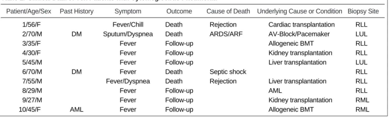
F: 4), who ranged in age from 27 to 70 (mean, 46) years, underwent CT examination. In all cases, CMV infection was confirmed by transbronchial lung biopsy using fiberop- tic bronchoscopy, and by immunohistochemical staining for CMV monoclonal antibody. All patients were immuno- compromised. Seven of the ten had received organ trans- plants [bone marrow (n = 2), liver (n = 2), kidney (n = 2), and heart (n = 1)]. Two had diabetes and one had acute myelogenous leukemia (Table 1).
In all patiets, high-resolution CT scanning using a HiSpeed Advantage scanner (GE Medical Systems, Milwaukee, WI) was performed throughout the thorax (1- mm collimation at 10-mm intervals; scans obtained at end inspiration). Both mediastinal (width, 400 H; level, 20 H) and lung (width, 1,500 H; level, -700 H) window images were printed. HRCT scans were analyzed by two chest ra- diologists and decisions regarding the findings were reached by consensus.
As seen on HRCT scans, patterns of parenchymal abnor- malities were subdivided into areas of ground-glass opaci- ty, consolidation, and small nodules (< 7 mm in diameter).
The presence of septal thickening, crazy paving, air trap- ping, and pleural effusion was also recorded, and zonal dis- tribution (upper/middle/lower) and laterality (unilateral vs.
bilateral) of the lesions were evaluated. Consolidation on CT indicated the area of opacity with obscuration of un- derlying vessels. Ground-glass opacity was defined as a hazy increase in lung attenuation without obscuration of underlying vessels. Lesions were considered to be centrally
distributed if located within one third of the lung from the mediastinum in transverse plane, and peripheral if located within the inner one third of the lung from the chest wall.
If they did not fit either of these categories, they were re- garded as random. If scattered widely they were consid- ered to be diffuse, and if inconsistent and variable, to be patchy. Additional findings including the presence of medi- astinal nodal enlargement (positive when short axis was greater than 10 mm in diameter), were also recorded.
Transbronchial lung biopsy specimens were reviewed by an experienced lung pathologist. Using a limited number of these, the findings of CT and of the pathologic findings ob- tained at preoperatively determined biopsy sites were cor- related.
RESULTS
The most frequent CT appearance was ground-glass opacity, seen in all patients (Fig. 1). It was mixed with oth- er patterns, mainly areas of small nodules (n = 9) and con- solidation (n = 7). Areas of ground-glass opacity were dis- tributed bilaterally in all patients, and were patchy in eight and diffuse in two. There was no zonal predominance (Table 2).
Small nodules were seen in nine patients (Figs. 2, 3), be- ing associated with ground-glass opacity in all and with consolidation in six. They were distributed bilaterally in eight patients and unilaterally in one, and in all nine were located in the centrilobular region of the secondary pul- monary lobule.
The area of consolidation, seen in seven patients, was bi- lateral in five (71%). The lesion was patchy and random in distribution, and was associated with ground-glass opacity in all patients and with small nodules in six of seven.
Bilateral areas of thickened interlobular septa were pre-
Table 1. Clinical Data of Ten Patients with Cytomeglovirus Pneumonia
Patient/Age/Sex Past HistorySymptom Outcome Cause of Death Underlying Cause or Condition Biopsy Site 01/56/F Fever/Chill Death Rejection Cardiac transplantation RLL 02/70/M DM Sputum/Dyspnea Death ARDS/ARF AV-Block/Pacemaker LUL
03/35/F Fever Follow-up Allogeneic BMT RLL
04/30/F Fever Follow-up Kidney transplantation RLL
05/45/M Fever Follow-up Liver transplantation LUL
06/70/M DM Fever Death Septic shock RLL
07/55/M Fever/Dyspnea Death Rejection Liver transplantation RLL
08/29/M Fever Follow-up AML RLL
09/27/M Fever Follow-up Kidney transplantation RML
10/45/F AML Fever Follow-up Allogeneic BMT RML
Note.─DM: diabetes mellitus, AML: acute myelogenous leukemia, ARDS: acute respiratory distress syndrome, ARF: acute renal failure, AV: atrioventricular, RML: right middle lobe, RLL: right lower lobe, LUL: left upper lobe, LLL: left lower lobe, BMT: bone marrow transplantation
sent in six patients (60%) (Fig. 3). Pleural effusion was seen in six, in three of whom it was bilateral (Fig. 1); in four of the six, the amount of was small. Additional find- ings were crazy paving (2/10; 20%) (Fig. 1), a small amount of pericardial effusion (2/10; 20%), air trapping in the right upper and lower lobes (1/10; 10%), and cystic le- sions in both upper lobes (1/10; 10%).
Limited correlation between the CT findings and the patho- logic findings of transbronchial lung biopsy revealed that ar- eas of ground-glass attenuation or consolidation seen on CT scans corresponded to areas of the exudative or proliferative phase of diffuse alveolar damage consisting of interstitial fi- broblastic proliferation and lymphocytic infiltration (Fig. 2B).
Type-2 pneumocyte hyperplasia, hyaline membrane forma- tion, and intra-alveolar exudate with or without organization were associated. Histopathologically, the poorly-defined cen- trilobular nodules seen on HRCT represented intra-alveolar collections of macrophages, red blood cells and fibrin (Fig.
3B). CMV-infected cells showing intra-nuclear inclusions were seen in all pathologic specimens.
Table 2. High-Resolution CT Findings of Cytomeglovirus Pneumonia
Laterality Zonal Distribution Transverse Plane Uniformity Uni Bi Upper Middle Low Central Peripheral Random PatchyDiffuse
Ground-glass opacity (n=10; 100%) 0 10 10 10 10 0 0 10 8 2
Small nodule (n=9; 90%) 1 08 09 08 08 0 0 09 9 0
Air-space consolidation (n=7; 70%) 2 05 04 03 04 0 0 07 7 0
Note.─Uni: unilateral, Bi: bilateral
Fig. 2. Cytomegalovirus pneumonia in a 35-year-old woman who underwent bone marrow transplantation 27 days earlier.
A. High-resolution (1.0-mm collimation) CT scan obtained at level of bronchus intermedius shows numerous small nodules predominant- ly in centrilobular regions (arrows). There are associated focal patchy areas of ground-glass opacity.
B. Photomicrograph of transbronchial lung biopsy specimen obtained from superior segment of right lower lobe shows diffuse alveolar damage consisting of interstitial fibroblastic proliferation and type-2 pneumocyte hyperplasia (arrows), in addition to characteristic cells (arrowheads) showing intra-nuclear inclusions (H & E, ×100).
A B
Fig. 1. Cytomegalovirus pneumonia in a 27-year-old man who underwent kidney transplantation. High-resolution (1.0-mm colli- mation) CT scan obtained at level of distal trachea 32 days after transplantation shows bilateral patchy areas of ground-glass opacity with crazy paving appearance. The presence of a few poorly-defined centrilobular nodules (arrows) and bilateral pleural effusion should also be noted.
DISCUSSION
Due to its physical characteristics, CMV is considered a member of the Herpetoviridae family. One of the major bi- ological characteristics of CMV (as with other herpes virus- es) is its ability to become latent in the human host, with the potential for reactivation (6). It is evident that in im- munocompromised patients, CMV may cause severe symp- tomatic pulmonary disease (13, 14, 17-22).
It has been suggested that CMV pneumonia in allogeneic transplant recipients is caused by immune mechanisms me- diated by a T-cell response to virally-induced antigens ex- pressed in the lungs. Severe necrotizing pneumonia may occur in spite of the suppression of virus replication during antiviral therapy. AIDS patients with more profound im- mune deficiency than transplant recipients may be unable to initiate the immune response necessary to cause CMV pneumonia by this mechanism. In AIDS patients, lung damage may be directly due to the cytopathogenic effects of CMV, related to the extent of active virus replication (23). These differences in the pathogenic mechanisms of pulmonary CMV infection may result in different histopathologic features between AIDS patients and trans- plant recipients. In transplant recipients with CMV pneu- monia, necrotizing inflammation is dominant, and relative- ly few CMV-infected cells are revealed by histopathologic examination. In AIDS patients, there is a high density of CMV inclusion bodies, and this leads to severe sympto-
matic pneumonia (23). In AIDS patients, the cytopathogen- ic effect of CMV may well cause diffuse alveolar damage more frequently than in non-AIDS patients.
The CT findings of CMV pneumonia are diverse and have been described without distinction between AIDS and non-AIDS patients (9, 10). Common findings are mixed alveolar-interstitial infiltrates such as ground-glass opacity, consolidation, bronchiectasis and interstitial reticu- lation. McGuinness et al. (9) noted that masses or mass-like infiltrates were more frequent in AIDS patients with CMV pneumonia (12/21) than in non-AIDS patients. Kang et al.
(16) reported the CT findings of CMV pneumonia in ten transplant patients in whom diagnosis was confirmed by open lung biopsy in nine and autopsy in one. In their study, parenchymal abnormalities were revealed by CT in nine of ten patients, while in one, CT scans were normal.
In those nine patients, the findings were as follows: small nodules (n = 6), consolidation (n = 4), ground-glass opacity (n = 4), and irregular linear opacity (n = 1). The nodules were bilaterally and symmetrically distributed and in- volved all lung zones. Consolidation was prominent mostly in the lower zones, while ground-glass opacity was com- bined only with nodules and/or consolidation. The distribu- tion of ground-glass opacity was not specifically men- tioned.
In our study, the CT findings of CMV pneumonia in im- munocompromised non-AIDS patients were somewhat dif- ferent from those described by Kang et al. (16). Bilateral patchy areas of ground-glass opacity were the commonest
Fig. 3. Cytomegalovirus pneumonia in a 30-year-old woman who presented with fever two months after kidney transplantation.
A. High-resolution (1.0-mm collimation) CT scan obtained at level of right inferior pulmonary vein shows mixed areas of ground-glass opacity and small poorly-defined centrilobular nodules (arrows). There is associated interlobular septal thickening (arrowheads).
B. Photomicrograph of transbronchial lung biopsy specimen obtained from superior segment of right lower lobe shows intra-alveolar col- lection of macrophages, fibrin and red blood cells (arrows) that corresponds to the centrilobular nodules seen on CT scans. There is as- sociated alveolar wall thickening, with interstitial inflammatory cell infiltration (H & E, ×200).
A B
pattern, seen in all patients. These differences may be due to the fact that in our study, HRCT images were available, while Kang et al. used the conventional technique in seven and the HRCT technique in only three. In our study, poor- ly-defined centrilobular small nodules (90%) and areas of consolidation (70%) were also common. Kim et al. (24) re- cently reported the HRCT findings of CMV pneumonia in eleven immunocompromised patients. Their findings were very similar to ours and included ground-glass opacity (n = 11), consolidation (n = 7), reticular opacity (n = 10), multi- ple small nodules or mass (n = 6), and bronchiectasis or bronchial wall thickening (n = 5).
In cases of CMV pneumonia, pathologic examination re- veals cytomegalic cells within areas of alveolar damage, the alveolar-filling process being caused by hemorrhage and fibrinous exudate. These areas may correspond to the areas of ground-glass opacity and/or consolidation seen on HRCT scans. Kang et al. (16) reported small nodules, re- vealed by CT seen in six of ten patients, which correspond- ed histopathologically to areas of inflammatory or hemor- rhagic nodules or organizing pneumonia. In our study, the poorly-defined centrilobular nodules seen on HRCT scans represented histopathologically areas of intra-alveolar col- lection of macrophages, red blood cells, and fibrin (hemor- rhagic and inflammatory nodule).
In their study of 21 AIDS patients with CMV pneumo- nia, McGuinness et al. (9) suggested that patterns of dis- ease, revealed by CT closely correspond to the pathologic changes seen in CMV pneumonia. Diffuse alveolar dam- age, which is probably caused by the cytopathogenic effect of CMV, involves pneumocyte injury and reactive prolifer- ation, as well as neutrophilic and fibrinous exudates, and hyaline membrane formation and hemorrhage fill the alve- olar spaces. Interstitial infiltrates are comprised mainly of lymphocytes and result in alveolar wall or interlobular sep- tal thickening. Interspersed throughout these changes there may be areas of hemorrhage, fibrin deposition, and -rarely- necrosis. These pathologic changes, depending on their severity, distribution, and relative proportions of each dis- ease process, may result in ground-glass opacity, dense consolidation, mass-like infiltrates and irregular linear opacity on CT scans (9).
Our investigation suffers from two limitations. First, it was retrospective. Because true CMV infection is not easily diagnosed, we might not have included all patients with pulmonary CMV infection. It may be that only patients di- agnosed by means of transbronchial lung biopsy, and in whom, therefore, apparent abnormalities were revealed by CT scanning, were included. Second, because we had limit- ed pathologic specimens, obtained only by means of trans- bronchial lung biopsy, pathological correlation was incom-
plete.
In conclusion, the most common HRCT findings of CMV pneumonia in non-AIDS immunocompromised patients are bilateral mixed areas of ground-glass opacity, poorly-de- fined centrilobular small nodules, and consolidation.
Interlobular septal thickening and pleural effusion are fre- quently associated.
References
1. Rubin RH, Cosimi AB, Tolkoff-Rubin NE, Russel PS, Hirsch MS.
Infectious disease syndromes attributable to cytomegalovirus and their significance among renal transplantation recipients.
Transplantation 1977;24:458-464
2. Abdallah PS, Mark JBD, Merigan TC. Diagnosis of cy- tomegalovirus pneumonia in compromised hosts. Am J Med 1976;61:326-332
3. Ravin CE, Smith GW, Ahern MJ, et al. Cytomegaloviral infec- tion presenting as a solitary pulmonary nodule. Chest 1977;
71:220-222
4. Krowka MJ, Rosenow EC, Hoagland HC. Pulmonary complica- tions of bone marrow transplantation. Chest 1985;87:237-246 5. Schulman LL. Cytomegalovirus pneumonitis and lobar consoli-
dation. Chest 1987;91:558-561
6. Klotman ME, Hamilton JD. Cytomegalovirus pneumonia.
Semin Respir Infect 1987;2:95-103
7. Wallace JM, Hannah J. Cytomegalovirus pneumonitis in pa- tients with AIDS. Findings in an autopsy series. Chest 1987;
92:198-203
8. Johnson PC, Hogg KM, Sarosi GA. The rapid diagnosis of pul- monary infections in solid organ transplant recipients. Semin Respir Infect 1990;5:2-9
9. McGuinness G, Scholes JV, Garay SM, Leitman BS, McCauley DI, Naidich DP. Cytomegalovirus pneumonitis: spectrum of parenchymal CT findings with pathologic correlation in 21 AIDS patients. Radiology 1994;192:451-459
10. Aafedt BC, Halvorsen RA, Tylen U, Hertz M. Cytomegalovirus pneumonia: computed tomography findings. J Can Assoc Radiol 1990;41:276-280
11. Northfelt DW, Sollitto RA, Miller TR, Hollander H. Cytome- galovirus pneumonitis. An unusual cause of pulmonary nodule in a patient with AIDS. Chest 1993;103:1918-1920
12. Berger LA. Imaging in the diagnosis of infections in immuno- compromised patients. Curr Opin Infect Dis 1998;11:431-436 13. Austin JH, Schulman LL, Mastrobattista JD. Pulmonary infec-
tion after cardiac transplantation: clinical and radiologic correla- tions. Radiology 1998;72:259-265
14. Shreeniwas R, Schulman LL, Berkmen YM, McGregor CC, Austin JHM. Opportunistic bronchopulmonary infections after lung transplantation: clinical and radiographic findings.
Radiology 1996;200:349-356
15. Afessa B, Gay PC, Plevak DJ, et al. Pulmonary complications of orthotopic liver transplantation. Mayo Clin Proc 1993;68:427- 434
16. Kang EY, Patz EF, Mu¨ller NL. Cytomegalovirus pneumonia in transplant patients: CT findings. J Comput Assist Tomogr 1996;
20:295-299
17. Zantel J, Leij L, Prop J, Harmsen MC. The Human cytome- galovirus: a viral complication in transplantation. Clin Trans- plant 1998;12:145-158
18. Moore EH, Webb WR, Amend WJC. Pulmonary infections in renal transplantation patients treated with cyclosporine.
Radiology 1988;167:97-103
19. Leung AN, Grosselin MV, Napper CH, et al. Pulmonary infec- tions after bone marrow transplantation: clinical and radi- ographic findings. Radiology 1999;210:699-710
20. Husni RN, Gordon SM, Longworth DL, et al. Cytomegalovirus infection is a risk factor for invasive aspergillosis in lung trans- plant recipients. Clin Infect Dis 1998;26:753-755
21. Chien J, Chan CK, Chamberlain D, et al. Cytomegalovirus pneumonia in allogeneic bone marrow transplantation. An im-
munopathologic process? Chest 1990;98:1034-1037
22. Worthy SA, Flint JD, Mu¨ller NL. Pulmonary complications af- ter bone marrow transplantation: high-resolution CT and patho- logic findings. RadioGraphics 1997;17:1395-1371
23. Aukrust P, Farstad IN, Froland SS, Holter E. Cytomegalovirus (CMV) pneumonitis in AIDS patients: the result of intensive CMV replication? Eur Respir J 1992;5:362-364
24. KIM HS, LEE JS. Cytomegalovirus pneumonia in immunocom- promised patients: HRCT findings. J Korean Radiol Soc 1999;
41:1133-1138