INTRODUCTION
Non-alcoholic fatty liver disease (NAFLD) has been increas-
ingly recognized as the most common chronic liver disease owing to excessive lipid accumulation in hepatocytes, leading to hepatocyte apoptosis, fat denaturalization, and the progres- sion of hepatic fibrosis.1 For some cases, this disease often in- cluded a spectrum of histological changes, from simple steatosis to non-alcoholic steatohepatitis (NASH), which may develop into liver fibrosis, cirrhosis, and even hepatocellular carcino- ma.2 With great changes in lifestyle and diet, as well as im- provement of people’s living standards, NAFLD incidence is gradually rising with a trend in younger ages, which has be- come one of the most prevalent chronic diseases in China after chronic viral hepatitis.3 Generally, NAFLD, accompanied with obesity, diabetes, hyperlipidemia, hypertension, and meta- bolic syndrome, is well-regarded as the hepatic manifestation of metabolic syndrome.4 Although effective therapies are ab-
S100A4 Gene is Crucial
for Methionine-Choline-Deficient Diet-Induced Non-Alcoholic Fatty Liver Disease in Mice
Yin-Hua Zhang1*, De-Qiang Ma1*, De-Ping Ding1, Juan Li2, Lin-Li Chen1, Kang-Jian Ao1, and You-You Tian1
1Department of Infectious Diseases, Taihe Hospital, Hubei University of Medicine, Shiyan, Hubei;
2Maternal and Child Health-Care Hospital, Shiyan, Hubei, China.
Purpose: To explore the influence of S100 calcium binding protein A4 (S100A4) knockout (KO) on methionine-choline-deficient (MCD) diet-induced non-alcoholic fatty liver disease (NAFLD) in mice.
Materials and Methods: S100A4 KO mice (n=20) and their wild-type (WT) counterparts (n=20) were randomly divided into KO/
MCD, Ko/methionine-choline-sufficient (MCS), WT/MCD, and WT/MCS groups. After 8 weeks of feeding, blood lipid and liver function-related indexes were measured. HE, Oil Red O, and Masson stainings were used to observe the changes of liver histopa- thology. Additionally, expressions of S100A4 and proinflammatory and profibrogenic cytokines were detected by qRT-PCR and Western blot, while hepatocyte apoptosis was revealed by TUNEL staining.
Results: Serum levels of aminotransferase, aspartate aminotransferase, triglyceride, and total cholesterol in mice were increased after 8-week MCD feeding, and hepatocytes performed varying balloon-like changes with increased inflammatory cell infiltra- tion and collagen fibers; however, these effects were improved in mice of KO/MCD group. Meanwhile, total NAFLD activity scores and fibrosis were lower compared to WT+MCD group. Compared to WT/MCS group, S100A4 expression in liver tissue of WT/
MCD group was enhanced. The expression of proinflammatory (TNF-α, IL-1β, IL-6) and profibrogenic cytokines (TGF-β1, CO- L1A1, α-SMA) in MCD-induced NAFLD mice were increased, as well as apoptotic index (AI). For MCD group, the expressions of proinflammatory and profibrogenic cytokines and AI in KO mice were lower than those of WT mice.
Conclusion: S100A4 was detected to be upregulated in NAFLD, while S100A4 KO alleviated liver fibrosis and inflammation, in addition to inhibiting hepatocyte apoptosis.
Key Words: S100 calcium-binding protein A4, non-alcoholic fatty liver disease, fibrosis, inflammation, apoptosis
pISSN: 0513-5796 · eISSN: 1976-2437
Received: April 18, 2018 Revised: August 6, 2018 Accepted: August 24, 2018
Corresponding author: De-Ping Ding, MD, Department of Infectious Diseases, Taihe Hospital, Hubei University of Medicine, No. 32, South Renmin Road, Shiyan, Hubei 442000, China.
Tel: 86-0719-8801492, Fax: 86-0719-8801669, E-mail: [email protected]
*Yin-Hua Zhang and De-Qiang Ma contributed equally to this work.
•The authors have no financial conflicts of interest.
© Copyright: Yonsei University College of Medicine 2018
This is an Open Access article distributed under the terms of the Creative Com- mons Attribution Non-Commercial License (https://creativecommons.org/licenses/
by-nc/4.0) which permits unrestricted non-commercial use, distribution, and repro- duction in any medium, provided the original work is properly cited.
Yonsei Med J 2018 Nov;59(9):1064-1071 https://doi.org/10.3349/ymj.2018.59.9.1064
sent due to inadequate elucidation of NAFLD pathogenesis, many factors such as immunity, genetics, metabolism, and en- vironment are linked to the presence of NAFLD.5
S100 protein, a low molecular weight acidic protein (10–12 kDa), is reported to play a crucial role in many human diseas- es, and it controls multiple processes like apoptosis, inflamma- tion, and cell movement.6 A total of 25 calcium-binding S100 family members, namely S100A1–18, hair hyaluronin, keratin fibrin, repetin, S100B, S100P, S100Z, and S100G, have been currently identified.7 As indicated by Mukai, et al.,8 S100A8 could induce the upregulation of tumor necrosis factor-alpha (TNF-α) in CXCR2-expressing CD11b+Gr-1high cells, thereby aggravating hepatitis in mice. In addition, Liu, et al.9 also found that expression levels of S100A9 was significantly elevated in patients with non-alcoholic fatty liver, which were suggested to be even higher in patients with NASH, highlighting the in- volvement of S100 protein in NAFLD progression. S100 calci- um binding protein A4 (S100A4), another member of the S100 protein family, also known as Mts1, metastasin, p9Ka, pEL98, CAPL, and calvasculin, Fsp-1, placental calcium-binding pro- tein, is a polypeptide comprised of 101 amino acids with a mo- lecular weight of 11.5 kDa.10 Until now, existing studies on S100A4 mainly focused on its effects on tumors, like reducing intercellular adhesion, remodeling extracellular matrix, and promoting abnormal cell proliferation and angiogenesis, ulti- mately enhancing tumor cell invasion and migration.11 More- over, S100A4 is known as an inducer of inflammatory processes, and its expression is strongly upregulated in various inflamma- tory diseases such as rheumatoid arthritis,12 idiopathic inflam- matory myopathies13 and so on. In addition, there has been evidence stating that S100A4 levels in liver tissues were posi- tively correlated with liver fibrosis.14 All factors mentioned above showed a possible role of S100A4 in NAFLD, which has been increasingly recognized as an inflammatory disease with different degrees of liver fibrosis.15 However, it remains unclear whether S100A4 is associated with the pathology of NAFLD.
Methionine-choline-deficient (MCD) diet impaired the secre- tory process of very low-density lipoprotein from the liver, which could induce hepatic lipid accumulation, aminotransferase elevation, and hepatic histological changes including steato- sis, hepatic inflammation, and fibrosis.16,17 These histological changes were similar to those of human NAFLD pathology.18 Therefore, MCD diet has been used as an internationally rec- ognized animal model of NAFLD.19,20 In our study, we intend- ed to observe the influences of S100A4 knockout (KO) on NAFLD by feeding S100A4 KO mice and their wild-type (WT) counterparts with either MCD diet or methionine-choline-suf- ficient (MCS) control diet, which were identical to MCD but sufficient in choline chloride (2 g/kg) and DL-methionine (3 g/kg).
MATERIALS AND METHODS
Ethics statement
A total of 100 male SPF C57BL/6 mice (age: 6–8 weeks; weigh- ing: 18–20 g) were purchased from Shanghai Experimental Ani- mal Center of Chinese Academy of Sciences (Shanghai, China).
The current study was consistent with the Laboratory Animal Use Convention published by the National Institutes of Health,21 and all animal experimental procedures were conducted and supervised by the Medical Laboratory Animal Ethics Commit- tee of Taihe Hospital.
Mice model construction
The study on S100A4 KO mice was conducted with a germline inactivation of S100A4 gene, as described from the previous study.22 S100A4 KO mice (n=20) and their WT counterparts (n=
20) were randomly divided into model and control groups. The mice in model group were fed with MCD diet, namely KO/
MCD and WT/MCD groups with 10 mice in each group, and control group mice were treated with MCS diet, namely KO/
MCS and WT/MCS groups with another 10 mice in each group.
The composition of MCS was identical to MCD but sufficient in choline chloride (2 g/kg) and DL-methionine (3 g/kg). Both MCS and MCD were obtained from MP Biomedicals (Solon, OH, USA).
Specimen preparation
Mice in each group were executed after 8 weeks of feeding, and peripheral blood was obtained after removal of eye-balls.
Then, serum was collected after centrifugation and stored at -20°C. The blood biochemical parameters including alanine aminotransferase (ALT), aspartate aminotransferase (AST), triglyceride (TG), and total cholesterol (TC) levels in each group were measured by an automatic biochemical analyzer 7180 (Hitachi Ltd, Tokyo, Japan). Mice were fixed on the oper- ating table, and their skin and peritoneum were cut open using surgical scissors, exposing and removing liver tissues. A part of acquired liver tissues was stabilized in 4% paraformaldehyde for 24 h to make regular paraffin embedded slices, while the other part was fixed in 4% paraformaldehyde for 2–4 h and soaked in 30% sucrose solution overnight at 4°C, which was stored in a refrigerator at -80°C for subsequent tests after opti- mal cutting temperature embedded.
Histological analysis
Hematoxylin and eosin (HE) staining: Slices of liver tissues were dewaxed in xylene twice for 5 min, dehydrated with gra- dient alcohol, and washed with distilled water for 5 min. Then, slices were stained with hematoxylin stain for 5 min and dif- ferentiated with 1% hydrochloric acid for 30 s, followed by 1%
eosin-alcohol dyeing for 5 min, which could be observed un- der a microscope after regular gradient alcohol dehydration and mounting.
Oil Red O (ORO) staining: Tissue sections were placed on slide sat room temperature for 30 min, fixed in 10% ice para- formaldehyde for 10 min, and then washed three times by dis- tilled water. After drying for several minutes, oil red and de- ionized water were diluted in a 3:2 ratio and placed at room temperature for 10 min. Following that, slices experienced ORO staining for 8 min, 85% propylene glycol solution differ- entiation for 2 min, washed twice, hematoxylin counterstained for 30 s, flushed with water for 3 min, and then mounted for mi- croscope observation.
Masson staining: Paraffin section of mice was observed after a series of procedures including routine dewaxing rehydration, ponceaufuchs in acid solution staining for 5–10 min followed by washing, 1% phosphomolybdic acid solution differentiation for 5 min, aniline blue solution counterstain for 5 min, treat- ment of 1% glacial acetic acid for 1 min, alcohol gradient de- hydration, transparent through dimethylbenzene xylene, and mounting.
NAFLD was diagnosed according to NAFLD activity scores (NAS) including steatosis (0–3), lobular inflammation (0–3), and hepatocyte ballooning (0–2),23 while liver fibrosis was cal- culated as grade 0 (none), grade 1 (zone perisinusoidal fibro- sis), grade 2 (as above with portal fibrosis), grade 3 (as above with bridging fibrosis), and grade 4 (cirrhosis).24
qRT-PCR
Total RNA was extracted by using total RNA extraction kit (Bei- jing Tian Enze Gene Technology Co., Ltd., Beijing, China), and cDNA was synthesized under the support of reverse transcrip- tion kit (Hangzhou Bori Technology Co., Ltd., Hangzhou, Chi- na). RT-PCR mixture was obtained from Bio-Rad (Hercules, CA, USA), and the reaction was carried out on ABI 7500 Quan- titative PCR instrument (Appplied Biosystems, Foster City, CA, USA) under the following conditions: pre-denaturation at 94°C for 5 min, followed by 40 cycles of denaturation at 94°C for 30 s, annealing at 58°C for 30 s, and extension at 72°C for 20 s. GAP- DH was selected as a reference gene, and formula 2-ΔΔCT was used to compare and analyze the differences in gene expres- sions. The experiment was repeated three times.
Western blot
Liver tissue was dissolved in a pre-cooled (2 mL) phosphate- buffered saline (PBS) solution (pH 7.4). Total protein concen- tration of supernatant was quantified by Bradford method after ultrasonic disintegration. Protein was separated by polyacryl- amide gel electrophoresis, and then was transferred to polyvi- nylidene difluoride (PVDF) membrane semi-dry membrane apparatus (Bio-Rad, USA). The transferred PVDF membrane was blocked by skimmed milk powder at room temperature, and washed with PBS with Tween-20 (PBST) buffer. Afterwards, 1 μg/mL anti-S100A4 antibody (ab41532) and 1 μg/mL anti-β- actin antibody (ab8227), purchased from Abcam (Cambridge, MA, USA), were added for hybridization at room temperature
for 1 h. After being washed with PBST buffer for five times×3 min, membrane was incubated with the second antibody, and washed with PBST again. At last, target protein was detected with horseradish peroxidase (HRP) substrate (Bio-Rad). The relative content of target protein was expressed by the ratio of gray-value to the corresponding internal reference (S100A4/β- actin). We conducted every experiment three times for mean value.
TUNEL staining
Paraffin sections of liver tissue were routinely dewaxed and rehydrated, and 5 µL TdT and 45 µL fluoresce in-labeled dUTP were added for incubation at 37°C for 60 min, and rinsed with PBS for 3 min×three times. Then, sections with an additional 50 μL converter-PODs were incubated at 37°C for 30 min, with PBS washing for 3 min in triplicate. After that, moderate DAB (3, 3'-diaminobenzidine) substrate was added for coloration, and hematoxylin was used for counterstaining. Subsequently, sections were dehydrated and mounted, and positive apoptotic cells appeared reddish brown under a light microscope. Apop- totic-positive cells in a total of 1000 cells were counted by micro- scopic examination in 5–10 random fields, according to apop- totic index (AI).
Statistical analysis
All statistical data were analyzed using SPSS 22.0 (IBM Corp., Armonk, NY, USA). Measured data in this study were expressed as mean±standard deviation. Differences between the two groups were compared using an independent sample t-test.
Among groups, differences were analyzed by one-way ANO- VA, and the least significant difference test was applied for in- ter-group analyses. p<0.05 was considered significant.
RESULTS
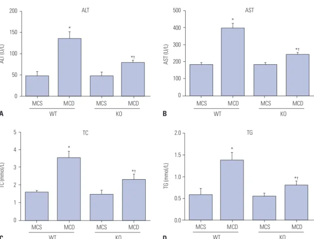
S100A4 knockout improves liver function and blood lipid levels in MCD diet-induced NAFLD mice
After feeding with MCD or MCS for 8 weeks, no significant dif- ferences were found in liver function (ALT and AST) and lipid- related parameters (TG and TC) between mice of WT/MCS group and KO/MCS group (all p>0.05). However, serum levels of the above parameters were obviously higher in MCD diet mice compared to MCS-diet mice; specifically, these factors were relatively lower in mice from KO/MCD group than those of WT/MCD group (all p<0.05) (Fig. 1).
Influences of S100A4 knockout on liver morphology in MCD diet-induced NAFLD mice
As shown in Fig. 2, livers in MCS-diet fed mice exhibited dark red color, smooth and shiny surface, and sharp edges. On the other hand, livers of mice in WT/MCD group had khaki-yellow color, while the edges became blunt with increased volume
and tense capsule, and even coagulation and yellow-white focal degeneration occurred. Livers in mice from KO/MCD group showed slightly yellow color and larger size, along with slightly tough and smooth surface.
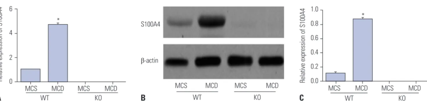
Comparison of S100A4 expressions in mice liver tissue of each group
According to the detection of qRT-PCR and Western blot, S100 A4 mRNA and protein expressions in liver tissues of mice from WT/MCD group were found to be highly upregulated com- pared to those of mice in WT/MCS group (all p<0.05), as shown in Fig. 3, but were not detected in mice from both KO/MCS and KO/MCD groups.
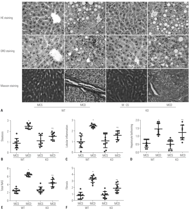
S100A4 knockout attenuates liver tissue injury in MCD diet-induced NAFLD mice
In order to determine the effect of S100A4 KO on liver tissue in- jury in MCD diet-induced NAFLD mice, we performed HE staining, ORO staining, and Masson staining to assess hepatic inflammation and fibrosis by using NAS, to visualize lipid drop- lets, and to observe the distribution of collagen fibers in mice liver tissue of each group, respectively. As illustrated in Fig. 4A, MCS-fed-induced mice had regular structure of hepatic lob- ule, clearly visible hepatic sinus, as well as normal structure and morphology of hepatic cells, without lipid droplets deposi- tion and collagen fibers. However, a large number of fat vacu- oles accumulated in liver tissues of mice in WT/MCD group and hepatocytes showed balloon-like changes accompanied by increased inflammatory cell and collagen fibers. Other 5
4
3
2
1
0
TC (mmol/L)
*†
*
MCS MCD MCS MCD WT KO
TC 200
150
100
50
0
ALT (U/L) *†
*
MCS MCD MCS MCD WT KO
ALT
2.0
1.5
1.0
0.5
0.0
TG (mmol/L)
*†
*
MCS MCD MCS MCD WT KO
TG 500
400
300
200
100
0
AST (U/L)
*†
*
MCS MCD MCS MCD WT KO
AST
A
C
B
D
Fig. 1. S100A4 KO improves liver function and blood lipid-related parameters in MCD diet-induced non-alcoholic fatty liver disease mice. Influences of S100A4 KO on serum ALT (A), AST (B), TC (C), and TG (D) levels in mice. *p<0.05 compared to MCS group, †p<0.05 compared to WT/MCD group. S100A4, S100 calcium binding protein A4; MCD, methionine-choline-deficient; MCS, methionine-choline-sufficient; KO, knockout; WT, wild-type; ALT, amino- transferase; AST, aminotransferase; TG, triglyceride; TC, total cholesterol.
Fig. 2. Effect of S100A4 KO on liver morphology in MCD diet-induced non- alcoholic fatty liver disease mice. S100A4, S100 calcium binding protein A4; KO, knockout; WT, wild-type; MCD, methionine-choline-deficient;
MCS, methionine-choline-sufficient.
WT mice
KO mice
MCS diet MCD diet
than that, reduced liver cell damage and inflammatory cell in- filtration, coupled with a small amount of collagen fibers, were observed in liver tissues of mice in KO/MCD group. Moreover, steatosis, inflammatory infiltration, ballooning, total NAS, and liver fibrosis in KO/MCD group were significantly lower com- pared to those in WT/MCD group (all p<0.05) (Fig. 4B-F).
S100A4 knockout inhibits expressions
of proinflammatory-/profibrogenic cytokines in MCD diet-induced NAFLD mice
qRT-PCR was performed to detect the expression of pro-in- flammatory (including TNF-α, IL-1β, and IL-6) and profibro- genic cytokines (including TGF-β1, COL1A1, and α-SMA) in liv- er tissues of mice, as demonstrated in Fig. 5. As a result, mRNA levels of TNF-α, IL-1β, IL-6, TGF-β1, COL1A1, and α-SMA were significantly higher in mice fed with MCD than those fed with MCS (all p<0.05). As for mice in MCD groups, expressions of proinflammatory-/profibrogenic cytokines in KO mice were significantly lower than those in WT group (all p<0.05).
S100A4 knockout reduces hepatocyte apoptosis in MCD diet-induced NAFLD mice
As evaluated by TUNEL staining in Fig. 6, increased number of TUNEL-positive hepatocytes were discovered in mice of WT+MCD group compared to mice in MCS group, which ex- hibited less and scattered positive hepatocytes, while mice in KO+MCD group showed less positive hepatocytes than those in WT+MCD group. Furthermore, statistical analysis demon- strated that AI of mice in WT/MCD group was markedly ele- vated compared to mice in KO/MCS and WT/MCS groups, but was obviously decreased in mice from KO/MCD group when compared to those in WT/MCD group (all p<0.05).
DISCUSSION
“First hit” refers to several processes, including the digestion and absorption of exogenous lipids, lipoprotein metabolism, as well as conversion and decomposition of cholesterol in liver, to serve as a part of NAFLD pathogenesis.25 Concerning MCD diet-fed mice, liver function indexes (ALT and AST) and blood
lipid parameters (TG and TC) were significantly lower in KO mice than in WT mice, suggesting that S100A4 deletion en- hanced liver function and blood lipid levels in NAFLD mice.
Similarly, patients with type 2 diabetes also had higher serum S100A4 concentrations, which were related to the metabolic pathways.1 This demonstrates an important role of S100A4 in lipid metabolism, possibly since S100A4 could activate the ex- pression of receptor of advanced glycation end products in he- patocytes and hepatic stellate cells (HSCs), and thereby exerting functions in development of NAFLD.26,27
In addition, “second hit” has been widely acknowledged as the pathophysiological model of NAFLD, which is defined as increased oxidative stress and initiation of lipid peroxidation, resulting in the formation of inflammatory mediators and ac- tivation of HSCs to produce irreversible lesions in hepato- cytes.28,29 A recent study demonstrated a significant role of S100A4 in the inflammatory response of diseases.30 In our study, S100A4 KO mice fed with MCD showed noticeably de- creased liver steatosis, inflammation, and ballooning scores, with reduced total NAS and downregulated pro-inflammatory cytokines (including TNF-α, IL-1β, and IL-6) expressions in liv- er tissues, compared to WT mice fed with MCD. As document- ed, TNF-α is a firstly appearing cytokine in the process of liver injury, which could facilitate the release of IL-1β and IL-6, act- ing as a crucial factor in the progress from NAFLD to NASH,31 indicating S100A4 deletion weakened NAFLD inflammation which could be possibly related to the inflammatory response mediated by TLR4 signaling.32 In addition, S100A4 was credit- ed as a fibroblast-specific marker in liver fibrosis. For example, S100A4 was found to be secreted by a subpopulation of macro- phages in fibrotic liver,33 and its increased levels in liver tissue and serum of hepatitis patients were positively correlated with liver fibrosis.34 In our study, less amount of collagen fiber was observed in MCD diet-fed KO mice compared to WT mice, and expressions of pro-fibrogenic cytokines (including TGF-β1, CO- L1A1, and α-SMA) were also significantly reduced, which might have resulted from the overexpression of α-SMA stimulated by S100A4 through c-Myb in HSCs to promote liver fibrosis,33 or a common mediator of S100A4 in epithelial-mesenchymal transi- tion,35 thus contributing to the cirrhosis progression in NAFLD.36 In recent years, “third hit” of NAFLD was pointed out, name- 6
4
2
0
1.0 0.8 0.6 0.4 0.2
Relative expression of S100A4 Relative expression of S100A4 0.0
*
MCS MCD MCS MCD WT KO
S100A4
β-actin
MCS MCD MCS MCD WT KO
MCS MCD MCS MCD WT KO
*
A B C
Fig. 3. Relative S100A4 mRNA and protein expressions in liver tissues of mice detected by qRT-PCR (A) and Western blot (B and C). *p<0.05 compared to WT/MCS group. S100A4, S100 calcium binding protein A4; MCD, methionine-choline-deficient; MCS, methionine-choline-sufficient; KO, knockout; WT, wild-type.
ly hepatocyte apoptosis, which could accelerate the transfor- mation of NAFLD into liver cirrhosis.4 In NAFLD patients, he- patocyte proliferation was blocked, and apoptotic cells was replaced by proliferation and differentiation of hepatic progeni-
tor cells, leading to hepatic fibrosis and inflammatory cell infil- tration, and so on.37 In our research, S100A4 KO reduced hepa- tocyte apoptosis in MCD diet-induced mice, showing that S100A4 deficiency might play a protective role in NAFLD via
Fig. 4. S100A4 KO reduces liver tissue damage and inflammatory cell infiltration in MCD diet-induced NAFLD mice. (A) HE staining, ORO staining, and Masson staining were used to observe pathological changes of liver tissues in each group (×400); (B-E) Comparison of steatosis (B), lobular inflammation (C), hepatocyte ballooning (D), total NAS (E), and fibrosis (F) in liver tissues of mice in each group. *p<0.05 compared to MCS group, †p<0.05 compared to WT/MCD group. S100A4, S100 calcium binding protein A4; MCD, methionine-choline-deficient; MCS, methionine-choline-sufficient; KO, knockout;
WT, wild-type; HE, hematoxylin and eosin; ORO, Oil Red O; NAFLD, non-alcoholic fatty liver; NAS, NAFLD activity scores.
3
2
1
0
8
6
4 2
0
5 4 3 2 1 0 3
2
1
0
2.0
1.5 1.0
0.5
0.0
SteatosisTotal NAS FibrosisLobular inflammation Hepatocyte ballooning
ORO staining HE staining
Masson staining
MCS MCD M CS MCD WT KO
MCS MCD MCS MCD MCS MCD MCS MCD MCS MCD MCS MCD
MCS MCD MCS MCD MCS MCD MCS MCD
WT KO WT KO WT KO
WT KO WT KO
*† *†
*†
*†
*†
* *
*
* *
A
B
E F
C D
inhibiting hepatocyte apoptosis. A possible reason could be correlated with elevated levels of WT p53,38 which enhances TGF-β-induced p66Shc signaling, ROS accumulation, and he- patocyte apoptosis.39 On the contrary, S100A4 KO could also lead to the stabilization of p53 protein in two p53 WT cell lines (A549 and HeLa), which implies that S100A4 could accelerate p53 degradation.40 Therefore, whether S100A4 can affect hepa- tocyte apoptosis in NAFLD mice via regulation of p53 should be further explored, and we plan to deeply investigate this mat- ter in our subsequent and future studies. In summary, S100A4 was upregulated in NAFLD mice, and S100A4 KO significantly improved liver function and blood lipid levels, contributing to reduced hepatic fibrosis and inflammation as well as inhibited hepatocyte apoptosis in MCD diet-induced NAFLD mice, which provided new clues for the treatment of NAFLD.
ORCID
De-Ping Ding https://orcid.org/0000-0002-4481-5514
REFERENCES
1. Arner P, Petrus P, Esteve D, Boulomié A, Näslund E, Thorell A, et al.
Screening of potential adipokines identifies S100A4 as a marker of pernicious adipose tissue and insulin resistance. Int J Obes (Lond) 2018 Jan 30 [Epub]. https://doi.org/10.1038/s41366-018-0018-0.
2. Afonso MB, Rodrigues PM, Simão AL, Castro RE. Circulating mi- croRNAs as potential biomarkers in non-alcoholic fatty liver dis- ease and hepatocellular carcinoma. J Clin Med 2016;5:30.
3. Nair S. Nonalcoholic Fatty liver disease from the perspective of an internist. Ochsner J 2002;4:92-7.
4. Patel A, Harrison SA. Hepatitis C virus infection and nonalcoholic steatohepatitis. Gastroenterol Hepatol (N Y) 2012;8:305-12.
5. Dongiovanni P, Valenti L. Genetics of nonalcoholic fatty liver dis- ease. Metabolism 2016;65:1026-37.
6. Saleh A, Kamel L, Ghali A, Ismail A, El Khayat H. Serum levels of astroglial S100-beta and neuron-specific enolase in hepatic en- cephalopathy patients. East Mediterr Health J 2007;13:1114-23.
7. Dempsey BR, Rintala-Dempsey AC, Shaw GS. S100 proteins. In:
Choi S, editor. Encyclopedia of signaling molecules. New York (NY):
Springer; 2012. p.1711-7.
8. Mukai K, Miyagi T, Nishio K, Yokoyama Y, Yoshioka T, Saito Y, et al.
S100A8 production in CXCR2-expressing CD11b+Gr-1high cells aggravates hepatitis in mice fed a high-fat and high-cholesterol diet. J Immunol 2016;196:395-406.
MCS MCD MCS MCD WT KO WT mice
KO mice
MCS diet MCD diet
8
6
4
2
0
Apoptotic index (%)
*
*† 8
6
4
2
0
8
6
4
2
0
Relative mRNA expression Relative mRNA expression
*†
*†
*†
*† *† *†
*
*
* *
* *
TNF-α IL-1β IL-6 TGF-β1 COL1A1 α-SMA WT/MCS
WT/MCD KO/MCS KO/MCD
WT/MCS WT/MCD KO/MCS KO/MCD
A B
Fig. 5. S100A4 KO on expressions of proinflammatory cytokines (TNF-α, IL-1β, and IL-6) (A) and profibrogenic cytokines (TGF-β1, COL1A1 and α-SMA) (B) in liver tissues of MCD diet-induced non-alcoholic fatty liver disease mice detected by qRT-PCR. *p<0.05 compared to MCS group, †p<0.05 compared to WT/MCD group. S100A4, S100 calcium binding protein A4; MCD, methionine-choline-deficient; MCS, methionine-choline-sufficient; KO, knockout;
WT, wild-type.
Fig. 6. S100A4 KO on hepatocyte apoptosis in MCD diet-induced non-alcoholic fatty liver disease mice evaluated by TUNEL staining (×400). Black arrows indicate hepatocyte apoptosis. *p<0.05 compared to MCS group, †p<0.05 compared to WT/MCD group. S100A4, S100 calcium binding protein A4; MCD, methionine-choline-deficient; MCS, methionine-choline-sufficient; KO, knockout; WT, wild-type.
9. Liu X, Wang Y, Ming Y, Song Y, Zhang J, Chen X, et al. S100A9: a potential biomarker for the progression of non-alcoholic fatty liver disease and the diagnosis of non-alcoholic steatohepatitis. PLoS One 2015;10:e0127352.
10. Malashkevich VN, Dulyaninova NG, Ramagopal UA, Liriano MA, Varney KM, Knight D, et al. Phenothiazines inhibit S100A4 func- tion by inducing protein oligomerization. Proc Natl Acad Sci U S A 2010;107:8605-10.
11. Hou S, Tian T, Qi D, Sun K, Yuan Q, Wang Z, et al. S100A4 promotes lung tumor development through β-catenin pathway-mediated autophagy inhibition. Cell Death Dis 2018;9:277.
12. Oslejsková L, Grigorian M, Gay S, Neidhart M, Senolt L. The me- tastasis associated protein S100A4: a potential novel link to inflam- mation and consequent aggressive behaviour of rheumatoid ar- thritis synovial fibroblasts. Ann Rheum Dis 2008;67:1499-504.
13. Cerezo LA, Kuncová K, Mann H, Tomcík M, Zámecník J, Lukanidin E, et al. The metastasis promoting protein S100A4 is increased in idiopathic inflammatory myopathies. Rheumatology (Oxford) 2011;
50:1766-72.
14. Louka ML, Ramzy MM. Involvement of fibroblast-specific protein 1 (S100A4) and matrix metalloproteinase-13 (MMP-13) in CCl4- induced reversible liver fibrosis. Gene 2016;579:29-33.
15. Wree A, Broderick L, Canbay A, Hoffman HM, Feldstein AE. From NAFLD to NASH to cirrhosis-new insights into disease mecha- nisms. Nat Rev Gastroenterol Hepatol 2013;10:627-36.
16. Okubo H, Kushiyama A, Sakoda H, Nakatsu Y, Iizuka M, Taki N, et al. Involvement of resistin-like molecule β in the development of methionine-choline deficient diet-induced non-alcoholic steato- hepatitis in mice. Sci Rep 2016;6:20157.
17. Rizki G, Arnaboldi L, Gabrielli B, Yan J, Lee GS, Ng RK, et al. Mice fed a lipogenic methionine-choline-deficient diet develop hyper- metabolism coincident with hepatic suppression of SCD-1. J Lip- id Res 2006;47:2280-90.
18. Rinella ME, Elias MS, Smolak RR, Fu T, Borensztajn J, Green RM.
Mechanisms of hepatic steatosis in mice fed a lipogenic methio- nine choline-deficient diet. J Lipid Res 2008;49:1068-76.
19. Wang Y, Li J, Zhuge L, Su D, Yang M, Tao S, et al. Comparison be- tween the efficacies of curcumin and puerarin in C57BL/6 mice with steatohepatitis induced by a methionine- and choline-defi- cient diet. Exp Ther Med 2014;7:663-8.
20. Kim SB, Kang OH, Lee YS, Han SH, Ahn YS, Cha SW, et al. Hepato- protective effect and synergism of bisdemethoycurcumin against MCD diet-induced nonalcoholic fatty liver disease in mice. PLoS One 2016;11:e0147745.
21. National Research Council. Guide for the care and use of laboratory animals. 8th ed. Washington (DC): National Academies Press; 2011.
22. EL Naaman C, Grum-Schwensen B, Mansouri A, Grigorian M, Santoni-Rugiu E, Hansen T, et al. Cancer predisposition in mice deficient for the metastasis-associated Mts1(S100A4) gene. On- cogene 2004;23:3670-80.
23. Alam S, Alam M, Alam SMNE, Chowdhury ZR, Kabir J. Prevalence and predictor of nonalcoholic steatohepatitis (NASH) in nonalco- holic fatty liver disease (NAFLD). J Bangladesh Coll Phys Surg 2014;
32:71-7.
24. García-Galiano D, Sánchez-Garrido MA, Espejo I, Montero JL,
Costán G, Marchal T, et al. IL-6 and IGF-1 are independent prog- nostic factors of liver steatosis and non-alcoholic steatohepatitis in morbidly obese patients. Obes Surg 2007;17:493-503.
25. Ying LI, Hong ZF. Hypothesis of “two hits” in NAFLD. Medical Re- capitulate 2013;19:594-6.
26. Abu El-Asrar AM, Nawaz MI, De Hertogh G, Alam K, Siddiquei MM, Van den Eynde K, et al. S100A4 is upregulated in proliferative diabetic retinopathy and correlates with markers of angiogenesis and fibrogenesis. Mol Vis 2014;20:1209-24.
27. Takeuchi M, Takino JI, Sakasai-Sakai A, Takata T, Tsutsumi M.
Toxic AGE (TAGE) theory for the pathophysiology of the onset/
progression of NAFLD and ALD. Nutrients 2017;9:634.
28. Tilg H, Moschen AR. Evolution of inflammation in nonalcoholic fatty liver disease: the multiple parallel hits hypothesis. Hepatolo- gy 2010;52:1836-46.
29. Berlanga A, Guiu-Jurado E, Porras JA, Auguet T. Molecular path- ways in non-alcoholic fatty liver disease. Clin Exp Gastroenterol 2014;7:221-39.
30. Klingelhöfer J, Senolt L, Baslund B, Nielsen GH, Skibshøj I, Pavelka K, et al. Up-regulation of metastasis-promoting S100A4 (Mts-1) in rheumatoid arthritis: putative involvement in the pathogenesis of rheumatoid arthritis. Arthritis Rheum 2007;56:779-89.
31. Tilg H, Moschen AR, Szabo G. Interleukin-1 and inflammasomes in alcoholic liver disease/acute alcoholic hepatitis and nonalcohol- ic fatty liver disease/nonalcoholic steatohepatitis. Hepatology 2016;
64:955-65.
32. Sharifnia T, Antoun J, Verriere TG, Suarez G, Wattacheril J, Wilson KT, et al. Hepatic TLR4 signaling in obese NAFLD. Am J Physiol Gastrointest Liver Physiol 2015;309:G270-8.
33. Chen L, Li J, Zhang J, Dai C, Liu X, Wang J, et al. S100A4 promotes liver fibrosis via activation of hepatic stellate cells. J Hepatol 2015;
62:156-64.
34. Yan LB, Zhang QB, Zhu X, He M, Tang H. Serum S100 calcium binding protein A4 improves the diagnostic accuracy of transient elastography for assessing liver fibrosis in hepatitis B. Clin Res Hepatol Gastroenterol 2018;42:64-71.
35. Schneider M, Hansen JL, Sheikh SP. S100A4: a common mediator of epithelial-mesenchymal transition, fibrosis and regeneration in diseases? J Mol Med (Berl) 2008;86:507-22.
36. Syn WK, Jung Y, Omenetti A, Abdelmalek M, Guy CD, Yang L, et al.
Hedgehog-mediated epithelial-to-mesenchymal transition and fi- brogenic repair in nonalcoholic fatty liver disease. Gastroenterolo- gy 2009;137:1478-88.e8.
37. Maher JJ. Pathogenesis of NAFLD and NASH. In: Chalasani N, Sza- bo G, editors. Alcoholic and non-alcoholic fatty liver disease.
Cham: Springer; 2016. p.71-101.
38. Mazzucchelli L. Protein S100A4: too long overlooked by patholo- gists? Am J Pathol 2002;160:7-13.
39. Tomita K, Teratani T, Suzuki T, Oshikawa T, Yokoyama H, Shi- mamura K, et al. p53/p66Shc-mediated signaling contributes to the progression of non-alcoholic steatohepatitis in humans and mice. J Hepatol 2012;57:837-43.
40. Orre LM, Panizza E, Kaminskyy VO, Vernet E, Gräslund T, Zhivot- ovsky B, et al. S100A4 interacts with p53 in the nucleus and pro- motes p53 degradation. Oncogene 2013;32:5531-40.