• 검색 결과가 없습니다.

Comparison of mandibular arch forms of Korean and Vietnamese patients by using facial axis points on three-dimensional models

N/A
N/A
Protected

Academic year: 2022

Share "Comparison of mandibular arch forms of Korean and Vietnamese patients by using facial axis points on three-dimensional models"

Copied!
6
0
0

로드 중.... (전체 텍스트 보기)

전체 글

(1)

Comparison of mandibular arch forms of Korean and Vietnamese patients by using facial axis points on three-dimensional models

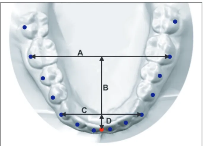
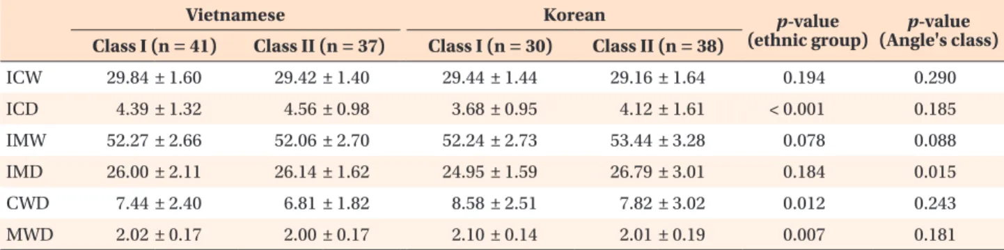
Objective: This study was aimed at comparing the mandibular arch forms of Korean and Vietnamese patients by using facial axis (FA) points on three- dimensional (3D) models. Methods: Mandibular casts of 68 Korean (Class I malocclusion, 30; Class II malocclusion, 38) and 78 Vietnamese (Class I malocclusion, 41; Class II malocclusion, 37) patients were scanned in their occluded positions and grouped according to arch form (tapered, ovoid, and square). The FA point of each tooth was digitized on the 3D mandibular models.

The measurements and frequency distributions of the arch forms were compared between the ethnic groups. Results: The Vietnamese patients had significantly greater intercanine depth and intercanine and intermolar width-to-depth ratios than the Korean patients (p < 0.05). The frequency distributions of the arch forms were also significantly different (p = 0.038), but no sexual dimorphism was found. Conclusions: Vietnamese people tend to have deeper and wider arches than Korean people. The three arch forms are evenly distributed in Korean people, but Vietnamese people frequently have square arches. Clinicians should identify the correct arch form of an ethnic group before initiating orthodontic treatment.

[Korean J Orthod 2013;43(6):288-293]

Key words: Arch form, Digital models, Ethnic norms, Vietnamese, Korean Kil-jun Lee

a

Vu Thi Thu Trang

b

Mohamed Bayome

c,d

Jae Hyun Park

e,f

Yong Kim

g

Yoon-Ah Kook

h

a

Graduate School of Clinical Dental Science, The Catholic University of Korea, Seoul, Korea

b

Graduate School of The Catholic University of Korea, Seoul, Korea

c

The Catholic University of Korea, Seoul, Korea

d

Department of Postgraduate Studies, Universidad Autonoma del Paraguay, Asuncion, Paraguay

e

Postgraduate Orthodontic Program, Arizona School of Dentistry and Oral Health, A.T. Still University, Mesa, AZ, USA

f

Graduate School of Dentistry, Kyung Hee University, Seoul, Korea

g

Private Practice, Seoul, Korea

h

Department of Orthodontics, Seoul St. Mary’s Hospital, The Catholic University of Korea, Seoul, Korea

Received June 3, 2013; Revised July 15, 2013; Accepted July 16, 2013.

Corresponding author: Yoon-Ah Kook.

Professor, Department of Orthodontics, Seoul St. Mary’s Hospital, The Catholic University of Korea, 222 Banpo-daero, Seocho-gu, Seoul 137-701, Korea.

Tel +82-2-2258-1776 e-mail [email protected]

*This study was partly supported by the alumni fund of the Department of Dentistry and Graduate School of Clinical Dental Science, The Catholic University of Korea.

© 2013 The Korean Association of Orthodontists.

The authors report no commercial, proprietary, or financial interest in the products or companies described in this article.

This is an Open Access article distributed under the terms of the Creative Commons Attribution Non-Commercial License (http://creativecommons.org/licenses/by-nc/3.0) which permits unrestricted non-commercial use, distribution, and reproduction in any medium, provided the original work is properly cited.

pISSN 2234-7518 • eISSN 2005-372X

http://dx.doi.org/10.4041/kjod.2013.43.6.288

(2)

INTRODUCTION

Nowadays, clinicians tend to apply lighter forces during orthodontic treatment by means of self-ligating brackets and small superelastic archwires. However, because superelastic archwires are preformed, they may be dif- ficult to adapt to a patient’s unique arch form. Failure to maintain the arch form increases the probability of relapse.

1-6

In this regard, correct identification of the arch form is very important for achieving a stable, functional, and esthetic outcome.

Several classification methods using incisal edges and cuspal tips as landmarks have been developed to eva- lu ate arch forms.

1-7

Clinical bracket points have also been used as landmarks for mandibular arch form as- sess ment.

8,9

Unfortunately, these methods are not suffi- ciently accurate to allow proper placement of supe- relastic archwires. On the other hand, Camporesi et al.

10

evaluated the average arch form through three- dimen sional (3D) derived facial axis (FA) points, which cor respond to bracket points and are more relevant to clinical practice. Further, Bayome et al.

11

suggested a new classification of arch form based on FA points.

Arch size and arch form tend to differ among ethnic groups.

7-9,12-18

The distribution and dimensions of the arch forms of Korean subjects were found to be dif- ferent from those of other ethnic groups.

8,9,11,12,14

Never- theless, the average arch form of Vietnamese peo ple, who originated from a homogeneous ethnic back ground to Korean people in Southeast Asia, has not yet been assessed. Moreover, no comparative studies of arch forms have been conducted between these ethnic groups by using FA points as landmarks on 3D models.

The purpose of this study was to compare the mandi- bular arch forms of Korean and Vietnamese patients by using FA points on 3D models.

MATERIALS AND METHODS

Subjects

The Korean population consisted of 68 patients (mean

age, 22.6 years), with Class I (n = 30) and Class II (n = 38) malocclusion, from the Department of Orthodontics, Seoul St. Mary’s Hospital, The Catholic University of Korea (Seoul, Korea). The Vietnamese population comprised 78 patients (mean age, 17.3 years), with Class I (n = 41) and Class II (n = 37) malocclusion, from the Department of Orthodontic, Faculty of Odonto- stomatology Medicine, Hanoi University, and private clinics in Hanoi and Ho Chi Minh City. The institutional review board of The Catholic University of Korea ap- proved this study (MC12SSS10073).

The inclusion criteria were as follows: complete per- manent dentition excluding the third molars; normal tooth size and shape; absence of supernumerary teeth;

3 mm or less of arch length discrepancy; no restorations extending to the contact areas, cuspal tips (incisal edges), or cervical areas; and no previous orthodontic treatment.

Digitization

Mandibular casts of the subjects were scanned in their occluded relationships by using a 3D laser scanner (KOP- 300; Orapix Co. Ltd., Seoul, Korea) at 20-μm resolution.

The FA point

19

of each tooth was digitized on the 3D models by using Rapidform 2006 software (INUS Tech- nology Inc., Seoul, Korea) (Figure 1).

The transverse and anteroposterior directions were set as the X-axis and Y-axis, respectively, and the line per- pen dicular to these planes was marked as the Z-axis.

The contact point of the central incisors was considered the origin of the coordinates. The original X-axis was adjusted so that it was parallel to the mean inclinations of the lines connecting the bilateral contact points of the first and second premolars (line A) and the second premolars and first molars (line B). The Z-axis values were nullified (Figure 2).

Measurements

Four linear measurements were calculated for each arch form (Figure 3): intercanine width, the distance between the canine FA points; intermolar width, the

Figure 1. Facial axis (FA) points on a digitized mandibular model. The red dots represent the FA points of the (A) central

incisor and (B) first molar.

(3)

dis tance between the first molar FA points; intercanine depth, the shortest distance from the line connecting the canine FA points to the origin; and intermolar depth, the shortest distance from the line connecting the first molar FA points to the origin. The intercanine and intermolar width-to-depth ratios were also calculated.

In addition, arch form images were printed on a 1:1 scale and compared with arch form templates (OrthoForm; 3M Unitek, Monrovia, CA, USA) to select the arch form that best fit the eight FA points from the right to the left first premolars. The subjects were then grouped according to the arch form (tapered, ovoid, and square) and the frequency distributions of each arch form in both the ethnic groups were calculated.

To assess the measurement error, 10 randomly selec-

ted models from each group were analyzed after two weeks to evaluate intra-operator variability. The intra- class correlation (ICC) test revealed high reliability bet- ween the assessments (ICC > 0.8). The arch forms were also classified twice by the same examiner. The dif- ference between the evaluations was analyzed by the Wilcoxon signed-rank test and found to be negligible (p > 0.05). Therefore, the first classification was used.

For confirmation, a second examiner classified 30 arch forms from each ethnicity. The agreement between the examiners was high (kappa = 0.898). Both the examiners were blinded to the groups.

Statistical analysis

Statistical evaluation was performed with SPSS 16.0 software (SPSS Inc., Chicago, IL, USA). The chi-square test was used to analyze the association between arch form and ethnicity. Multivariate analysis of covariance (MANCOVA) was performed to compare the gender-ad- justed means of the arch measurements according to ethnicity, arch form, and Angle’s classification of malo- cclusion. All analyses were tested at the significance level of 0.05.

RESULTS

Square arches were the most common in the Korean and Vietnamese groups (44% and 62%, respectively), followed by ovoid (29% and 27%, respectively) and tapered (27% and 12%, respectively) arches (Figure 4).

The frequency distributions of the arch forms within the ethnic groups were significantly different (p = 0.038;

Table 1). Square arches were significantly more common in the Vietnamese group (p < 0.001). The Korean group showed even distribution of the three arch forms (p = Figure 2. The X-axis reorientation method. The X-axis was

adjusted so that it was parallel to the mean inclinations (C) of line A (the line connecting the bilateral contact points of the first and second premolars) and line B (the line connecting the bilateral contact points of the second premolars and first molars).

Figure 3. Arch measurements. A, Intermolar width; B, intermolar depth; C, intercanine width; D, intercanine depth. The blue dots represent FA points and the red dot represents the origin.

Figure 4. Frequency distributions of the arch forms

according to Angle’s classification of malocclusion and

ethnicity.

(4)

0.161).

In the multivariate analysis of the arch measurements according to ethnicity and Angle’s classification of malo cclusion, arch form (p < 0.001), ethnicity (p = 0.001), and type of malocclusion (p = 0.024) had signi- ficant main effects. Gender showed no significant effect (p = 0.19). Further, no significant interaction was noted between ethnicity and type of malocclusion (p = 0.056). Intercanine and intermolar widths showed

no significant differences according to ethnicity and type of malocclusion. However, intercanine depth and intercanine and intermolar width-to-depth ratios demon strated significant differences between the ethnic groups. Intermolar depth was significantly different according to Angle’s classification of malocclusion (Table 2).

In the multivariate analysis of the arch measurements according to arch form and ethnicity, both arch form (p

< 0.001) and ethnicity (p = 0.002) had significant main effects. Gender (p = 0.16) and Angle's classification of malocclusion (p = 0.23) had no significant effects. No significant interaction was observed between ethnicity and arch form (p = 0.054). Further, intercanine width showed no significant difference, but intercanine depth and intercanine and intermolar width-to-depth ratios again demonstrated significant differences. Intermolar depth and width were also significantly different: the Vietnamese group had significantly narrower intermolar width in the tapered and ovoid arches, and the square Table 1. Frequency distributions of the arch forms in the

Korean and Vietnamese groups Arch form, n (%)

Total Tapered Ovoid Square

Vietnamese 9 (11.5) 21 (26.9) 48 (61.5) 78 Korean 18 (26.5) 20 (29.4) 30 (44.1) 68 Pearson chi-square = 6.52; p = 0.038.

Table 2. Comparison of the arch measurements according to ethnicity and Angle’s classification of malocclusion

Vietnamese Korean p-value

(ethnic group) p-value (Angle's class) Class I (n = 41) Class II (n = 37) Class I (n = 30) Class II (n = 38)

ICW 29.84 ± 1.60 29.42 ± 1.40 29.44 ± 1.44 29.16 ± 1.64 0.194 0.290

ICD 4.39 ± 1.32 4.56 ± 0.98 3.68 ± 0.95 4.12 ± 1.61 < 0.001 0.185

IMW 52.27 ± 2.66 52.06 ± 2.70 52.24 ± 2.73 53.44 ± 3.28 0.078 0.088

IMD 26.00 ± 2.11 26.14 ± 1.62 24.95 ± 1.59 26.79 ± 3.01 0.184 0.015

CWD 7.44 ± 2.40 6.81 ± 1.82 8.58 ± 2.51 7.82 ± 3.02 0.012 0.243

MWD 2.02 ± 0.17 2.00 ± 0.17 2.10 ± 0.14 2.01 ± 0.19 0.007 0.181

Values are presented as mean ± standard deviation.

Gender and arch form were covariates in multivariate analysis of covariance.

ICW, Intercanine width; ICD, intercanine depth; IMW, intermolar width; IMD, intermolar depth; CWD, intercanine width-to- depth ratio; MWD, intermolar width-to-depth ratio.

Table 3. Comparison of the arch measurements according to ethnicity and arch form

Vietnamese Korean p-value

(ethnic group)

p-value (arch form) Tapered

(n = 9) Ovoid

(n = 21) Square

(n = 48) Tapered

(n = 18) Ovoid

(n = 20) Square (n = 29)

ICW 29.30 ± 1.92 29.45 ± 1.47 29.79 ± 1.46 29.34 ± 1.51 29.04 ± 1.40 29.36 ± 1.71 0.385 0.846 ICD 4.84 ± 1.30 4.65 ± 1.01 4.32 ± 1.20 4.85 ± 1.61 3.99 ± 0.93 3.44 ± 1.03 0.007 0.001 IMW 50.15 ± 2.65 50.75 ± 1.94 53.17 ± 2.48 53.28 ± 3.14 53.04 ± 3.53 52.56 ± 2.85 0.005 0.117 IMD 26.14 ± 3.01 26.36 ± 1.43 25.93 ± 1.83 27.18 ± 2.71 26.90 ± 2.79 24.69 ± 1.81 0.900 0.003 CWD 6.68 ± 2.83 6.64 ± 1.58 7.45 ± 2.22 6.70 ± 2.25 7.67 ± 1.80 9.40 ± 3.19 0.010 0.001 MWD 1.94 ± 0.21 1.93 ± 0.13 2.06 ± 0.16 1.97 ± 0.15 1.98 ± 0.14 2.14 ± 0.17 0.040 < 0.001 Values are presented as mean ± standard deviation.

Gender and Angle’s classification of malocclusion were covariates in multivariate analysis of covariance.

ICW, Intercanine width; ICD, intercanine depth; IMW, intermolar width; IMD, intermolar depth; CWD, intercanine width-to-

depth ratio; MWD, intermolar width-to-depth ratio.

(5)

arches of the Korean group showed significantly less intermolar depth (Table 3).

DISCUSSION

Great technologic strides in communication and trans- portation over the past few decades have contributed to rapid globalization and widespread flow of people.

Therefore, it is appropriate to examine and compare the morphologic features of ethnic groups from various parts of the world.

The population of Vietnam is reportedly 92.5 million.

20

The Vietnamese population in Korea has reached over 160,000 and is rapidly increasing.

21

However, until now, research on Vietnamese arch forms has not been conducted. Therefore, we aimed to identify the ave rage arch form of Vietnamese people and clarify the mor- phologic differences in the mandibular arches bet ween Vietnamese and Korean populations.

McLaughlin and Bennett

5

classified arch forms into 3 main types: tapered, ovoid, and square. Several mathe- matical methods have been applied to depict arch forms.

1,10,11

However, the accuracy of arch form iden ti fi- cation by using incisal edges and cuspal tips

1,2,7-9,22-28

or clinical bracket points as landmarks on two-dimensional photocopies of casts is doubtful because of the failure to digitize FA points. In our study, FA points were used on 3D models for evaluating and classifying arch forms because of their high accuracy.

29,30

In this study, the Vietnamese population had greater arch depth but not significantly different arch width from the Korean population. Kook et al.

8

showed that Cau casians have a narrow arch but not significantly dif- ferent arch depth compared with Korean people, which suggests that Vietnamese people have the deepest arches and Caucasians have the narrowest ones among the three ethnic groups.

Previous studies demonstrated that the most common arch form is tapered in Caucasians (43.8%), square in Korean and Japanese people (46.7% and 45.6%, res- pec tively), and ovoid in Israeli people (50.7%).

8,9,15

Meanwhile, Bayome et al.

12

reported an even distribution of the arch forms in Egyptians. In our study, square arches were the most common in the Vietnamese group (62%), followed by the ovoid and tapered arches; on the other hand, the Korean subjects showed an even distribution of the arch forms. These differences are attributable to genetic and environmental factors. Class III malocclusion cases were not included in this study;

this type of malocclusion might have decreased the percentage of the square arch form.

This study was the first comparison of Vietnamese and Korean arch forms. The sample size was not large enough to evaluate the frequency distributions of

the arch forms in Class I and II malocclusion cases.

Further, Class III malocclusion cases are difficult to find in Vietnamese people. Further research using cone- beam computed tomography in a larger sample size including Class III malocclusion cases is recommended for evaluating the clinical and basal arch forms.

CONCLUSION

Vietnamese people tend to have deeper and wider arches than Korean people. The three arch forms are evenly distributed in Korean people, but Vietnamese people frequently have square arches. Clinicians should identify the arch form of each ethnic group before ini- tiating orthodontic treatment.

ACKNOWLEDGEMENTS

The authors thank Orapix Co., Ltd. (Seoul, Korea) for their assistance with the 3D digital technology.

REFERENCES

1. Ferrario VF, Sforza C, Miani A Jr, Tartaglia G. Mathe- matical definition of the shape of dental arches in human permanent healthy dentitions. Eur J Orthod 1994;16:287-94.

2. Merz ML, Isaacson RJ, Germane N, Rubenstein LK.

Tooth diameters and arch perimeters in a black and a white population. Am J Orthod Dentofacial Orthop 1991;100:53-8.

3. de la Cruz A, Sampson P, Little RM, Artun J, Sha piro PA. Long-term changes in arch form after ortho- dontic treatment and retention. Am J Orthod Den- tofacial Orthop 1995;107:518-30.

4. Felton JM, Sinclair PM, Jones DL, Alexander RG.

A computerized analysis of the shape and stability of mandibular arch form. Am J Orthod Dentofacial Orthop 1987;92:478-83.

5. McLaughlin RP, Bennett JC. Arch form conside- rations for stability and esthetics. Rev Esp Ortod 1999;29:216-33.

6. Shapiro PA. Mandibular dental arch form and di- men sion. Treatment and postretention changes. Am J Orthod 1974;66:58-70.

7. Nummikoski P, Prihoda T, Langlais RP, McDavid WD, Welander U, Tronje G. Dental and mandibular arch widths in three ethnic groups in Texas: a radio- graphic study. Oral Surg Oral Med Oral Pathol 1988;

65:609-17.

8. Kook YA, Nojima K, Moon HB, McLaughlin RP,

Sinclair PM. Comparison of arch forms between

Korean and North American white populations. Am

J Orthod Dentofacial Orthop 2004;126:680-6.

(6)

9. Nojima K, McLaughlin RP, Isshiki Y, Sinclair PM.

A comparative study of Caucasian and Japanese mandibular clinical arch forms. Angle Orthod 2001;

71:195-200.

10. Camporesi M, Franchi L, Baccetti T, Antonini A.

Thin-plate spline analysis of arch form in a Sou- thern European population with an ideal natural occlusion. Eur J Orthod 2006;28:135-40.

11. Bayome M, Han SH, Choi JH, Kim SH, Baek SH, Kim DJ, et al. New clinical classification of dental arch form using facial axis points derived from three- dimensional models. Aust Orthod J 2011;27:117-24.

12. Bayome M, Sameshima GT, Kim Y, Nojima K, Baek SH, Kook YA. Comparison of arch forms between Egyptian and North American white populations.

Am J Orthod Dentofacial Orthop 2011;139:e245-52.

13. Burris BG, Harris EF. Maxillary arch size and shape in American blacks and whites. Angle Orthod 2000;

70:297-302.

14. Collins BP, Harris EF. Arch form in American blacks and whites with malocclusions. J Tenn Dent Assoc 1998;78:15-8.

15. Gafni Y, Tzur-Gadassi L, Nojima K, McLaughlin RP, Abed Y, Redlich M. Comparison of arch forms between Israeli and North American white popu- lations. Am J Orthod Dentofacial Orthop 2011;

139:339-44.

16. Kasai K, Kanazawa E, Aboshi H, Richards LC, Mat- suno M. Dental arch form in three Pacific popu la- tions: a comparison with Japanese and Austra lian aboriginal samples. J Nihon Univ Sch Dent 1997;

39:196-201.

17. Koyoumdjisky-Kaye E, Zilberman Y, Zeevi Z. A comparative study of tooth and dental arch dimen- sions in Jewish children of different ethnic descent.

I. Kurds and Yemenites. Am J Phys Anthropol 1976;44:437-43.

18. Othman SA, Xinwei ES, Lim SY, Jamaludin M, Mohamed NH, Yusof ZY, et al. Comparison of arch form between ethnic Malays and Malaysian Abori- gines in Peninsular Malaysia. Korean J Orthod 2012;

42:47-54.

19. Andrews L. Straight wire - the concept and appli- ance. San Diego: LA Wells; 1989.

20. Country Comparison: Population [Internet]. Wa-

shington, D.C: Central Intelligence Agency. 2013 [cited Jul, 2013]. Available from: https://www.

cia.gov/ library/publications/the-world-factbook/

rankorder/ 2119rank.html?countryName=Vietnam&c ountryCode=vm&regionCode=eas&rank=14#vm.

21. More than 1.4 million foreign residents living in Korea [Internet]. Seoul, Korea: Ministry of Security and Public Administration. 2012 [cited Jul, 2013].

Ava il able from: http://www.mospa.go.kr/gpms/ns/

mogaha/user/userlayout/bulletin/userBtView.ac- tion?userBtBean.bbsSeq=1022589&userBtBean.

ctxCd=1012&userBtBean.ctxType=21010002&

userBtBean.categoryCd.

22. Cordato MA. A simple mathematical study of an- te rior dental relations: Part I. Aust Orthod J 1995;

13:249-52.

23. Cordato MA. A mathematical study of anterior dental relations: Part II, Incisor and canine overjet.

Aust Orthod J 1996;14:143-9.

24. Cordato MA. A simple mathematical study of an- terior dental relations. Part III: incisor and canine over bite. Aust Orthod J 1998;15:75-84.

25. Ferrario VF, Sforza C, Miani A Jr, Tartaglia G.

Maxillary versus mandibular arch form differences in human permanent dentition assessed by Eucli- dean-distance matrix analysis. Arch Oral Biol 1994;

39:135-9.

26. Kim SC. A study on the configurations of Korean normal dental arches for preformed arch wire.

Korean J Orthod 1984;14:93-101.

27. Lee YC, Park YC. A study on the dental arch by occlusogram in normal occlusion. Korean J Orthod 1987;17:279-87.

28. Yun YK, Kook YA, Kim SH, Mo SS, Cha KS, Kim JG, et al. Mandibular clinical arch forms in Koreans with normal occlusions. Korean J Orthod 2004;34:481-7.

29. Kook YA, Bayome M, Park SB, Cha BK, Lee YW, Baek SH. Overjet at the anterior and posterior segments:

three-dimensional analysis of arch coordination.

Angle Orthod 2009;79:495-501.

30. Kim BI, Bayome M, Kim Y, Baek SH, Han SH, Kim

SH, et al. Comparison of overjet among 3 arch

types in normal occlusion. Am J Orthod Dentofacial

Orthop 2011;139:e253-60.

수치

Figure 1. Facial axis (FA) points on a digitized mandibular model. The red dots represent the FA points of the (A) central  incisor and (B) first molar.
Figure  4.  Frequency  distributions  of  the  arch  forms  according to Angle’s classification of malocclusion and  ethnicity
Table 3. Comparison of the arch measurements according to ethnicity and arch form

참조

관련 문서