◈ 원 저 ◈
중재적 시술 시 팬텀을 이용한 환자의 피폭선량 분석
강병삼1·동경래2,3
1신구대학교 방사선과ㆍ2광주보건대학교 방사선과ㆍ3조선대학교 원자력공학과
4)
TA Study on Patient Exposure Dose Used the Phantom for Interventional Procedure
Byung Sam Kang1ㆍKyung Rae Dong2,3
1Department of Radiological Technology, Shingu University·
2Department of Radiological Technology, Gwangju Health College University·
3Department of Nuclear Engineering, Chosun University
Abstract
Because interventional procedure operates looking at premier as real time when perate intervention enemy, by patient is revealed during suitableness time in radiation, side effect such as radiation injury of skin is apt to happen. It established by purpose of study that measure exposure dose that patient receives about these problem, and find solution for radiation injury and repletion method. In this study, we used Rando phantom of identical structure with the human body which becomes accomplished with 4 branch ingredient of the attempt and system equivalent material them and absorbed dose were measured by TLD. According to the laboratory, it shows that operations such as TFCA procedure or uterine myoma embolization are more dangerous than TACE procedure. If both operations are inspected during a short time, it is not affected in being bombed . However, it can lead to palliative agenesis or depilate, definitive agenesis only if operations are repeated more than three times. Dose distibution based on experiment, to reduce radiation exposure to patients result from reduction of scatter ray as we control field size of radiation and protection of side organs except for tumor. also we knew that we can protect patients form radiation exposure, if we increas SOD and decrease SID.
Key Words : Rando phantom, TLD, TFCA, TACE
Received March 26, 2011/ 1st Revised April 10, 2011/ 2nd Revised April 17, 2011/ Accepted for Publication April 27, 2011 Corresponding Author: 동경래
(506-701) 광주광역시 광산구 신창동 683-3번지 광주보건대학교 방사선과
Tel: 062) 958-7668 Fax: 062) 958-7669 E-mail: [email protected]
Ⅰ. 서 론
여러 해 동안 국제방사선방호위원회(International Commission on Radiological Protection, ICRP)가 발 간한 간행물의 거의 절반이 의료 관련 피폭에 관련된 보고서이다. 이 중 ICRP 85는 중재적 시술 시 방사선 상해 예방에 대해 권고하고 있다. 이와 같은 관점에서 환자의 방사선량을 최적화 할 필요성이 있기 때문에 구 체적 실천 방안이 제시되고 있다.1~2 1960년대 후반부 터 의학계에서 방사선 의학을 이용하는 중재적시술이 꾸준하게 증가했으며 일부 국가에서는 그 빈도가 2~4 년 마다 두 배로 증가해 왔다. 중재적시술은 주로 실시 간으로 영상을 보면서 시술하므로 환자가 방사선에 상 당 시간 노출된다. 이로인해 피부에 방사선 상해와 같 은 부작용이 발생하기 쉽기 때문에 중재적 시술에서 환 자가 받는 피폭 선량을 최소화 해야 한다.3~4 방사선으 로 인한 피부 상해는 부적절한 장비, 열악한 운영 기술 때문에 발생하고 있다. 환자의 급성 방사선량이 2 Gy 이면 홍반이나 백내장을, 7 Gy이면 영구 탈모를, 그리 고 12 Gy이면 지발성 조직 괴사를 초래할 수 있다.
눈의 분할된 피폭(직업상 피폭 포함)은 선량을 3개월 이내에 받는 경우 4 Gy에서, 3개월 이상의 기간에 받 는 경우 5.5 Gy에서 백내장을 야기할 수 있다. 이에 따 라 본 논문은 중재적시술 시 인체의 주요 장기가 받는 흡수 선량에 대하여 인체등가팬텀 내에 열형광선량계 (Thermo Luminescence Dosimeter, TLD)를 삽입하여 측정하고 피폭선량 감소 방안을 연구해 보고자 한다.
Ⅱ. 연구대상 및 방법
1. 측정 기구
1) 모의 피폭체(Humanoid phantom)
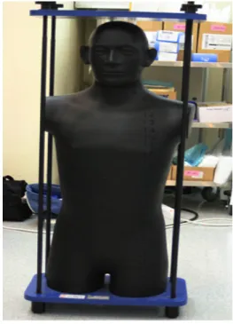
측정에 사용된 인체 모형의 Rando phantom은 건조 시킨 뼈, 폐, 기도, 그리고 조직등가물질의 4가지 성분 으로 이루어져 있어 인체와 동일 구조를 이루고 있다.
이 Rando phantom은 163 cm의 길이와 54 kg의 체중 과 35개의 단면으로 구성되어 있으며 각 단면의 두께는 2.5 cm이다. 각 단면은 5 mm의 직경을 가진 구멍이 15 mm 간격으로 배열되어 있어 열형광 선량계의 삽입 이 가능하도록 제조되었고 또한 조직등가물질의 유효 원자 번호는 인체의 유효 원자 번호 7.4와 유사한 7.3 을 가지고 있다(Fig. 1).
Fig. 1. Rando phantom
2) TLD
실험에 사용된 TLD(GR 200)의 열형광물질(Thermo Luminescence, TL)은 유효 원자 번호가 인체의 원자 번호와 비슷한 인체등가물질이므로 인체에서의 흡수 선 량을 직접 얻을 수 있다. LiF : Mg, Cu, P로 이루어진 TL소자는 인체 팬텀에 장착하기 용이한 형태로써 직경 1 mm, 길이 6 mm의 막대 형태의 소자를 사용하였다.
눈으로 구별이 가능하도록 하기 위해 고유 번호를 부여 하여 각 TLD chip 마다 투명테이프를 붙여 사용하였다 (Fig. 2).
3) TLD시스템
HARSHAW TLD 3500 Dosimeter system을 사용하 였다.
Fig. 2. TLD chip
4) 실험 장비
(1) TACE : Pgilips Allura Xper FD 20 (2) UAE : Pgilips Allura Xper FD 20 (3) TFCA : Philips Integris Allura
2. 측정 이론
1) TL현상은 방사선이 조사된 후 가열이 되면 가시 광선을 방출하는 현상이다.
2) TTP(Time Temperature Profile)는 135℃를 10초 간 예열하고 최대 240℃까지 초당 10℃씩 올려 30초 동안 가열한다
3) RCF(Reader Calibration Factor)는 TL소자에서 방출된 전하량에 대한 정보를 판독자가 읽을 수 있는 선량에 대한 정보로 전환하기 위해 정의된 값이다.
4) ECC(Element Correction Coeffcient)는 모든 선 량계가 한 시스템에서 주어진 방사선에 대해 동일 한 반응도를 가지게 하기 위함이다.
3. 측정 방법
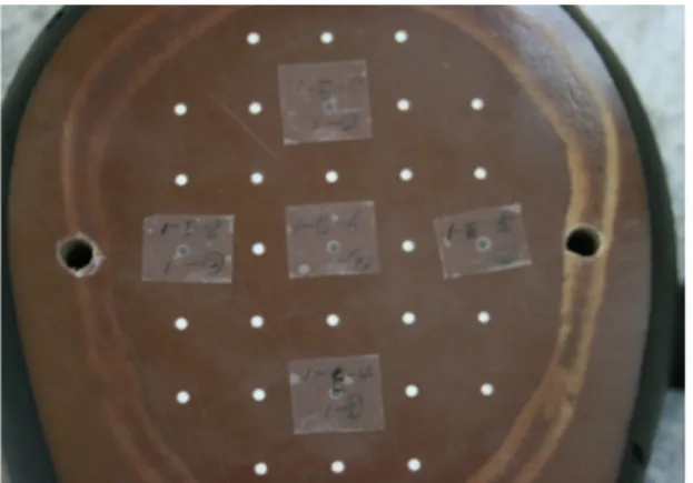
1) 장기 내 TLD 부착(Table 1)(Fig. 3, 4)
TACE TFCA
Right eye skin Right eye skin Thyroid skin Thyroid skin
Chest skin Occipital region skin Abdomen skin Anterior of EAM 1 cm,
Right skin
Gonad skin Anterior of EAM 1 cm, Left skin
Liver organ(9 slice, 17~23
slice, 30 slice) Vertex skin
Table 1. TLD Chip attach Region of TACE and TFCA Procedure
Fig. 3. Rando phantom skin TLD chip attach
Fig. 4. Rando phantom organ TLD chip attach
2) 촬영 조건 및 변화 인자
Rando phantom을 환자 테이블 위에 위치시킨 후에 조사야의 중심과 간의 중심부를 일치시키고 SID를 110 cm로 고정하고 조사야 크기는 복부에서는 42 cm, 두 부에서는 25 cm로 고정 하였다. 이 때 조사 시간은 5 분으로 설정하며 경동맥화학색전요법(Transarterial Chemoembolization : TACE)은 전면상을 조사하고 뇌 혈관조영술(Transfemoral Cerebral Angiography : TFCA) 은 전면상과 측면상을 조사한다(Fig. 5, 6).
Fig. 5. For five minutes X-ray exposure
Fig. 6. APㆍLat X-ray exposure
Skin Liver organ
TLD attach region Dose rate TLD attach region Dose rate
Rt. eye 0.02 9 slice(Thyroid) 0.08
Thyroid 0.074 17 slice 2.67
Chest 0.564 18 slice 5.508
Abdomen 47.092 19 slice 5.792
Gonad 0.03 20 slice 6.532
21 slice(Center) 7.274
22 slice 6.824
23 slice 6.056
30 slice(Gonad) 0.27
Table 2. Result of TACE measurement. (Exposure condition : 76 kVp, 17.2 mA, Unit : mGy/min)
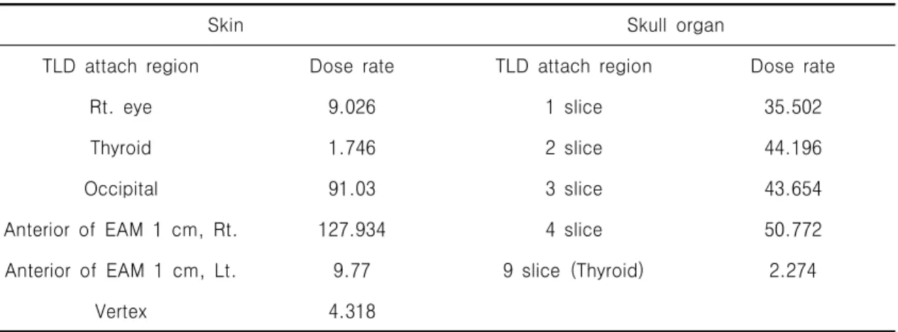
Skin Skull organ
TLD attach region Dose rate TLD attach region Dose rate
Rt. eye 9.026 1 slice 35.502
Thyroid 1.746 2 slice 44.196
Occipital 91.03 3 slice 43.654
Anterior of EAM 1 cm, Rt. 127.934 4 slice 50.772
Anterior of EAM 1 cm, Lt. 9.77 9 slice (Thyroid) 2.274
Vertex 4.318
Table 3. Result of TFCA measurement. (Exposure condition : 81 kVp, 16 mA, Unit : mGy/min)
Ⅲ. 결 과
측정 결과 TACE 시술 시에 각 장기가 받는 선량은 수정체가 0.02 mGy/min로 가장 작게 나타났고, 복부 는 47.092 mGy/min로 가장 높게 나타났다. 간의 중심 부인 21 절편은 7.274 mGy/min이고 5 cm 위쪽인 19 절편은 5.792 mGy/min, 5 cm 아래인 23 절편은 6.056 mGy/min로 중심부보다 낮게 나타났다(Table 2). 뇌혈관조영술(Transfemoral Cerebral Angiography, TFCA) 시 각 장기가 받는 선량 중 갑상선이 1.746 mGy/min로 낮게 나타났으나, 외이도 앞 1 cm 오른쪽 은 127.934 mGy/min로 가장 높고 외이도 앞 1 cm 왼 쪽은 9.77 mGy/min로 오른쪽보다 현저히 낮게 나타났
다. 두개부 장기 내에 부착한 TLD의 선량값은 4 절편 이 50.772 mGy/min로 가장 높고 9 절편인 갑상선은 2.274 mGy/min로 나타났다(Table 3). 그리고 자궁근 종색전술 시 1분당 난소가 받는 선량은 20.68 mGy으 로 타나났다(Table 4).
Ovary organ
TLD attach region Dose rate
30 slice (Ovary) 20.68
Table 4. Result of uterine myoma embolization
measurement. (Exposure condition : 76 kVp, 17.2
mA, Unit : mGy/min)
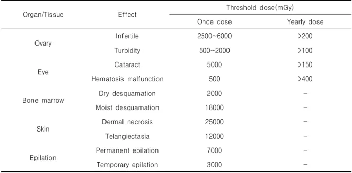
Organ/Tissue Effect
Threshold dose(mGy)
Once dose Yearly dose
Ovary
Infertile 2500~6000 >200
Turbidity 500~2000 >100
Eye
Cataract 5000 >150
Hematosis malfunction 500 >400
Bone marrow
Dry desquamation 2000 -
Moist desquamation 18000 -
Skin
Dermal necrosis 25000 -
Telangiectasia 12000 -
Epilation
Permanent epilation 7000 -
Temporary epilation 3000 -
Table 6. Radiation-induced skin injuries
Ⅳ. 고 찰
의료용 방사선 피폭은 검사를 받은 사람에게는 항상 어떤 확률적 영향을 발생시킬 수 있는 가능성을 지니고 있고 중재적시술을 받는 환자의 경우는 주기적으로 장 시간 방사선 피폭을 받기 때문에 시술자는 최소의 선량 으로 진단적 가치가 있는 영상을 구현하도록 노력해야 한다. 이 실험에서 TACE 시술 시 선량은 조사중심 부 위인 복부 표면과 21 절편에서 선량이 높은 것으로 나 타났다.5~6 우리나라의 TACE 시술 평균 시간은 12.8분 으로 1회 시술 할 경우 578 mGy의 선량을 받게 된다.
여러 번 반복 시술을 하지 않을 경우에는 피폭에 의한 피해는 거의 없다고 할 수 있다. 자궁근종 색전술 시 1 회 단기 조사 할 경우 2시간 이상 시술하게 되면 불임 을 초래할 수 있다. TFCA시술 시 선량은 4 절편과 외 이도 앞 1 cm 오른쪽과 후두부에서 선량이 가장 높은 것으로 확인된다.7~10 외이도 앞 1 cm 오른쪽이 선량이 높게 나온 이유는 왼쪽 측면 방향으로 조사를 했기 때 문이고, 후두부와 4 절편은 조사 중심이기 때문이다.11 TFCA의 평균 시술 시간은 12.7분으로 1회 시술 했을 경우 피폭에 영향은 거의 없다고 할 수 있다. 하지만 뇌혈관중재술에서의 평균 시술 시간은 72.1분으로 약 70분간 선량을 받게 된다면 외이도 앞 1 cm 오른쪽이 8890 mGy, 4 절편이 3500 mGy, 오른쪽 안구가 1100 mGy의 선량을 받게 된다. 이 수치는 영구탈모, 홍반,
가시적 홍탁을 일으킬 수 있다(Table. 6).
즉, 실험에서 TACE 시술 시보다 TFCA, 자궁근종색 전술, 뇌혈관중재술 시에 더 큰 위험이 따르는 것으로 나타났다. 이러한 위험을 최소화하기 위해 선량을 줄일 수 있는 다음과 같은 방법이 있다. 영상 증배관 장비와 비교하여 디지털 디텍터 장비를 사용함으로써 43% 선 량을 감소시킬 수 있고 영상의 확대 정도에 따라 선량 이 5~20% 증가되며, SID를 100 cm를 기준으로 10 cm 감소시키면 선량은 20% 감소되고 반대로 10 cm증 가시키면 30%, 20 cm 증가시키면 57% 증가된다. SOD 를 20 cm 증가 시켰을 때 31% 감소하고 조사야를 30%
감소시키면 선량은 3%, 50% 감소시키면 24%의 선량 감쇄 효과가 있다고 보고된 바 있다.4
Ⅴ. 결 론
TACE는 반복 시행되는 시술이며, 부가적으로 컴퓨터 단층촬영(Computer Tomography, CT)과 같이 시행될 경우 선량은 더욱 증가하게 된다. 또한 뇌혈관중재술은 단 한번의 시술로도 방사선 상해를 일으킬 수 있는 선 량이 발생되고 있다. 이러한 문제를 줄이기 위해 검사 시 여러 가지 기하학적 요소를 변화시켜 선량을 줄이 며, 정확한 시술의 시행으로 환자를 방사선으로부터 보 호해야 한다.
참고문헌
1. International commission on radiation protec- tion. Avoidance of radiation injuries from medical interventional procedures. 2000. Cont- ract No.: ICRP Publication 80.
2. Kim TH, Shin JH, Oh SJ, Park IK, Woo CW, Han KH, et al. Inhibition of Neointimal Hyperplasia after Stent Placement with Rhenium 188-filled Balloon Dilation in a Canine Iliac Artery Model. J Vasc Interv Radiol 2010; 21: 1066-70.
3. Oh SJ, Moon DH, Ha HJ, Park SW, Hong MK, Park SJ, et al. Automation of the synthesis of highly concentrated 188Re-MAG3 for intraco- ronary radiation therapy. Appl Radiat Isot 2001; 54: 419–27.
4. Kandarpa K, Becker GJ, Hunink MG, McNamara TO, Rundback JH, Trost DW, et al. Transca- theter interventions for the treatment of peripheral atherosclerotic lesions: part I. J Vasc Interv Radiol 2001; 12: 683–95.
5. International commission on radiation protec- tion. Avoidance of radiation injuries from medical interventional procedures. 2001. Cont- ract No.: ICRP Publication Supporting Guidance 2.
6. Wolfram RM, Budinsky AC, Pokrajac B, Potter R, Minar E. Vascular brachytherapy with 192Ir after femoropopliteal stent implantation in high risk patients: twelve month follow up results from the Vienna 5 trial. Radiology 2005; 236: 343–51.
7. Hang CL, Fu M, Hsieh BT, Leung SW, Wu CJ, Yip HK, et al. Intracoronary beta irradiation with liquid rhenium-188: results of the Taiwan radiation in prevention of post pure balloon angioplasty restenosis study. Chest 2003; 124:
1284–93.
8. Choi Y, Kang BS, Min JW. A study on the isodose distribution in a vascular characteri- zation room. Korean J Digit Imaging Med 2011; 13: 7-11.
9. Han EO, Kwon DM, Dong KR, Han SM. A Model for Protective Behavior against the Harmful Effects of Radiation based on Medical Institution Classifications. J Korea Asso Radiat Prot 2010; 35: 157-62.
10. Dong KR, Kweon DC, Chung WK, Goo EH, Kevin. D, Choe JH. Study on the angular dependence of personal exposure dosimeter (Focus on thermoluminescent dosimeter and photoluminescent dosimeter). Annals of Nuclear Energy 2011; 38: 383-8.
11. Jung Y, Dong KR, Kweon DC, Kevin D, Goo EH, Ahn SY. A Study on the Effects of Scattering Dose on Eyes and Thyroid for Panoramagraphy (Focus on TLD and PLD). J Korea Asso Radiat Prot 2010; 35: 1-5.